Jennifer E. Phillips-Cremins (upper left) and members of her lab.
Each year, the National Institutes of Health (NIH) recognizes exceptionally creative scientists through its High-Risk, High-Reward Research Program. The four awards granted by this program are designed to support researchers whose “out of the box” and “trailblazing” ideas have the potential for broad impact.
Jennifer E. Phillips-Cremins, Associate Professor and Dean’s Faculty Fellow in Penn Engineering’s Department of Bioengineering and the Perelman School of Medicine’s Department of Genetics, is one such researcher. As a recipient of an NIH Director’s Pioneer Award, she will receive $3.5 million over five years to support her work on the role that the physical folding of chromatin plays in the encoding of neural circuit and synapse properties contributing to long-term memory.
Phillips-Cremins’ award is one of 106 grants made through the High-Risk, High-Reward program this year, though she is only one of 10 to receive the Pioneer Award, which is the program’s largest funding opportunity.
“The science put forward by this cohort is exceptionally novel and creative and is sure to push at the boundaries of what is known,” said NIH Director Francis S. Collins.
Phillips-Cremins’ research is in the general field of epigenetics, the molecular and structural modifications that allow the genome — an identical copy of which is found in each cell — to express genes differently at different times and in different parts of the body. Within this field, her lab focuses on higher-order folding patterns of the DNA sequence, which bring distant sets of genes and regulatory elements into close proximity with one another as they are compressed inside the cell’s nucleus.
Previous work from the Cremins lab has investigated severe genome misfolding patterns common across a class of genetic neurological disorders, including fragile X syndrome, Huntington’s disease, ALS and Friedreich’s ataxia.
With the support of the Pioneer Award, she and the members of her lab will extend that research to a more fundamental question of neuroscience: how memory is encoded over decades, despite the rapid turnover of the relevant proteins and RNA sequences within the brain’s synapses.
“Our long-term goals are to understand how, when and why pathologic genome misfolding leads to synaptic dysfunction by way of disrupted gene expression,” said Phillips-Cremins, “as well as to engineer the genome’s structure-function relationship to reverse pathologic synaptic defects in debilitating neurological diseases.”
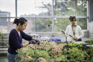
Sophomores Linda Wu and Nova Meng spent the summer studying coevolution among plants, mutualistic bacteria, and parasitic nematodes in Corlett Wood’s biology lab.
To study coevolution, the responsibilities of Nova Meng and Linda Wu included caring for plants in the Penn greenhouse. (Image: From July 2021, when masks were not required)
Coevolution is all around us. Think of the elongated blooms that perfectly accommodate a hummingbird’s slender mouth parts. But not all examples of species influencing one another’s evolutionary course accrue benefits to all parties. Tradeoffs are part of the game.
This summer, sophomores Linda Wu of Annandale, Virginia, and Nova Meng of Akron, Ohio, researched an coevolutionary scenario with benefits as well as costs for the species involved. Their work, supported by the Penn Undergraduate Research Mentoring Program (PURM) and conducted in the lab of biology professor Corlett Wood, has examined the relationship among plants in the genus Medicago, beneficial bacteria that dwell in their roots, and parasitic nematodes that try to steal the plants’ nutrients.
The Center for Undergraduate Research & Fellowships provides students in the PURM program awards of $4,500 during the 10-week summer research internship. Wu and Meng stayed busy through those weeks. Whether evaluating plants in a soybean field in Michigan or tending to hundreds—even thousands—of plants in the greenhouse at Penn, these aspiring researchers built a foundation for future scientific endeavors with hands-on practice.
“It’s been an amazing experience,” says Wu. “I’ve always been interested in genetics and evolution and have found parasitic relationships in particular really interesting. I like reading about weird parasites. This summer I’ve gotten to participate in lab meetings, read books about coevolution, and expand my knowledge about the topic.”
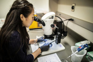
Mentored by Ph.D. student McCall Calvert, Wu spent the summer focused on the parasites in the Medicago model system the Wood lab uses. “I’m trying to see if those nematodes are specialists or generalists, if they’re locally adapted to their host plant or open to parasitizing on different species,” Wu says.
To do so, she’s grown pots and pots of plants in the Penn greenhouse, experimentally infecting Medicago plants as well as other species, such as carrot and daisy plants, with nematodes, to measure the degree to which the parasites flourish.
Meng, who is pursuing a bioengineering major, is examining how bacteria that dwell in plant roots affect the plants’ susceptibility to parasites.
Meng’s project looked at the bacterial side of the coevolutionary relationship. Overseen by lab manager and technician Eunnuri Yi, Meng looked at four strains of bacteria, known as rhizobia. Two strains are nitrogen-fixing, giving their associated plants a crucial nutrient to promote growth, while the other two do not seem to contribute nitrogen to the plants, and instead exist as parasites in the plants’ roots. “I’m looking at what happens when we infect the plants with nematode parasites,” Meng says, “to see if the plants that are open to mutualistic rhizobia are more susceptible to the nematode parasites.”
Linda Wu is a sophomore pursuing an uncoordinated dual degree in business, energy, environment, and sustainability in the Wharton School and in biology with a concentration in ecology and evolution in the College of Arts and Sciences at the University of Pennsylvania.
Nova Meng is a sophomore majoring in bioengineering in the School of Engineering and Applied Science at Penn.
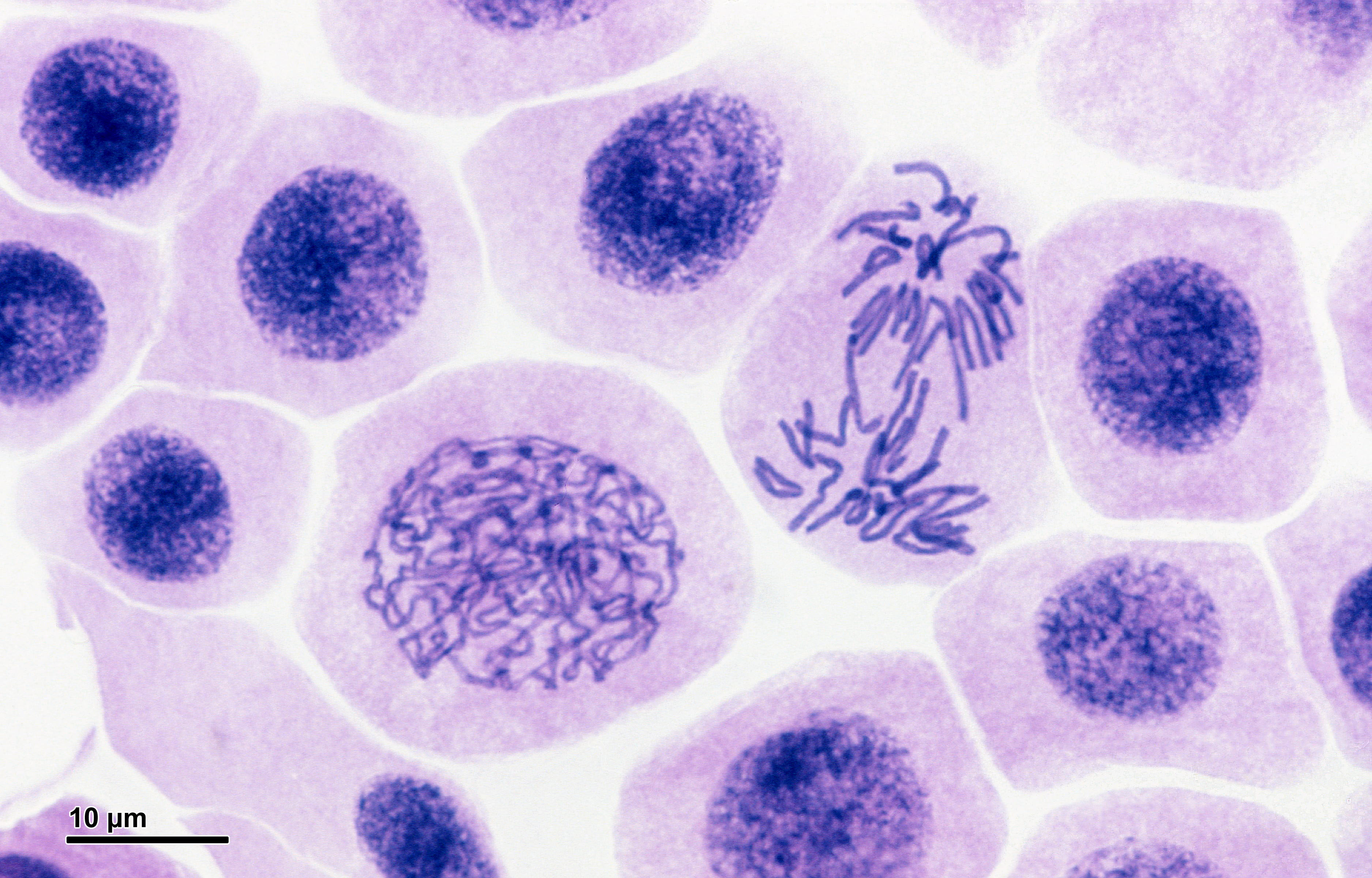
A collaborative study conducted by researchers at the Children’s Hospital of Philadelphia (CHOP), Penn Engineering and Pennsylvania State University has uncovered new information about how chromosomal material in cell nuclei reorganizes itself after cell division.
While a deep understanding of the cell cycle is a cornerstone of biology and health sciences, research into the complex relationship between three-dimensional chromatin structure and gene transcription is still in its infancy. The results of this study will contribute to a more robust understanding of chromatin rebuilding after mitosis and potentially aid in the treatment of genetic diseases.
Jennifer E. Phillips-Cremins, Ph.D.
Jennifer E. Phillips-Cremins, Assistant Professor in the Department of Bioengineering, contributed to the study alongside Gerd A. Blobel, Frank E. Weise III Endowed Chair in Pediatric Hematology at CHOP and Ross C. Hardison, an expert in gene regulation at Penn State.
Phillips-Cremins’ research uses genetic engineering approaches to discover the mechanisms regulating chromatin organizing principles in cells, as well as computational approaches to investigate cellular function. Her lab’s techniques provide ways of mapping the three-dimensional organization of genes while they are folded together in the genome and how those spatial relationships impact gene expression.
The research team performed their experiments in blood-forming cells from a well-established mouse model. They used sophisticated techniques called high throughput chromosome conformation capture (Hi-C) that detect and map interactions across three-dimensional space between specific sites in chromosomal DNA. These maps also allowed the scientists to measure such interactions at different time points in the cell cycle. In all, the tools detected roughly 2 billion interactions during mitosis and thereafter, when the daughter nuclei are rebuilt.
Members of the Cremins Lab, Daniel J. Emerson, Thomas G. Gilgenast and Katelyn R. Titus, also contributed to the study, which was published in Nature.
Bio-inspiration Informs New Football Helmet Design from IUPUI Students
Art, design, biology, and engineering all interact with each other in a recent design for a football helmet from two students— one of media arts and the other of engineering — at the Indiana University – Purdue University Indianapolis. Directed by Lecturer in Media Arts and Science Zebulun Wood, M.S., and Associate Professor of Mechanical and Energy Engineering and Assistant Professor of Biomedical Engineering Andres Tovar, Ph.D., the students found inspiration in biological structures like a pomelo peel, nautilus shell, and woodpecker skull to create energy-absorbing helmet liners. The resulting design took these natural concussion-reducing structures and created compliant mechanism lattice-based liners the replace the foam traditionally placed in between two harder shells of a typical helmet. Their work not only exemplifies the benefits of bio-inspiration, but demonstrates the way that several different domains of study can overlap in the innovation of a new product.
Study of Mechanical Properties of Hyaluronic Acid Could Help Inform Current Debates Over Treatment Regulation for Osteoarthritis
Arthritis is an extremely common condition, especially in older patients, in which inflammation of the joints can cause high amounts of stiffness and pain. Osteoarthritis in particular is the result of the degradation of flexible tissue between the bones of a joint, which increases friction in joint motion. A common treatment of this form of arthritis is the injection of hyaluronic acid, which is meant to provide joint lubrication, and decreases this friction between bones. Recently, however, there has been a debate over hyaluronic acid’s classification by the FDA and whether it should remain based on the knowledge of the mechanical actions of the acid in treatment for osteoarthritis or if potential chemical action of the acid should be considered as well.
Because of limited ways of testing the mechanical properties of the acid, many researchers felt that there could be more to hyaluronic acid’s role in pain relief for arthritic patients. But Lawrence Bonassar, Ph.D., the Daljit S. and Elaine Sarkaria Professor in Biomedical Engineering at the Meinig School of Bioengineering of Cornell University, had another idea. With his lab, he created a custom-made tribometer to measure the coefficient of friction of a given lubricant by rubbing a piece of cartilage back and forth across a smooth glass plate. The research demonstrated that hyaluronic acid’s ability to reduce the coefficient of friction aligned with patients’ pain relief. Bonassar and his team hope that these results will demonstrate the heavy contribution of mechanical action that hyaluronic acid has in osteoarthritis treatment, and help bring an end to the debate over its FDA classification.
A New Way of Mapping the Heart Could Lead to Better Understanding of Contractile Activity
Though reduced contractions in certain regions of the heart can be an indicator of a certain condition, there is currently no way to directly measure contractile activity. This is why Cristian Linte, Ph.D., an Associate Professor of Biomedical Engineering in the Kate Gleason College of Engineering at the Rochester Institute of Technology (RIT), hopes to create a map of the heart that can quantify contraction power. In collaboration with Niels Otani, Ph.D., an Associate Professor in the School of Mathematics at RIT, Linte plans to use an $850,000 grant from the National Science Foundation to achieve a more comprehensive understanding of the heart through both medical imaging and mechanical modeling. The group hopes that their approach will lead to not only a better way to diagnose certain heart conditions and diseases, but also open up understanding of active contraction, passive motion, and the stresses within the heart walls that underlie each.
Celebrity Cat Lil Bub Helps Penn and German Researchers Draw Public Attention to Genetics
Lil Bub’s unique appearance has garnered millions of online fans, and now, an avenue for researchers to talk about genetics. (Photo Courtesy of Mike Bridavsky)
In 2015, a group of curious researchers set out to sequence the genome of a celebrity cat named Lil Bub. They were hoping to understand the genetics behind Lil Bub’s extra toes and unique skeletal structure, which contribute to her heart-warming, kitten-like appearance. However, an equally important goal of their “LilBUBome” project was to invite the general public into the world of genetics.
Because of Lil Bub’s online fame, the project garnered attention from her fans and the media, all hoping to discover the secret to Lil Bub’s charm. As early as 2015, Gizmodo’s Kiona Smith-Strickland reported on the team’s intentions to sequence Lil Bub’s genome, and, since then, many have been awaiting the results of the LilBUBome.
The Alfred P. Sloan Foundation awarded a six-year grant to Barnard College and Columbia University’s School of Engineering and Applied Science to support graduate education for women in engineering. The funding will go towards a new five-year program that enables Barnard students to attain both a B.A. and M.S. in one year after their traditional four years of undergraduate education. The program will offer M.S. degrees in chemical engineering, biomedical engineering, and industrial engineering and operations research, and is one of the first of its kind for women’s colleges.
We would like to congratulate Jean Paul Allain, Ph.D., on being named the first head of the new Ken and Mary Alice Lindquist Department of Nuclear Engineering at Penn State. Allain, who is currently a Professor and head of graduate programs in the University of Illinois at Urbana-Champaign’s Department of Nuclear, Plasma, and Radiological Engineering, conducts research in models of particle-surface interactions. In addition to being head of the new department at Penn State, Allain will also hold a position as a Professor of Biomedical Engineering at the university.
We would also like to congratulate Andrew Douglas, Ph.D., on his appointment as the Vice Provost for Faculty Affairs at Johns Hopkins University. Douglas currently holds the position of Vice Dean for Faculty at the Whiting School of Engineering, and has joint appointments in Mechanical and Biomedical Engineering. Douglas’s research at Hopkins focuses on mechanical properties and responses of compliant biological tissue and on the nonlinear mechanics of solids, with a focus on soft tissues and organs like the heart and tongue.
Online Tool for 3D Visualization of Gene Mutations
DNA inside cell nuclei undergoing the process of mitosis.
Fifteen years ago marked a major milestone in the Human Genome Project: scientists successfully sequenced all of the base pairs in our 23 sets of chromosomes. Following this accomplishment, researchers assembled generations of mathematical models to understand how gene mutations result in disease. A key barrier in developing these models is the size of genome itself: a single human genome requires approximately 2 GB of storage, and many studies examine thousands of genomes to detect changes in a small number of patients. Both processing these large datasets and efficiently storing them create challenges. Making these model predictions accurate and complete is another challenge.
Scientists collaborating among several universities on three continents developed an online computational tool to help overcome these barriers. The scientists, who include Bernhard Palsson, Ph.D., Galletti Professor of Bioengineering at University of California, San Diego, as one of the lead authors, report on the resource in a recent issue of Nature Biotechnology.
Called Recon3D, the new resource provides a metabolic network model using approximately 17% of known human genes. The model combines data on the genes, metabolites, proteins, and metabolic reactions for human metabolism. In addition, as the model’s name implies, Recon3D accounts for the physical structure of model components, imporving significantly on past models that relied on linear, two-dimensional models. Although the model still has 83% of genes left to incorporate, it could ultimately unravel some of the mysteries underlying virtually any disease with a genetic cause, from inborn errors of metabolism to cancer.
Bioengineering for Refugees
As the war in Syria enters its seventh year, at least five million refugees have left the country to seek asylum elsewhere. Roughly 20% of the refugees are now in Lebanon, where many reside in refugee camps. Although these refugees are now much safer than before, even in the best of circumstances, the conditions in refugee camps can compromise health and wellness.
Engineering can offer relief for some of these conditions. A three-week course offered in January at the American University of Beirut, co-designed and taught by Muhammad Zaman, Ph.D., Professor of Biomedical Engineering at Boston University, and entitled “Humanitarian Engineering: Designing Solutions for Health Challenges in Crises,” had students devising solutions to the issues facing these refugees.
Among the ideas generated by the students was “3D Safe Water” – a device designed to detect the contamination of water, decontaminate it, and deploy the technology in low resource settings. The device uses sensors to detect contamination and chlorine to decontaminate. With water-borne diseases taking an especially hard toll on camps like these, the device could significantly improve living conditions for refugees.
Placenta on a Chip
Organ-on-chip technologies use microfluidics to model organs or organ systems. So far, engineers have developed chip-based models of the lungs, heart, and kidneys, as well as the circulatory system.
The most recent addition to the organ-on-chip family is the placenta-on-a-chip, developed by Dan Huh, Ph.D., Wilf Family Term Assistant Professor of Bioengineering at the University of Pennsylvania. Modeling the organ that mediates and communicates between a pregnant woman and the fetus, Dr. Huh created a chip to study how drugs move from the bloodstream of the mother to the fetus. With this knowledge, one could determine more safely and more accurately how drugs taken by the mother can affect a pregnancy.
People and Places
Two colleges have announced new biomedical engineering programs. George Fox University, a Christian college in Oregon, will offer a BME concentration for engineering majors starting this fall. On the other side of the country, Springfield Technical Community College in western Massachusetts will offer a two-year associate’s degree in BME technology.
The University of Arizona, in cooperation with the City of Phoenix, will launch a new medical technology accelerator program, to be called InnoVention. It will be located on UofA’s Phoenix Biomedical Campus. Frederic Zenhausern, PhD, MBA, Professor of Basic Medical Sciences and Director of the Center for Applied NanoBioscience and Medicine at Arizona, is among the people leading the effort.
Finally, Distinguished Professor Craig Simmons of the University of Toronto’s Institute of Biomaterials and Biomedical Engineering is among 10 awardees sharing a $3.5 million grant (approximately $2.7 million in U.S. currency) for the development of medical devices and technologies. Dr. Simmons, a former postdoc at Penn, will use his funding to investigate the use of stem cells to repair congenital heart defects in infants.
by Meagan Ita, Ph.D. Student in Bioengineering and GABE Co-President
Cancer is a disease that affects millions, and over the last several decades, researchers have delved deeply into the biological underpinnings of the disease in the hopes of finding a cure. One major discovery is that mistakes in your DNA “instructions” can lead to cancer by crossing the wires in your cellular circuitry, and researchers have developed amazing new drugs that can cause tumors to melt away by targeting these broken components. The problem though is that, most of the time, the tumors come back, and this is a huge barrier to cures.
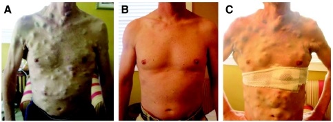
Picture of patient treated with vemurafenib and then developing resistance. Courtesy of the American Society of Clinical Oncology
For a long time, everyone assumed that the reason the tumors came back was DNA mistakes on top of the original mistakes, with these new mistakes blocking the activity of the anti-cancer drug. However, new work led by Sydney Shaffer from the Arjun Raj Lab at Penn Bioengineering, published this week in Nature, challenges this view by looking all the way down at individual cancer cells and seeing how they respond to these drugs on a cell-by-cell basis.
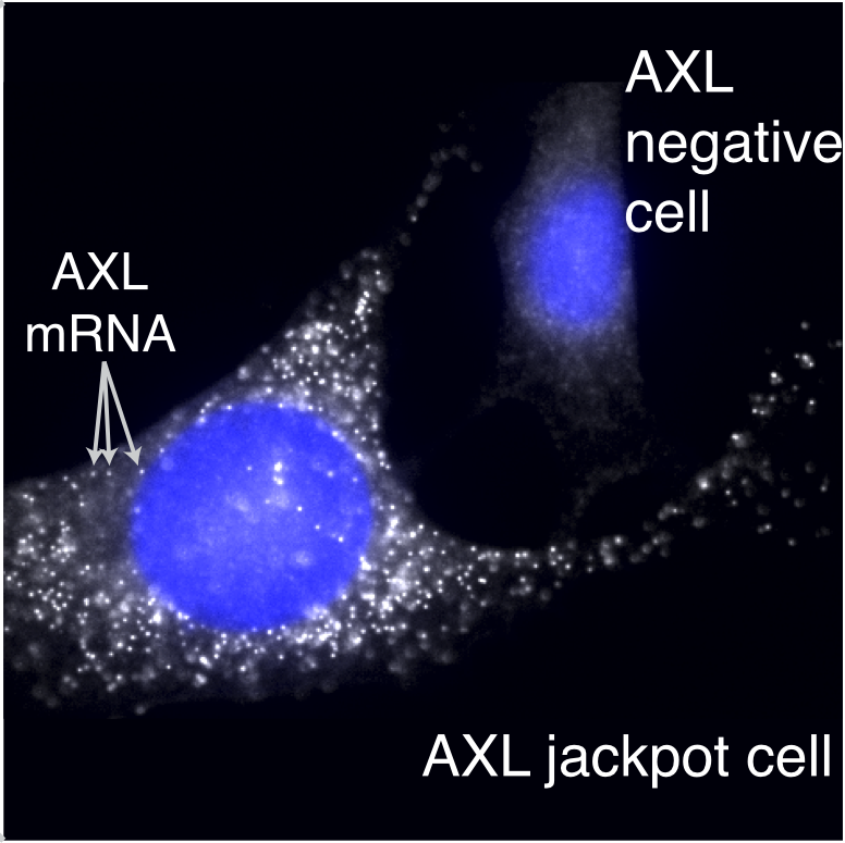
Sydney found that in melanoma, contrary to what researchers thought, it need not be a DNA mistake that leads a cell to become resistant to the drug, but rather a change in cellular identity. Just like your body has cells of all different types, like skin cells and brain cells, cancer cells appear to change between different types, but unlike in the body, cancer cells do it in a seemingly random and uncontrolled way, and the cells exploit this variability to allow those rare cells that have changed their type to survive the drug.
Here, we talk with Sydney about the inspiration, triumphs, and challenges she faced in her research.
What was the initial inspiration for looking at drug resistance in melanoma?
For the first two years of working on this project, we actually didn’t have a clear question in mind. I was just trying a bunch of different experiments with melanoma cells, and I noticed something that we found thought-provoking. Whenever we gave the melanoma cells a particular drug, they would become resistant at exactly the same point in time. At first, this may not seem unusual, but for example, if everyone showed up at a restaurant to eat lunch at exactly noon, you would guess this was not happening purely by chance. Maybe classes let out right beforehand? Or a big meeting? For the melanoma cells, we would similarly expect there to be a range of different times for the cells to become resistant, but instead it all happened at once.
This observation helped us figure out that the drug-resistant cells probably already exist before we treat them. It also got us curious about the particular processes that make the cells resistant, and we spent many lab meetings discussing this observation until one postdoc, Gautham Nair, suggested trying some experiments based upon the classical molecular biology experiments of Luria and Delbrück.
Who were Luria and Delbrück, and how did they influence your work?
Max Delbrück and Salvador Luria (below) were scientists who, in the 1940s, performed a clever experiment that demonstrated that bacteria become resistant to viruses through random DNA mutations. According to Wikipedia, Luria actually had the idea for these experiments while watching slot machines!
Delbrück (left) and Luria. Courtesy of the Genetics Society of America.
Their experiment was super simple: it was basically a statistical way to see whether cells “sense and respond” to a challenge, or whether they just passively get a mutation that lets some fraction of them survive the challenge, basically like Darwinian evolution. The idea is that, in the first scenario, there is no history: every cell has an equal chance to respond when challenged.
But in the second scenario, history matters in that if your great-grandparent was a survivor, then all your relatives would be too. If you could redo history over and over, then sometimes maybe your great-great-great-great-grandparent would be a survivor, and so you would get a whole bunch of survivors when the challenge came. Luria and Delbrück’s results showed that this second scenario was what happened with bacteria, providing the first evidence for genetic mutations in bacteria occurring in the absence of selection, and they both went on to win a Nobel prize in 1969.
Arjun actually had just lectured about these experiments in our graduate course on modeling biological systems. We adapted the same strategy and theory as Luria and Delbrück’s experiments for our work but applied it to melanoma and actually found a different result. Our experiments showed that resistance in melanoma does not arise through a heritable DNA mutation.
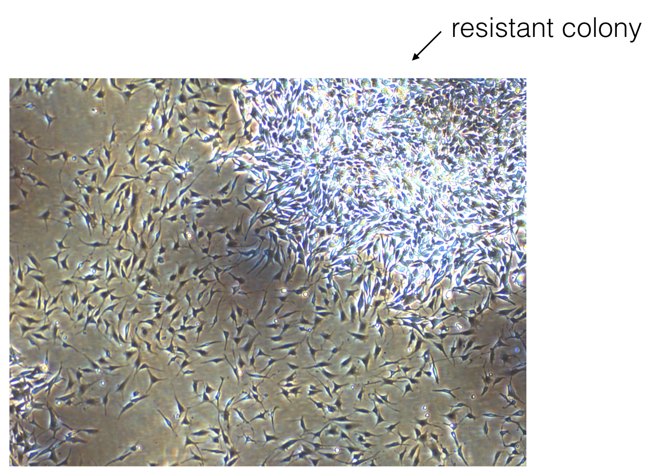
Picture of a resistant colony growing in the Raj lab.
Based upon this work, do you have any ideas for how we might prevent resistance in patients?
Yes. The recommended dosing for many of these drugs is daily. Our work would suggest that something like interval therapy might be more effective, for instance, if you gave the drug for a few days, killed many of the tumor cells, and then stopped the drug. During the time that the drug is stopped, the cells that initially survived the drug (we call these cells pre-resistant) could then transition out of this cell state and back to a sensitive state. Then, when the patient takes the drug again, it would be more effective at killing the remaining tumor cells. Another idea would be to find drugs that are specific to the pre-resistant cells and give these drugs in combination with other targeted therapies.
Were there any “Aha!” moments while working on this project?
One of the most exciting moments of this research was when we first found the pre-resistant cells. Hidden among thousands of pictures of empty cells, we were shocked to actually see the rare cells full of brightly tagged resistance genes (below).
Resistant cells growing in the Raj lab.
What were some low points in working on the project? Do you recall any specific moments that you just felt intellectually and/or emotionally stumped? How did you get through them?
Oh yes, there were definitely low points during this project. One that stands out to me specifically was this one Friday afternoon where I presented at lab meeting. At the time, I only had a little bit of preliminary data. One of the members of the lab asked me a series of questions about resistance: How many different drug doses had I tried? Could I just give a lot and kill them all? What dose of drug is relevant for patients? What about drug resistance? Was I really interested in? All reasonable questions to ask. However, this was really overwhelming to a first-year graduate student because it made me realize that I didn’t have a clearly defined project that I was working on yet. There were just so many different questions that I didn’t know where to start.
Ultimately, with Arjun’s guidance, I came to realize that this was part of the process of figuring out what my thesis project would be, and the vagueness of our ideas at this time was a great thing because it left me open to find a problem that I found really interesting.
At another point in working on this: I remember that we were clearly conceptually stuck. We had identified the rare cells, but it wasn’t clear how to find out if these were the same cells that become resistant to drug. I had an entire lab meeting where we discussed this concern and came to the conclusion that, without some connection between the cells in this state and resistance, the work would be very speculative, which felt unsatisfying to me. Unfortunately, there wasn’t a quick fix to this problem. We just ended up trying a whole bunch of different ideas and eventually one of our strategies worked out.
Were there any funny moments that stand out to you?
Yeah! I was 40 weeks pregnant as we were finishing off our first submission of the paper! As my due date passed, I was really feeling the pressure to finish everything. Each day, I was coming into lab and just hoping I wouldn’t go into labor yet! Actually, the members of our lab had placed bets on when the baby would be born. Fortunately, those who bet on a late arrival ended up winning, and we submitted the paper the day before my daughter, Julien, was born. I was actually still at the hospital when I got the e-mail that the paper went to review.
So even though it might seem like this project is checked off the list with a kick-ass publication, there are probably a bunch of unfinished ideas you have. So,what are you working on next? Will this project ever be “done?”
For sure. The list of unfinished ideas is very long, and some of the questions that came from this work are now being pursued by other people in the lab. Right now, I’m working on ways of measuring the length of time that individual cells remain in these different cell states.
Interested in sharing your research in Penn BE? Contact penngabe@gmail.com for an interview by GABE (Graduate Association of Bioengineers) and let us know!


 Art, design, biology, and engineering all interact with each other in a