The effectiveness of CAR T cell therapy against a variety of cancers, including solid tumors, could be boosted greatly by using CRISPR-Cas9 technology to knock out the gene for CD5, a protein found on the surface of T cells, according to a preclinical study from investigators at the University of Pennsylvania’s Perelman School of Medicine and Abramson Cancer Center.
CAR T cells are T cells that have been engineered to attack specific targets found on cancer cells. They have had remarkable results in some patients with blood cancers. But they have not performed well against other cancers including solid-tumor cancers, such as pancreatic cancer, prostate cancer, and melanoma. Researchers have been searching for techniques to boost the effectiveness of CAR T cell therapy.
The study, published today in Science Immunology, suggests that knocking out CD5 could be a prime technique. Illuminating the protein’s previously murky role, the researchers found that it works as a powerful immune checkpoint, reining in T cell effectiveness. Removing it, they showed, dramatically enhanced CAR T cell anticancer activity in a variety of preclinical cancer models.
“We’ve discovered in preclinical models that CD5 deletion greatly enhances the function of CAR T cells against multiple cancers,” said senior author Marco Ruella, MD, an assistant professor of Hematology-Oncology, researcher with the Center for Cellular Immunotherapies and the scientific director of Penn Medicine’s Lymphoma Program. “The striking effects we observed across preclinical models suggest that CD5 knockout could be a general strategy for enhancing CAR T cell function.”
The study’s first author is Ruchi Patel, PhD, a recent graduate student from the Ruella Laboratory.
Sherry (Xue) Gao, Presidental Penn Compact Associate Professor in Chemical and Biomolecular Engineering and in Bioengineering
Sherry (Xue) Gao, Presidential Penn Compact Associate Professor in Chemical and Biomolecular Engineering (CBE), always knew she had a future in the lab. “I grew up in China, and when I was little, maybe six or seven,” she recalls, “my teacher asked me, ‘What do you want to be when you grow up?’ I said, ‘I want to be a scientist!’”
Neither of her parents had studied beyond high school; when Gao finished her training as a chemical engineer, she became the first person in her family to graduate from college. “One of my greatest motivations is to help first-generation college students,” Gao says.
Now, as the newest faculty member in CBE, Gao is prepared to do just that: support the next generation of chemical engineers, while also conducting groundbreaking research in the development of small molecules to edit genes, pushing the boundaries of precision medicine.
The Presidential Penn Compact Professorships were created by former Penn President Amy Gutmann specifically to recruit and support faculty like Gao: transformative leaders working at the intersection of multiple fields with “a yen for collaboration,” as Gutmann told the Penn Gazette in 2021.
Gao joins Penn Engineering from Rice University, where she collected numerous accolades, including the 2024 BMES-CMBE Rising Star Award, a 2022 NSF CAREER Award, the 2022 Outstanding Young Faculty at Rice School of Engineering Award and the 2020 NIH MIRA R35 Award.
As a member of the Center for Precision Engineering for Health (CPE4H), Gao will partner with colleagues from across the School to develop technologies that bridge disciplines, all in the interest of advancing health care. “We are very excited to have Sherry as a new member of the Center,” says Daniel Hammer, Alfred G. and Meta A. Ennis Professor and inaugural Director of CPE4H. “Gene editing is an important new tool that can precisely alter cell behavior by deleting or redirecting cell pathways, as well as enhancing and suppressing gene expression. She will have significant interactions with other members of the Center, such as Mike Mitchell and myself, as well as the broader Penn community, especially with the CAR therapists.”
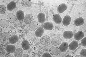
A group of bacteriophages, viruses that infect bacteria, imaged using transmission electron microscopy. New research sheds light on how bacteria fight off these invaders without triggering an autoimmune response. (Image: ZEISS Microscopy, CC BY-NC-ND 2.0)
During the last few years, CRISPR has grabbed headlines for helping treat patients with conditions as varied as blindness and sickle cell disease. However, long before humans co-opted CRISPR to fight genetic disorders, bacteria were using CRISPR as an immune system to fight off viruses.
In bacteria, CRISPR (Clustered Regularly Interspaced Short Palindromic Repeats) works by stealing small pieces of DNA from infecting viruses and storing those chunks in the genes of the bacteria. These chunks of DNA, called spacers, are then copied to form little tags, which attach to proteins that float around until they find a matching piece of DNA. When they find a match, they recognize it as a virus and cut it up.
Now, a paper published in Current Biology by researchers from the University of Pennsylvania Department of Physics and Astronomy shows that the risk of autoimmunity plays a key role in shaping how CRISPR stores viral information, guiding how many spacers bacteria keep in their genes, and how long those spacers are.
Ideally, spacers should only match DNA belonging to the virus, but there is a small statistical chance that the spacer matches another chunk of DNA in the bacteria itself. That could spell death from an autoimmune response.
“The adaptive immune system in vertebrates can produce autoimmune disorders. They’re very serious and dangerous, but people hadn’t really considered that carefully for bacteria,” says Vijay Balasubramanian, principal investigator for the paper and the Cathy and Marc Lasry Professor of Physics in the School of Arts & Sciences.
Balancing this risk can put the bacteria in something of an evolutionary bind. Having more spacers means they can store more information and fend off more types of viruses, but it also increases the likelihood that one of the spacers might match the DNA in the bacteria and trigger an autoimmune response.
Vijay Balasubramanian is the Cathy and Marc Lasry Professor of Physics at the Department of Physics and Astronomy of the University of Pennsylvania, a visiting professor at Vrije Universiteit Brussel, and a member of the Penn Bioengineering Graduate Group.
Speaker: Weihua Guan, Ph.D.
Assistant Professor
Department of Electrical Engineering & Department of Biomedical Engineering (courtesy)
Pennsylvania State University, University Park
Date: Thursday, April 8, 2021
Time: 3:00-4:00 PM EDT
Zoom – check email for link or contact ksas@seas.upenn.edu
Abstract:
Due to their conceptual simplicity, the nanopore sensors have attracted intense research interest in electronic single molecule detection. While considerable success has been achieved, the solid-state nanopores still face three significant challenges, including repeatable nanopore size control, introduction sensing specificity, and prolonged sensor response time at low concentrations. In this talk, I will discuss a calibration-free solid-state nanopore counting method and two representative applications in nucleic acid testing. One is an isothermal amplification-coupled nanopore counting for malaria analysis. The other is the CRISPR-cas12a-coupled nanopore counting for HIV analysis. Finally, I will also discuss how we can develop a fully integrated ‘sample-to-result’ nucleic acid testing device using the solid-state counting strategy. I believe the reaction-coupled solid-state nanopore digital counting could open a new avenue towards compact, robust, low-cost electronic nucleic acid testing at the point of care.
Weihua Guan Bio:
Weihua Guan received his Ph.D. degree in Electrical Engineering from Yale University in 2013 and did his postdoctoral training at Johns Hopkins University from 2013 to 2014. He joined the Department of Electrical Engineering at Pennsylvania State University in Jan 2015. He also held a courtesy appointment in the Department of Biomedical Engineering at Penn State. Dr. Guan’s research interests are in the multidisciplinary areas of micro- and nanotechnology, micro/nanofluidics, bioMEMS, lab-on-a-chip devices, and point-of-care devices. Dr. Guan’s research group at Penn State focuses on developing micro and nanoscale devices as well as novel sensing principles towards advanced medical diagnostics and testing. Dr. Guan is a member of IEEE, Engineering in Medicine and Biology Society, Biophysical Society, and American Physics Society. Among other honors, Dr. Guan is a recipient of the HHMI International Research Fellowship and NSF CAREER award.
Jennifer Phillips-Cremins, Ph.D., was recently promoted to the tenured position of Associate Professor in Penn’s Department of Bioengineering. Cremins, leads a lab on campus in 3D Epigenomes and Systems Neurobiology.
In a recent piece profiling top technologies to watch in 2020, Cremins spoke to Nature about which technological trends she saw as being important for the year to come. In the panel, which highlighted perspectives from a panel of researchers across several fields, Cremins discussed the increasing relevance of innovations that would allow researchers to study the way that folding patterns within the human genome can influence how genes are expressed in healthy individuals and misregulated in human disease.
One such innovation is actually employed by the Cremins Lab: light-activated dynamic looping (LADL). This technique uses both CRISPR/Cas9 and optogenetics to induce folding patterns into the genome on demand, using light as a trigger. In doing so, Cremins and her fellow researchers can more efficiently study the patterns of the human genome, and what effects certain folding patterns can have on the gene expression state of the cell.
Now, with her new promotion, Cremins can continue advancing her research in understanding the genetic and epigenetic mechanisms that regulate neural connections during brain development, with a focus on how that understanding can eventually lead to better treatments of neurological disease. Beyond the lab, she’ll now lead a new Spatial Epigenetics program, bringing together scientists across Penn’s campus to understand how the spatial connections between biomolecules influence biological behavior. She will also continue teaching her hallmark course for Penn Bioengineering undergraduate students, Biological Data Science, and her more advanced graduate-level course in epigenomics. Congratulations, Dr. Cremins!
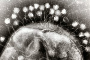
Transmission electron micrograph of multiple bacteriophages, viruses that infect bacteria, attached to a cell wall. New research describes how bacteria can optimize their “memory” of past viral infections in order to launch an effective immune response against a new invader. (Image: Graham Beards)
Before CRISPR became a household name as a tool for gene editing, researchers had been studying this unique family of DNA sequences and its role in the bacterial immune response to viruses. The region of the bacterial genome known as the CRISPR cassette contains pieces of viral genomes, a genomic “memory” of previous infections. But what was surprising to researchers is that rather than storing remnants of every single virus encountered, bacteria only keep a small portion of what they could hold within their relatively large genomes.
In recent years, CRISPR has become the go-to biotechnology platform, with the potential to transform medicine and bioengineering. In bacteria, CRISPR is a heritable and adaptive immune system that allows cells to fight viral infections: As bacteria come into contact with viruses, they acquire chunks of viral DNA called spacers that are incorporated into the bacteria’s genome. When the bacteria are attacked by a new virus, spacers are copied from the genome and linked onto molecular machines known as Cas proteins. If the attached sequence matches that of the viral invader, the Cas proteins will destroy the virus.
Bacteria have a different type of immune system than vertebrates, explains senior author Vijay Balasubramanian, but studying bacteria is an opportunity for researchers to learn more about the fundamentals of adaptive immunity. “Bacteria are simpler, so if you want to understand the logic of immune systems, the way to do that would be in bacteria,” he says. “We may be able to understand the statistical principles of effective immunity within the broader question of how to organize an immune system.”
This research was supported by the Simons Foundation (Grant 400425) and National Science Foundation Center for the Physics of Biological Function (Grant PHY-1734030).
Hammer will offer a course on COVID-19 and the coronavirus pandemic during Penn’s Summer II session, which will be held online this year. The course will be co-taught with Miriam Wattenbarger, senior lecturer in CBE.
The course, “Biotechnology, Immunology, and COVID-19,” will culminate with a case study of the coronavirus pandemic including the types of drugs proposed and their mechanism of action, as well as the process of vaccine development.
“Obviously, the pandemic has been a life-altering event, causing an immense dislocation for everyone in our community, especially the students. Between me and Miriam, who has been trumpeting the importance of vaccines for some time in her graduate-level CBE courses, we have the expertise to inform students about this disease and how we might combat it,” says Hammer.
For more than ten years, Wattenbarger has run courses and labs focused on drug delivery and biotechnology, key elements of the vaccine development process.
“I invite both researchers and industry speakers to meet with my students,” Wattenbarger says, “so that they learn the crucial role engineers play in both vaccine development and manufacturing.”
Beyond studying the interactions between the immune system and viruses — including HIV, influenza, adenovirus and coronavirus — students will cover a variety of biotechnological techniques relevant to tracking and defending against them, including recombinant DNA technology, polymerase chain reaction, DNA sequencing, gene therapy, CRISPR-Cas9 editing, drug discovery, small molecule inhibitors, vaccines and the clinical trial process.
Students will also learn the mathematical principles used to quantify biomolecular interactions, as well as those found behind simple epidemiological models and methods for making and purifying drugs and vaccines.
“We all have to contribute in the ways that we can. Having taught biotechnology to freshmen for the past decade, this is something that I can do that can both inform and build community,” says Hammer. “Never has it been more important to have an informed and scientifically literate community that can fight this or any future pandemic.”
Nature, one of the world’s most prestigious scientific journals, recently reached out to a panel of researchers from a variety of fields, asking them what technological trends they see as having the most impact on their disciplines in the coming year.
Jennifer Phillips-Cremins, assistant professor in the Department of Bioengineering, was among these panelists. As an expert in “3D epigenetics,” or the way the genome’s highly specific folding patterns influence how and when individual genes are expressed, she highlighted a slate of new techniques that will allow researchers to take a closer look at those relationships.
Positive results in first-in-U.S. trial of CRISPR-edited immune cells
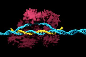
3D render of the CRISPR-Cas9 genome editing system
Genetically editing a cancer patient’s immune cells using CRISPR/Cas9 technology, then infusing those cells back into the patient appears safe and feasible based on early data from the first-ever clinical trial to test the approach in humans in the United States. Researchers from the Abramson Cancer Center have infused three participants in the trial thus far—two with multiple myeloma and one with sarcoma—and have observed the edited T cells expand and bind to their tumor target with no serious side effects related to the investigational approach. Penn is conducting the ongoing study in cooperation with the Parker Institute for Cancer Immunotherapy and Tmunity Therapeutics.
“This trial is primarily concerned with three questions: Can we edit T cells in this specific way? Are the resulting T cells functional? And are these cells safe to infuse into a patient? This early data suggests that the answer to all three questions may be yes,” says the study’s principal investigator Edward A. Stadtmauer, section chief of Hematologic Malignancies at Penn. Stadtmauer will present the findings next month at the 61st American Society of Hematology Annual Meeting and Exposition.
Tulane researchers join NIH HEAL initiative for research into opioid crisis
A Tulane University professor and researcher of biomedical engineering will join fellow researchers from over 40 other institutions in the National Institute of Health’s Help to End Addiction Long-Term (HEAL) Initiative. Of the $945 million that make up the project, Michael J. Moore, Ph.D. will receive a share of $1.2 million to advance research in modeling human pain through computer chips, with the help of fellow Tulane researchers Jeffrey Tasker, Ph.D., and James Zadina, Ph.D., each with backgrounds in neuroscience.
Because of the opioid epidemic sweeping the nation, Moore notes that there’s a rapid search going on to develop non-addictive painkiller options. However, he also sees a gap in adequate models to test those new drugs before human clinical trials are allowed to take place. Here is where he hopes to step in and bring some innovation to the field, by integrating living human cells into a computer chip for modeling pain mechanisms. Through his research, Moore wants to better understand not only how some drugs can induce pain, but also how patients can grow tolerant to some drugs over time. If successful, Moore’s work will lead to a more rapid and less expensive screening option for experimental drug advancements.
New machine learning-assisted microscope yields improved diagnostics
Researchers at Duke University recently developed a microscope that uses machine learning to adapt its lighting angles, colors, and patterns for diagnostic tests as needed. Most microscopes have lighting tailored to human vision, with an equal distribution of light that’s optimized for human eyes. But by prioritizing the computer’s vision in this new microscope, researchers enable it to see aspects of samples that humans simply can’t, allowing for a more accurate and efficient diagnostic approach.
Led by Roarke W. Horstmeyer, Ph.D., the computer-assisted microscope will diffuse light through a bowl-shaped source, allowing for a much wider range of illumination angles than traditional microscopes. With the help of convolutional neural networks — a special kind of machine learning algorithm — Horstmeyer and his team were able to tailor the microscope to accurately diagnose malaria in red blood cell samples. Where human physicians typically perform similar diagnostics with a rate of 75 percent accuracy, this new microscope can do the same work with 90 percent accuracy, making the diagnostic process for many diseases much more efficient.
Case Western Reserve University researchers create first-ever holographic map of brain
A Case Western Reserve University team of researchers recently spearheaded a project in creating an interactive holographic mapping system of the human brain. The design, which is believed to be the first of its kind, involves the use of the Microsoft HoloLens mixed reality platform. Lead researcher Cameron McIntyre, Ph.D., sees this mapping system as a better way of creating holographic navigational routes for deep brain stimulation. Recent beta tests with the map by clinicians give McIntyre hope that the holographic representation will help them better understand some of the uncertainties behind targeted brain surgeries.
More than merely providing a useful tool, McIntyre’s project also brings together decades’ worth of neurological data that has not yet been seriously studied together in one system. The three-dimensional atlas, called “HoloDBS” by his lab, provides a way of finally seeing the way all of existing neuro-anatomical data relates to each other, allowing clinicians who use the tool to better understand the brain on both an analytical and visual basis.
Implantable cancer traps reduce biopsy incidence and improve diagnostic
Biopsies are one of the most common procedures used for cancer diagnostics, involving a painful and invasive surgery. Researchers at the University of Michigan are trying to change that. Lonnie Shea, Ph.D., a professor of biomedical engineering at the university, worked with his lab to develop implants with the ability to attract any cancer cells within the body. The implant can be inserted through a scaffold placed under the patient’s skin, making it a more ideal option than biopsy for inaccessible organs like lungs.
The lab’s latest work on the project, published in Cancer Research, details its ability to capture metastatic breast cancer cells in vivo. Instead of needing to take biopsies from areas deeper within the body, the implant allows for a much simpler surgical procedure, as biopsies can be taken from the implant itself. Beyond its initial diagnostic advantages, the implant also has the ability to attract immune cells with tumor cells. By studying both types of cells, the implant can give information about the current state of cancer in a patient’s body and about how it might progress. Finally, by attracting tumor and immune cells, the implant has the ability to draw them away from the area of concern, acting in some ways as a treatment for cancer itself.
People and Places
Cesar de la Fuente-Nunez, PhD
The Philadelphia Inquirer recently published an article detailing the research of Penn’s Presidential Assistant Professor in Psychiatry, Microbiology, and Bioengineering, Cesar de la Fuente, Ph.D. In response to a growing level of worldwide deaths due to antibiotic-resistant bacteria, de la Fuente and his lab use synthetic biology, computation, and artificial intelligence to test hundreds of millions of variations in bacteria-killing proteins in the same experiment. Through his research, de la Fuente opens the door to new ways of finding and testing future antibiotics that might be the only viable options in a world with an increasing level of drug-resistant bacteria
Emily Eastburn, a Ph.D. candidate in Bioengineering at Penn and a member of the Boerckel lab of the McKay Orthopaedic Research Laboratory, recently won the Ashton fellowship. The Ashton fellowship is an award for postdoctoral students in any field of engineering that are under the age of 25, third-generation American citizens, and residents of either Pennsylvania or New Jersey. A new member of the Boerckel lab, having joined earlier this fall, Eastburn will have the opportunity to conduct research throughout her Ph.D. program in the developmental mechanobiology and regeneration that the Boerckel lab focuses on.
Cesar de la Fuente, Ph.D., a Presidential Assistant Professor in Psychiatry, Microbiology, and Bioengineering at Penn, recently published a literature review in Trends in Immunology entitled, “Emerging Frontiers in Microbiome Engineering.” The microbiome, in simple terms, consists of the genetic material of microorganisms in the gut, including bacteria, fungi, protozoa, viruses, and oral, vaginal, and skin microbiomes. Each human has a unique microbiome that depends both on predetermined factors like exposure to microorganisms within a mother’s birth canal or breastmilk in early life as well as environmental factors and diet in later life. The health of someone’s microbiome is extremely important, as an unhealthy microbiome with an imbalance of symbiotic and pathogenic microbes can make a person more susceptible to various diseases. The most common diseases or disorders associated with a problematic microbiome are rather far-reaching, including some of the most afflicting diseases of today like inflammatory bowel disease, diabetes, obesity, cardiovascular diseases, and neurological disorders.
In his recent literature review, de la Fuente provides an overview of microbiome engineering, and what the future might hold for the field. He defines microbiome engineering initially as a way of studying the “contribution of individual microbes and generating potential therapies against metabolic, inflammatory, and immunological diseases.” Currently, most treatments for issues with the microbiome are broad solutions like dietary adjustments to include more probiotics, antibiotics, or prebiotics, while more serious cases may require a fecal microbiota transplant. While these therapies may work for some patients, de la Fuente emphasizes the need for greater specificity in treatment targets and a need for precision in reprogramming existing microbial communities as an alternative to transplants.
De la Fuente highlights the current methods and tools in microbiome engineering such as the use of bacteriocins and bacteriophages to knock out specific bacteria within the microbiome. However, there are very few bacteriocins or bacteriophages commercially available on today’s market. Another common approach to microbiome engineering is in synthetic biology, or the use of “chassis” — a type of cell that maintains DNA constructs for different functions — to engineering interactions within the microbiome. De la Fuente continues his discussion of current methods by naming and describing several specific examples of these approaches, particularly in relation to synthetic biology options before moving on to examine future directions for these methods.
Before bringing up potential new frontiers for microbiome engineering, de la Fuente also outlines the way that microbiome engineering works in the first place, and dedicates sections of the review to the microbiome’s influence on its host’s immune system and how to engineer the microbiome to modulate that immune system. The main future methods for microbiome engineering that de la Fuente points out in his review include more precise regulation of gene expression through commensal organisms and the use of CRISPRi to find genes involved in bacterial maintenance. The conclusion of de la Fuente’s review brings up the notion of new personalized medicine or therapy for the microbiome that could come with further advances in the field. However, he also makes sure to bring up some still-outstanding questions about the human microbiome that require further research, most notably, what exactly makes a healthy human microbiome? Here’s hoping the research de la Fuente mentions can illuminate a path to the answer.