Top: Axons in female and male subject brains Bottom: damaged axons in male and female brains after injury (Credit: Penn Medicine)
Important brain structures that are key for signaling in the brain are narrower and less dense in females, and more likely to be damaged by brain injuries, such as concussion. Long-term cognitive deficits occur when the signals between brain structures weaken due to the injury. The structural differences in male and female brains might explain why females are more prone to concussions and experience longer recovery from the injury than their male counterparts, according to a preclinical study led by the Perelman School of Medicine at the University of Pennsylvania, published this week in Acta Neuropathologica.
Each year, approximately 50 million individuals worldwide suffer a concussion, also referred to as mild traumatic brain injury (TBI). However, there is nothing “mild” about this condition for the more than 15 percent of individuals who suffer persisting cognitive dysfunction, which includes difficulty concentrating, learning and remembering new information, and making decisions.
Although males make up the majority of emergency department visits for concussion, this has been primarily attributed to their greater exposure to activities with a risk of head impacts compared to females. In contrast, it has recently been observed that female athletes have a higher rate of concussion and appear to have worse outcomes than their male counterparts participating in the same sport.
“Clinicians have observed for a long time that females suffer from concussion at higher rates than males in the same sports, and that they take longer to recover cognitive function, but couldn’t explain the underlying mechanisms of this phenomenon,” said senior author Douglas Smith, MD, a professor of Neurosurgery and director of Penn’s Center for Brain Injury and Repair. “The variances in brain structures of females and males not only illuminate why this disparity exists, but also exposes biomarkers, such as axon protein fragments, that can be measured in the blood to determine injury severity, monitor recovery, and eventually help identify and develop treatments that help patients repair these damaged structures and restore cognitive function.”
The Office of the Provost awards the Penn Prize for Excellence in Teaching by Graduate Students in recognition of their profound impact on education across the University. Nominations come directly from undergraduate and graduate students in their courses and are narrowed down to ten awardees each year.
Anderson is a Ph.D. student who studies the computational modeling of injury in full-brain networks in the Molecular Neuroengineering Lab of David Meaney, Solomon R. Pollack Professor in Bioengineering and Senior Associate Dean of Penn Engineering. Anderson has served as a teaching assistant for Bioengineering Senior Design since Fall 2019. Senior Design (BE 495 & 496) is the Bioengineering Department’s two-semester capstone course in which students work in teams to conceive, design and pitch their final projects, and is taught by Meaney and Sevile Mannickarottu, Director of Educational Laboratories in Bioengineering. Anderson earned her B.S. in Bioengineering from Rice University in 2016. Her doctoral thesis focuses on how subconcussive head trauma affects subsequent concussion outcomes.
Colin Huber, a Ph.D. candidate in Bioengineering studying head impact biomechanics and concussion in sports at the Center for Injury Research and Prevention (CIRP) at the Children’s Hospital of Philadelphia (CHOP), recently published “Variations in Head Impact Rates in Male and Female High School Soccer” in Medicine & Science in Sports & Exercise with colleagues from CHOP’s Minds Matter Concussion Frontier Program and the CIRP.
Colin’s paper, the goal of which was to compare “to compare head impact exposure rates (head impacts/exposure period) in male and female high school soccer by using multiple methodological approaches,” was recently profiled in the Penn Engineering Research & Innovation Newsletter.
The Perelman School of Medicine has announced the winners of the 2020 Penn Medicine Awards of Excellence. The Office of the Dean says:
“These awardees exemplify our profession’s highest values of scholarship, teaching, innovation, commitment to service, leadership, professionalism and dedication to patient care. They epitomize the preeminence and impact we all strive to achieve. The awardees range from those at the beginning of their highly promising careers to those whose distinguished work has spanned decades.
Each recipient was chosen by a committee of distinguished faculty from the Perelman School of Medicine or the University of Pennsylvania. The contributions of these clinicians and scientists exemplify the outstanding quality of patient care, mentoring, research, and teaching of our world-class faculty.”
Two faculty members affiliated with Penn Bioengineering are among this year’s recipients.
Yale Cohen, PhD, Professor of Otorhinolaryngology with secondary appointments in Neuroscience and Bioengineering, is the recipient of the Jane M. Glick Graduate Student Teaching Award. Cohen is an alumnus of the Penn Bioengineering doctoral program and is currently the department’s Graduate Chair.
“Dr. Cohen’s commitment to educating and training the next generation of scientists exemplifies the type of scientist and educator that Jane Glick represented. His students value his highly engaging and supportive approach to teaching, praising his enthusiasm, energy, honesty, and compassion.”
Douglas H. Smith, MD, Robert A. Groff Endowed Professor of Research and Teaching in Neurosurgery and member of the Penn Bioengineering Graduate Group, is the recipient of this year’s William Osler Patient Oriented Research Award:
“Dr. Smith is the foremost authority on diffuse axonal injury (DAI) as the unifying hypothesis behind the short- and long-term consequences of concussion. After realizing early in his career that concussion, or mild traumatic brain injury (TBI), was a much more serious event than broadly appreciated, Dr. Smith and his team have used computer biomechanical modeling, in vitro and in vivo testing in parallel with seminal human studies to elucidate mechanisms of concussion.”
David F. Meaney, Solomon R. Pollack Professor of Bioengineering, has been named the Senior Associate Dean of Penn Engineering, effective January 1, 2020. This newly created leadership position will have oversight responsibilities in budget, space and infrastructure planning; facilities and research services; and will create and cultivate new interschool partnerships that will expand Penn Engineering’s footprint on campus.
Meaney is well known not only for his scholarship and innovation in neuroengineering and concussion science, but also for his leadership during his highly successful tenure as Chair of the Department of Bioengineering.
“Dave’s strong connections to the health schools will help strengthen Penn Engineering’s initiatives throughout campus,” says Vijay Kumar, Nemirovsky Family Dean of Penn Engineering. “He will have oversight of Penn Health-Tech, the Center for Engineering MechanoBiology and other efforts between engineering and the health schools, and Dave brings his unique creativity, energy and leadership experience to these collaborative efforts.”
Bio-inspiration Informs New Football Helmet Design from IUPUI Students
Art, design, biology, and engineering all interact with each other in a recent design for a football helmet from two students— one of media arts and the other of engineering — at the Indiana University – Purdue University Indianapolis. Directed by Lecturer in Media Arts and Science Zebulun Wood, M.S., and Associate Professor of Mechanical and Energy Engineering and Assistant Professor of Biomedical Engineering Andres Tovar, Ph.D., the students found inspiration in biological structures like a pomelo peel, nautilus shell, and woodpecker skull to create energy-absorbing helmet liners. The resulting design took these natural concussion-reducing structures and created compliant mechanism lattice-based liners the replace the foam traditionally placed in between two harder shells of a typical helmet. Their work not only exemplifies the benefits of bio-inspiration, but demonstrates the way that several different domains of study can overlap in the innovation of a new product.
Study of Mechanical Properties of Hyaluronic Acid Could Help Inform Current Debates Over Treatment Regulation for Osteoarthritis
Arthritis is an extremely common condition, especially in older patients, in which inflammation of the joints can cause high amounts of stiffness and pain. Osteoarthritis in particular is the result of the degradation of flexible tissue between the bones of a joint, which increases friction in joint motion. A common treatment of this form of arthritis is the injection of hyaluronic acid, which is meant to provide joint lubrication, and decreases this friction between bones. Recently, however, there has been a debate over hyaluronic acid’s classification by the FDA and whether it should remain based on the knowledge of the mechanical actions of the acid in treatment for osteoarthritis or if potential chemical action of the acid should be considered as well.
Because of limited ways of testing the mechanical properties of the acid, many researchers felt that there could be more to hyaluronic acid’s role in pain relief for arthritic patients. But Lawrence Bonassar, Ph.D., the Daljit S. and Elaine Sarkaria Professor in Biomedical Engineering at the Meinig School of Bioengineering of Cornell University, had another idea. With his lab, he created a custom-made tribometer to measure the coefficient of friction of a given lubricant by rubbing a piece of cartilage back and forth across a smooth glass plate. The research demonstrated that hyaluronic acid’s ability to reduce the coefficient of friction aligned with patients’ pain relief. Bonassar and his team hope that these results will demonstrate the heavy contribution of mechanical action that hyaluronic acid has in osteoarthritis treatment, and help bring an end to the debate over its FDA classification.
A New Way of Mapping the Heart Could Lead to Better Understanding of Contractile Activity
Though reduced contractions in certain regions of the heart can be an indicator of a certain condition, there is currently no way to directly measure contractile activity. This is why Cristian Linte, Ph.D., an Associate Professor of Biomedical Engineering in the Kate Gleason College of Engineering at the Rochester Institute of Technology (RIT), hopes to create a map of the heart that can quantify contraction power. In collaboration with Niels Otani, Ph.D., an Associate Professor in the School of Mathematics at RIT, Linte plans to use an $850,000 grant from the National Science Foundation to achieve a more comprehensive understanding of the heart through both medical imaging and mechanical modeling. The group hopes that their approach will lead to not only a better way to diagnose certain heart conditions and diseases, but also open up understanding of active contraction, passive motion, and the stresses within the heart walls that underlie each.
Celebrity Cat Lil Bub Helps Penn and German Researchers Draw Public Attention to Genetics
Lil Bub’s unique appearance has garnered millions of online fans, and now, an avenue for researchers to talk about genetics. (Photo Courtesy of Mike Bridavsky)
In 2015, a group of curious researchers set out to sequence the genome of a celebrity cat named Lil Bub. They were hoping to understand the genetics behind Lil Bub’s extra toes and unique skeletal structure, which contribute to her heart-warming, kitten-like appearance. However, an equally important goal of their “LilBUBome” project was to invite the general public into the world of genetics.
Because of Lil Bub’s online fame, the project garnered attention from her fans and the media, all hoping to discover the secret to Lil Bub’s charm. As early as 2015, Gizmodo’s Kiona Smith-Strickland reported on the team’s intentions to sequence Lil Bub’s genome, and, since then, many have been awaiting the results of the LilBUBome.
The Alfred P. Sloan Foundation awarded a six-year grant to Barnard College and Columbia University’s School of Engineering and Applied Science to support graduate education for women in engineering. The funding will go towards a new five-year program that enables Barnard students to attain both a B.A. and M.S. in one year after their traditional four years of undergraduate education. The program will offer M.S. degrees in chemical engineering, biomedical engineering, and industrial engineering and operations research, and is one of the first of its kind for women’s colleges.
We would like to congratulate Jean Paul Allain, Ph.D., on being named the first head of the new Ken and Mary Alice Lindquist Department of Nuclear Engineering at Penn State. Allain, who is currently a Professor and head of graduate programs in the University of Illinois at Urbana-Champaign’s Department of Nuclear, Plasma, and Radiological Engineering, conducts research in models of particle-surface interactions. In addition to being head of the new department at Penn State, Allain will also hold a position as a Professor of Biomedical Engineering at the university.
We would also like to congratulate Andrew Douglas, Ph.D., on his appointment as the Vice Provost for Faculty Affairs at Johns Hopkins University. Douglas currently holds the position of Vice Dean for Faculty at the Whiting School of Engineering, and has joint appointments in Mechanical and Biomedical Engineering. Douglas’s research at Hopkins focuses on mechanical properties and responses of compliant biological tissue and on the nonlinear mechanics of solids, with a focus on soft tissues and organs like the heart and tongue.
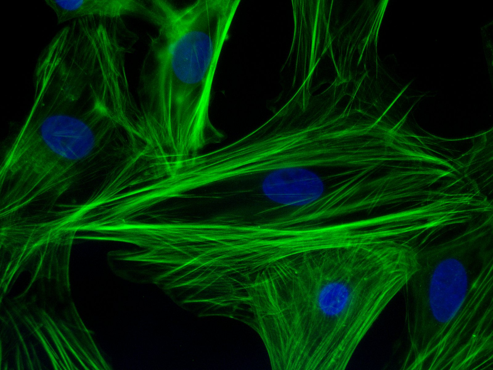
Immunofluorescent staining for F-actin filaments (green) and nuclei (blue) in neonatal cardiomyocyte
Two common diagnostic procedures in cardiology are intravascular ultrasound and cardiac angiography. These procedures are performed to quantify the amount of plaque affecting a patient’s blood vessels. This information is vital because it helps to determine how advanced heart degree is, as well as guiding treatment planning and even the course of bypass surgery. However, the current technologies used for these procedures have significant limitations. Although conventional angiography can help to quantify the plaque burden, it does not offer any information about how much of the diameter of a vessel is blocked. Intravascular ultrasound is very good at quantifying plaque burden, but it is poor at identifying smaller features of compromised blood vessels.
One solution suggested to these issues is the combination of these imaging technologies into a single multimodal technique. Scientists led by Laura Marcu, Ph.D., professor of biomedical engineering at the University of California, Davis, invented a method combining intravascular ultrasound with multispectral fluorescence lifetime imaging (FLIM). As published in Scientific Reports, the device resembles a typical cardiac catheter but contains an optical fiber within the catheter that emits fluorescent light to characterize the plaque components before treatment.
Dr. Marcu and her colleagues tested their new device in live pigs and in human coronary arteries obtained from cadavers. The fluorescence data acquired with the device were comparable to those acquired with traditional fluorescence angiography. Moreover, the device could acquire data without having to administer a contrast agent, which can be dangerous in some patients due to allergies or weakened kidneys. The authors are currently seeking FDA approval to test their combined catheter in humans.
In addition to treating vessels before a heart attack can occur, there is new work showing how to efficiently repair heart tissue after a heart attack. A team of scientists collaborating among Clemson University, the Medical University of South Carolina, the University of South Carolina, and the University of Chicago has received a $1.5 million grant from the National Institutes of Health to examine a treatment that combines stem cells with nanowires. The principal investigator on the grant is Ying Mei, Ph.D., who is assistant professor of bioengineering at Clemson. Dr. Mei’s team mixes stem cells with nanowires so that they form spheroids that are larger than single cells and thus less likely to wash away. In addition, the investigators hope that the spheroids will mitigate the issue of the transplanted cells and the recipient’s heart beating at different rhythms. If successful, the group’s treatment paradigm could be a major step forward in stem cell therapies and cardiology.
Look, Up in the Sky!
Drones became famous when deployed on battlefields for the first time a decade ago. Since then, they’ve been adopted as a technology for a variety of purposes. For example, Amazon introduced delivery drones almost a year ago, and it has plans to expand its drone fleet enormously in coming years. It was only a matter of time before engineers began to imagine medical applications for drones.
Engineers in Australia and Iraq recently investigated whether a drone could be used to monitor cardiorespiratory signals remotely. They reported their findings in BioMedical Engineering OnLine. The authors used imaging photoplethysmography (PPG), which employs a video camera to detect visual indications on the skin of heart activity. They also applied advanced digital processing technology due to the tendency of PPG to be affected by sound and movement in the area of detection. By testing the combined technologies in 15 healthy volunteers, the authors found that their data compared well with several traditional techniques for monitoring vital signs. Among the possible applications that the authors imagine for this technology is battlefield triage performed remotely using drones. In the meantime, they will seek to fine-tune the technology’s abilities.
Concussion Distressingly Common
A research letter published in a recent issue of JAMA reports that a study conducted in Canada found that one in five adolescents sustained a concussion on at least one occasion. Of the approximately 20% of the study respondents who had experienced concussions, one quarter had suffered more than one. The letter is particularly relevant to the United States because of the similar popularity in Canada of contact and semicontact sports such as ice hockey and football. In addition, the study included more than 13,000 teenagers, lending significantly reliability to the conclusions.
Ending the Time of Cholera
Although largely eradicated in the developed world, cholera remains a major public health issue in the Global South and other parts of the developing world. The disease is a bacterial infection that causes severe gastrointestinal distress. Because the disease is transmitted via water, effective public sanitation is a core requirement of an effective prevention campaign.
One technology being deployed in this fight is a smartphone microfluidics platform that can determine the presence of the pathogen that causes cholera in a sample and report the data almost immediately to public health authorities. This technology was produced by a company called PathVis, which was spun off at Purdue University based on science produced the laboratories of Tamara Kinzer-Ursem, Ph.D., and Jacqueline Linnes, Ph.D., both of whom are assistant professors in Purdue’s Weldon School of Biomedical Engineering. There are plans to test PathVis in Haiti and to expand it to detect other diseases in the future.
The Latest on CRISPR
CRISPR/Cas9 is the biggest bioengineering story to come along in some time — certainly the biggest in genetic engineering. But the mere fact that it’s here and already being used in animals and in human cell lines doesn’t mean that the story is over. For instance, the Cas9 protein, which CRISPR deploys as part of its gene editing process, is currently developed most often using a viral vector. However, this system of delivery has certain drawbacks, not the least of which is a host immune system response when levels of the deployed viral vector reach the levels necessary for CRISPR to work.
A recent study published in Nature Biomedical Engineering reports on the successful use of gold nanoparticles to deliver Cas9. The new delivery system, called CRISPR-Gold, could obviate the need to use a viral vector as part of the CRISPR induction process. So far, the authors, led by University of California, Berkeley, bioengineers Irina Conboy, Ph.D., and Niren Murthy, Ph.D., have only used CRISPR-Gold in mice, but their successful results indicate that nonviral delivery with CRISPR is possible, so CRISPR could be used for more than previously thought.
How can physicians and engineers help design athletic equipment and diagnostic tools to better protect teenaged athletes from concussions? A unique group of researchers with neuroscience, bioengineering and clinical expertise are teaming up to translate preclinical research and human studies into better diagnostic tools for the clinic and the sidelines as well as creating the foundation for better headgear and other protective equipment.
The five-year project focuses specifically on developing a suite of quantitative assessment tools to enhance accuracy of sports-related concussion diagnoses, with a focus on objective metrics of activity, balance, neurosensory processing, including eye tracking, and measures of cerebral blood flow. These could also provide prognoses of the time-to-recovery and safe return-to-play for youth athletes. Researchers will examine such factors such as repeated exposures and direction of head motion. In addition, they will also look at sex-specific data to see how prevention and diagnosis strategies need to be tailored for males and females.
The multidisciplinary research team believes this study will result in post-concussion metrics that can provide objective benchmarks for diagnosis, a preliminary understanding of the effect of sub-concussive hits, the magnitude and direction of head motion and sex on symptom time course, as well as markers in the bloodstream that relate to functional outcomes.
Knowing the biomechanical exposure and injury thresholds experienced by different player positions can help sports organizations tailor prevention strategies and companies to create protective equipment design for specific sports and even specific positions.
The study will enroll research participants from The Shipley School, a co-ed independent school in suburban Philadelphias, and from CHOP’s Concussion Care for Kids: Minds Matter program which annually sees more than 2,500 patients with concussion in the Greater Delaware Valley region.
The study is funded by the National Institutes of Health.


 Art, design, biology, and engineering all interact with each other in a