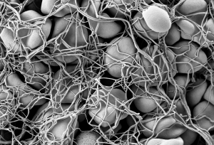
Modeling Hemostasis With Microfluidics

Hemostasis is the process by which blood stops flowing from damaged blood vessels. It is a complex process involving multiple molecules and forces, and our current understanding is limited by our inability to test these factors simultaneously in the laboratory. Some tests, for instance, can tell us much about clotting — a part of hemostasis — but little about the other elements at play. In particular, the role in hemostasis of the endothelium, which is the cell layer that lines the blood vessels, has generally been omitted from previous studies.
However, a new article in Nature Communications details the use of microfluidics technology, which is often used to model organ systems outside the body, to engineer a more complete model of hemostasis. Led by Wilbur A. Lam, M.D., Ph.D., Assistant Professor in the Wallace H. Coulter Department of Biomedical Engineering at Georgia Tech, the study authors fabricated microfluidics devices and then seeded vascular channels in the devices with human aortic and umbilical vein cells to simulate the endothelium.
Using the device, the authors were able to visualize hemostatic plug formation with whole blood and with blood from subjects with hemophilia. Although the authors concede that their model best represents capillary bleeding, rather than bleeding from larger vessels, they are confident that their model reliably represents the interaction of the endothelium with multiple varieties of blood cells.
Shedding Light on Cancer Response and Resistance
Penn’s founder, Benjamin Franklin, has a famous axiom: “an ounce of prevention is worth a pound of cure.” If Franklin were alive today, he would likely agree with two common axioms in cancer treatment: 1) if you can’t see it, you can’t treat it; and 2) if you treat it, treat all of it. Recent publications from investigators at Columbia and the University of Maryland reveal how imaging technologies can help predict to outcomes and how nanotechnology is offering a new therapeutic tools for fighting cancer.
Using diffuse optical tomography (DOT), which employs near-infrared spectroscopy to obtain three-dimensional images, scientists at Columbia have shown in an article in Radiology that treatment response could be predicted as early as two weeks after the start of therapy. The authors, led by Andreas H. Hielscher, PhD, Professor of Biomedical Engineering, Electrical Engineering, and Radiology at Columbia, applied DOT in 40 women with breast cancer undergoing chemotherapy. They found that DOT imaging features were associated, some very strongly, with treatment outcomes at 5 months. Given their positive findings, the authors intend to continue testing DOT in a larger cohort prospective study.
Another major issue in cancer chemotherapy is multidrug resistance (MDR), a highly frustrating complication resulting from lengthy treatment. In MDR, cancer types can find ways to overcome the effects of chemotherapy, resulting in relapse, often with deadly consequences. Therefore, among the challenges that oncologists face is the need to predict MDR, preferably before treatment even begins.
Based on the knowledge that adenosine triphosphate (ATP), a common organic molecule in energy generation, is involved in MDR, scientists at the University of Maryland engineered nanoparticles that could target cancer cells and, when exposed to near-infrared laser irradiation, reduce the amount of ATP in the cells . The scientists, led by Xiaoming Shawn He, Ph.D., Professor of Bioengineering at Maryland, published their findings in Nature Communications.
Dr. He’s team tested their nanoparticles in vitro and subsequently in mice and found decreased tumor sizes in mice treated with the particles, as well as more deaths of cancer cells. In addition, two of seven mice treated with the nanoparticles plus light experienced complete tumor eradication. The findings offer hope that MDR could be overcome with direct delivery of targeted treatment to resistant tumors.
Preserving the Tooth
A frustrating problem often encountered by dentists is the growth of new cavities around existing fillings. Microbes are often critical catalysts for these new cavities. Using antimicrobial agents at cavity-repair sites could make a real difference. However, mesoporous silica has proved suboptimal for this purpose.
However, help might be on the way. A study in a recent issue of Scientific Reports, written by a trio of authors led by Benjamin D. Hatton, Ph.D., Assistant Professor at the Institute of Biomaterials & Biomedical Engineering of the University of Toronto, reports the successful synthesis of 500-nm nanocomposite spheres combining silica with octenidine dihydrochloride, a common antiseptic. The newly synthesized nanospheres outperformed earlier attempts with mesoporous silica. The authors will continue to develop these nanoparticles to deliver other drugs for longer periods of time.
