The NEMO Prize Goes to Research Improving Soft-Tissue Transplant Surgeries

by Melissa Pappas

Daeyeon Lee (left), Oren Friedman (center) and Sergei Vinogradov (right)

Each year, the Nemirovsky Engineering and Medicine Opportunity (NEMO) Prize, funded by Penn Health-Tech, awards $80,000 to a collaborative team of researchers from the University of Pennsylvania’s Perelman School of Medicine and the School of Engineering and Applied Science for early-stage, interdisciplinary ideas.

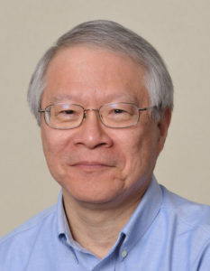
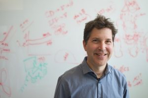
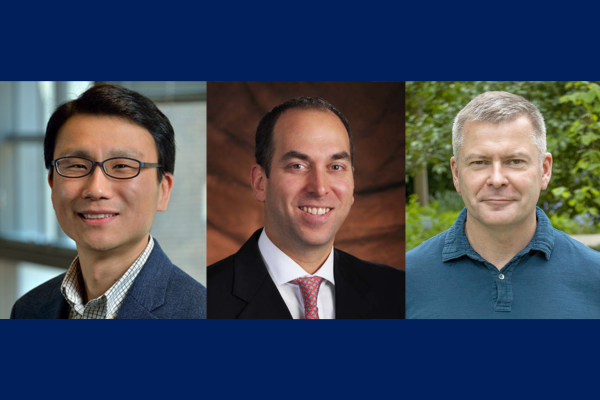
This year, the NEMO Prize has been awarded to Penn Engineering’s Daeyeon Lee, Russel Pearce and Elizabeth Crimian Heuer Professor in Chemical and Biomolecular Engineering, Oren Friedman, Associate Professor of Clinical Otorhinolaryngology in the Perelman School of Medicine, and Sergei Vinogradov, Professor in the Department of Biochemistry and Biophysics in the Perelman School of Medicine and the Department of Chemistry in the School of Arts & Sciences. Together, they are developing a new therapy that improves the survival and success of soft-tissue grafts used in reconstructive surgery.

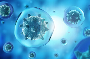
More than one million people receive soft-tissue reconstructive surgery for reasons such as tissue trauma, cancer or birth defects. Autologous tissue transplants are those where cells and tissue such as fat, skin or cartilage are moved from one part of a patient’s body to another. As the tissue comes from the patient, there is little risk of transplant rejection. However, nearly one in four autologous transplants fail due to tissue hypoxia, or lack of oxygen. When transplants fail the only corrective option is more surgery. Many techniques have been proposed and even carried out to help oxygenate soft tissue before it is transplanted to avoid failures, but current solutions are time consuming and expensive. Some even have negative side effects. A new therapy to help oxygenate tissue quickly, safely and cost-effectively would not only increase successful outcomes of reconstructive surgery, but could be widely applied to other medical challenges. 

The therapy proposed by this year’s NEMO Prize recipients is a conglomerate or polymer of microparticles that can encapsulate oxygen and disperse it in sustainable and controlled doses to specific locations over periods of time up to 72 hours. This gradual release of oxygen into the tissue from the time it is transplanted to the time it functionally reconnects to the body’s vascular system is essential to keeping the tissue alive. 

“The microparticle design consists of an oxygenated core encapsulated in a polymer shell that enables the sustained release of oxygen from the particle,” says Lee. “The polymer composition and thickness can be controlled to optimize the release rate, making it adaptable to the needs of the hypoxic tissue.” 

These life-saving particles are designed to be integrated into the tissue before transplantation. However, because they exist on the microscale, they can also be applied as a topical cream or injected into tissue after transplantation. 

“Because the microparticles are applied directly into tissues topically or by interstitial injection (rather than being administered intravenously), they surpass the need for vascular channels to reach the hypoxic tissue,” says Friedman. “Their micron-scale size combined with their interstitial administration, minimizes the probability of diffusion away from the injury site or uptake into the circulatory system. The polymers we plan to use are FDA approved for sustained-release drug delivery, biocompatible and biodegrade within weeks in the body, presenting minimal risk of side effects.”

The research team is currently testing their technology in fat cells. Fat is an ideal first application because it is minimally invasive as an injectable filler, making it versatile in remodeling scars and healing injury sites. It is also the soft tissue type most prone to hypoxia during transplant surgeries, increasing the urgency for oxygenation therapy in this particular tissue type.

Read the full story in Penn Engineering Today.

Daeyeon Lee and Sergei Vinogradov are members of the Penn Bioengineering Graduate Group.

Folding@Home: How You, and Your Computer, Can Play Scientist

by

Greg Bowman kneels, working on a server.
Folding@home is led by Gregory Bowman, a Penn Integrates Knowledge Professor who has appointments in the Departments of Biochemistry and Biophysics in the Perelman School of Medicine and the Department of Bioengineering in the School of Engineering and Applied Science. (Image: Courtesy of Penn Medicine News)

Two heads are better than one. The ethos behind the scientific research project Folding@home is that same idea, multiplied: 50,000 computers are better than one.

Folding@home is a distributed computing project which is used to simulate protein folding, or how protein molecules assemble themselves into 3-D shapes. Research into protein folding allows scientists to better understand how these molecules function or malfunction inside the human body. Often, mutations in proteins influence the progression of many diseases like Alzheimer’s disease, cancer, and even COVID-19.

Penn is home to both the computer brains and human minds behind the Folding@home project which, with its network, forms the largest supercomputer in the world. All of that computing power continually works together to answer scientific questions such as what areas of specific protein implicated in Parkinson’s disease may be susceptible to medication or other treatment.

Led by Gregory Bowman, a Penn Integrates Knowledge professor of Biochemistry and Biophysics in the Perelman School of Medicine who has joint appointments in the Department of Biochemistry and Biophysics in the Perelman School of Medicine and the Department of Bioengineering in the School of Engineering and Applied Science, Folding@home is open for any individual around the world to participate in and essentially volunteer their computer to join a huge network of computers and do research.

Using the network hub at Penn, Bowman and his team assign experiments to each individual computer which communicates with other computers and feeds info back to Philly. To date, the network is comprised of more than 50,000 computers spread across the world.

“What we do is like drawing a map,” said Bowman, explaining how the networked computers work together in a type of system that experts call Markov state models. “Each computer is like a driver visiting different places and reporting back info on those locations so we can get a sense of the landscape.”

Individuals can participate by signing up and then installing software to their standard personal desktop or laptop. Participants can direct the software to run in the background and limit it to a certain percentage of processing power or have the software run only when the computer is idle.

When the software is at work, it’s conducting unique experiments designed and assigned by Bowman and his team back at Penn. Users can play scientist and watch the results of simulations and monitor the data in real time, or they can simply let their computer do the work while they go about their lives.

Read the full story at Penn Medicine News.

Gregory Bowman Appointed Penn Integrates Knowledge University Professor

by Ron Ozio

Greg Bowman
Gregory Bowman, the Louis Heyman University Professor, has joint appointments in the Department of Biochemistry and Biophysics in the Perelman School of Medicine and the Department of Bioengineering in the School of Engineering and Applied Science. (Image: Courtesy of School of Engineering and Applied Sciences)

Gregory R. Bowman, a pioneer of biophysics and data science, has been named a Penn Integrates Knowledge University Professor at the University of Pennsylvania. The announcement was made today by President Liz Magill and Interim Provost Beth A. Winkelstein.

Bowman holds the Louis Heyman University Professorship, with joint appointments in the Department of Biochemistry and Biophysics in the Perelman School of Medicine and the Department of Bioengineering in the School of Engineering and Applied Science.

His research aims to combat global health threats such as COVID-19 and Alzheimer’s disease by better understanding how proteins function and malfunction, especially through new computational and experimental methods that map protein structures. This understanding of protein dynamics can lead to effective new treatments for even the most seemingly resistant diseases.

“Delivering the right treatment to the right person at the right time is vital to sustaining—and saving—lives,” Magill said. “Greg Bowman’s novel work holds enormous promise and potential to advance new forms of personalized medicine, an area of considerable strength for Penn. A gifted researcher and consummate collaborator, we are delighted to count him among our distinguished PIK University Professors.”

Bowman came to Penn from the Washington University School of Medicine’s Department of Biochemistry and Molecular Biophysics, where he served on the faculty since 2014. He previously completed a three-year postdoctoral fellowship at the University of California, Berkeley.

Bowman’s research utilizes high-performance supercomputers for simulations that can better explain how mutations and disease change a protein’s functions. These simulations are enabled in part through the innovative Folding@home project, which Bowman directs. Folding@home empowers anyone with a computer to run simulations alongside a consortium of universities, with more than 200,000 participants worldwide.

His research has been supported by the National Science Foundation, National Institutes of Health, National Institute on Aging, and Packard Foundation, among others, and he has received a CAREER Award from the NSF, Career Award at the Scientific Interface from the Burroughs Wellcome Fund, and Thomas Kuhn Paradigm Shift Award from the American Chemical Society. He received a Ph.D. in biophysics from Stanford University and a B.S. (summa cum laude) in computer science, with a minor in biomedical engineering, from Cornell University.

“Greg Bowman’s highly innovative work,” Winkelstein said, “exemplifies the power of our interdisciplinary mission at Penn. He brings together supercomputers, biophysics, and biochemistry to make a vital impact on public health. This brilliant fusion of methods—in the service of improving people’s lives around the world—will be a tremendous model for the research of our faculty, students, and postdocs in the years ahead.”

The Penn Integrates Knowledge program is a University-wide initiative to recruit exceptional faculty members whose research and teaching exemplify the integration of knowledge across disciplines and who are appointed in at least two schools at Penn.

The Louis Heyman University Professorship is a gift of Stephen J. Heyman, a 1959 graduate of the Wharton School, and his wife, Barbara Heyman, in honor of Stephen Heyman’s uncle. Stephen Heyman is a University Emeritus Trustee and member of the School of Nursing Board of Advisors. He is Managing Partner at Nadel and Gussman LLC in Tulsa, Oklahoma.

This story originally appeared in Penn Today.

Dr. Bowman is Penn Bioengineering’s third PIK Professor after Kevin Johnson and Konrad Kording. See the full list of University PIK Professors here.

More Cancers May be Treated with Drugs than Previously Believed

by Alex Gardner

3D illustration of cancer cells
nucleus and membrane of pathogen micro organisms in blue background

Up to 50 percent of cancer-signaling proteins once believed to be immune to drug treatments due to a lack of targetable protein regions may actually be treatable, according to a new study from the Perelman School of Medicine at the University of Pennsylvania. The findings, published this month in Nature Communications, suggest there may be new opportunities to treat cancer with new or existing drugs.

Researchers, clinicians, and pharmacologists looking to identify new ways to treat medical conditions—from cancer to autoimmune diseases—often focus on protein pockets, areas within protein structures to which certain proteins or molecules can bind. While some pockets are easily identifiable within a protein structure, others are not. Those hidden pockets, referred to as cryptic pockets, can provide new opportunities for drugs to bind to. The more pockets scientists and clinicians have to target with drugs, the more opportunities they have to control disease.

The research team identified new pockets using a Penn-designed neural network, called PocketMiner, which is artificial intelligence that predicts where cryptic pockets are likely to form from a single protein structure and learns from itself. Using PocketMiner—which was trained on simulations run on the world’s largest super computer—researchers simulated single protein structures and successfully predicted the locations of cryptic pockets in 35 cancer-related protein structures in thousands of areas of the body. These once-hidden targets, now identified, open up new approaches for potentially treating existing cancer.

What’s more, while successfully predicting the cryptic pockets, the method scientists used in this study was much faster than previous simulation or machine-learning methods. The network allows researchers to nearly instantaneously decide if a protein is likely to have cryptic pockets before investing in more expensive simulations or experiments to pursue a predicted pocket further.

“More than half of human proteins are considered undruggable due to an apparent lack of binding proteins in the snapshots we have,” said Gregory R. Bowman, PhD, a professor of Biochemistry and  Biophysics and Bioengineering at Penn and the lead author of the study. “This PocketMiner research and other research like it not only predict druggable pockets in critical protein structures related to cancer but suggest most human proteins likely have druggable pockets, too. It’s a finding that offers hope to those with currently untreatable diseases.”

Read the full story in Penn Medicine News.

Penn Bioengineering Alumna Cynthia Reinhart-King is President Elect of BMES

Dr. Cynthia Reinhart-King, Engineering, BME, Photo by Joe Howell

Penn Bioengineering alumna Cynthia Reinhart-King, Cornelius Vanderbilt Professor of Engineering and Professor of Biomedical Engineering at Vanderbilt University, was elected the next President of the Biomedical Engineering Society (BMES), the largest professional society for biomedical engineers. Her term as president-elect started at the annual BMES meeting in October 2021.

Reinhart-King graduated with her Ph.D. from Penn Bioengineering in 2006. She studied in the lab of Daniel Hammer, Alfred G. and Meta A. Ennis Professor in Bioengineering and Chemical and Biomolecular Engineering as a Whitaker Fellow and went on to complete postdoctoral training as an Individual NIH NRSA postdoctoral fellow at the University of Rochester. Prior to joining Vanderbilt, she was on the faculty of Cornell University and received tenure in the Department of Biomedical Engineering. The Reinhart-King lab at Vanderbilt “uses tissue engineering, microfabrication, novel biomaterials, model organisms, and tools from cell and molecular biology to study the effects of mechanical and chemical changes in tissues during disease progression.”

Reinhart-King gave the 2019 Grace Hopper Distinguished Lecture, sponsored by the Department of Bioengineering. This lecture series recognizes successful women in engineering and seeks to inspire students to achieve at the highest level. She is a recipient of numerous prestigious awards, including the Rita Schaffer Young Investigator Award in 2010, an NSF CAREER Award, and the Mid-Career Award in 2018 from BMES.

In a Q&A on the BMES Blog, Reinhart-King said that:

“BMES is facing many challenges, like many societies, as we deal with the hurdles associated with COVID-19 and inequities across society. We must continue to address those challenges. However, we are also in a terrific window of having robust membership, many members who are eager to get involved with the society’s activities, and a national lens on science and scientists. One of my goals will be to identify and create opportunities for our members to help build the reach of the society and its member.”

Read “Cynthia Reinhart-King is president-elect of the Biomedical Engineering Society” in Vanderbilt News.

BE Seminar: “Dynamics of 3D Cell Migration and Organ Formation” (Kenneth Yamada)

Our next Penn Bioengineering seminar will be held on zoom next Thursday.

Kenneth Yamada, MD, PhD

Speaker: Kenneth Yamada, M.D., Ph.D.
NIH Distinguished Investigator
Cell Biology Section
National Institute of Dental and Craniofacial Research, National Institutes of Health (NIH)

Date: Thursday, September 9, 2021
Time: 3:30-4:30 PM EDT
Zoom – check email for link or contact ksas@seas.upenn.edu
Location: Moore Room 216, 200 S. 33rd Street

Abstract: Real-time microscopy of the dynamics of cells and tissues in 3D environments is opening new windows to understanding the biophysical mechanisms of complex biological processes. Direct visualization is allowing us to explore fundamental questions in more depth that include: How do cells migrate in 3D? How do cancer cells invade? How is the extracellular matrix assembled? How are organs formed? Visualizing how cells move and organize into tissues is not only providing descriptive insights, but is also leading to the identification of novel, unexpected physical and mechanical mechanisms relevant to tissue engineering. Cells can use varying combinations of cell adhesion to adjacent cells and to the surrounding extracellular matrix with localized cellular contractility to migrate, invade, and produce the complex tissue architecture needed for organ formation.

Kenneth Yamada Bio: Kenneth Yamada has been an NIH Distinguished Investigator since 2011. He received MD and PhD degrees from Stanford. He was a Section Chief at the National Cancer Institute for 10 years and has been a Section Chief at NIDCR since 1990. He is an elected Fellow of the AAAS and American Society for Cell Biology. His research focuses on discovering novel mechanisms and regulators of cell interactions with the extracellular matrix and their roles in embryonic development and cancer. His research group focuses on the mechanisms by which three-dimensional (3D) extracellular matrix mediates key biological events, including cell migration, tissue morphogenesis, and cancer cell invasion. His research places particular emphasis on characterizing the dynamic movements of cells and their extracellular matrix as tissues are remodeled in 3D in real time. The biological systems they study include human primary cells migrating in 3D, human tumor cells and tissues, and mouse organ development. He places particularly high priority on developing future independent research leaders.

Seminar: “The Coming of Age of De Novo Protein Design” (David Baker)

David Baker, Ph.D.

Speaker: David Baker, Ph.D.
Professor
Biochemistry
University of Washington

Date: Thursday, March 18, 2021
Time: 3:00-4:00 PM EDT
Zoom – check email for link or contact ksas@seas.upenn.edu

Title: “The Coming of Age of De Novo Protein Design”

This seminar is jointly hosted by the Department of Bioengineering and the Department of Biochemistry & Biophysics.

Abstract:

Proteins mediate the critical processes of life and beautifully solve the challenges faced during the evolution of modern organisms. Our goal is to design a new generation of proteins that address current day problems not faced during evolution. In contrast to traditional protein engineering efforts, which have focused on modifying naturally occurring proteins, we design new proteins from scratch based on Anfinsen’s principle that proteins fold to their global free energy minimum. We compute amino acid sequences predicted to fold into proteins with new structures and functions, produce synthetic genes encoding these sequences, and characterize them experimentally. I will describe the de novo design of fluorescent proteins, membrane penetrating macrocycles, transmembrane protein channels, allosteric proteins that carry out logic operations, and self-assembling nanomaterials and polyhedra. I will also discuss the application of these methods to COVID-19 challenges.

Bio:

David Baker is the director of the Institute for Protein Design, a Howard Hughes Medical Institute Investigator, a professor of biochemistry and an adjunct professor of genome sciences, bioengineering, chemical engineering, computer science, and physics at the University of Washington. His research group is a world leader in protein design and protein structure prediction. He received his Ph.D. in biochemistry with Randy Schekman at the University of California, Berkeley, and did postdoctoral work in biophysics with David Agard at UCSF. Dr. Baker is a member of the National Academy of Sciences and the American Academy of Arts and Sciences. Dr. Baker is a recipient of the Breakthrough Prize in Life Sciences, Irving Sigal and Hans Neurath awards from the Protein Society, the Overton Prize from the ISCB, the Feynman Prize from the Foresight Institute, the AAAS Newcomb Cleveland Prize, the Sackler prize in biophysics, and the Centenary Award from the Biochemical society. He has also received awards from the National Science Foundation, the Beckman Foundation, and the Packard Foundation. Dr. Baker has published over 500 research papers, been granted over 100 patents, and co-founded 11 companies. Seventy-five of his mentees have gone on to independent faculty positions.

BE Seminar: “Imaging and Sequencing Single Cells” (Aaron Streets, UC Berkeley)

The Penn Bioengineering virtual seminar series continues on October 8th.

Aaron Streets, PhD


Speaker: Aaron Streets, Ph.D.
Associate Professor of Bioengineering
University of California, Berkeley

Date: Thursday, October 8, 2020
Time: 2:00-3:00 pm (note the change from our regular seminar time)
Zoom – check email for link or contact ksas@seas.upenn.edu

Title: “Imaging and Sequencing Single Cells”

Abstract:

Recent advances in microfluidics and high-throughput sequencing technology have enabled rapid profiling of genomic material in single cells. Valve- and droplet-based microfluidic platforms can precisely and efficiently manipulate, sort, and process cells to generate indexed sequencing libraries, allowing for high-throughput single-cell analysis of the genome, transcriptome, proteome, and epigenome. Such technology has been instrumental in the global effort to create a human cell atlas, with the ambitious goal of identifying and cataloging all human cell types and cell states in health and disease. However, not all cell phenotypes are directly encoded in the genome and high-throughput sequencing cannot probe the full space of cellular identity. Therefore, microscopy remains one of the most powerful and versatile tools for characterizing cells. Fluorescent imaging and quantitative non-linear optical imaging can reveal morphological characteristics, protein localization, chromatin organization, and chemical composition in single cells. Both single-cell genomics and microscopy can uncover heterogeneity in cellular populations that would otherwise be obscured in ensemble measurement. In this talk, I will discuss a suite of new microfluidic platforms for coupling genomic measurements and optical measurements of the same single cell, and some novel computational approaches to grapple with these new datasets. With a combination of new hardware and software, our goal is to converge on a quantitative and comprehensive understanding of cellular identity.

Bio:

Aaron received a Bachelor of Science in Physics and a Bachelor of Arts in Art at UCLA. He completed his PhD in Applied Physics at Stanford with Dr. Stephen Quake. Aaron then went to Beijing, China as a Whitaker International Postdoctoral Fellow and a Ford postdoctoral fellow and worked with Dr. Yanyi Huang in the Biodynamic Optical Imaging Center (BIOPIC) at Peking University. Aaron joined the faculty of UC Berkeley as an Assistant Professor in Bioengineering in 2016 and is currently a core member of the Biophysics Program and the Center for Computational Biology and he is a Chan Zuckerberg Biohub investigator. Aaron has received the NSF Early Career award and was recently named a Pew Biomedical Scholar.

See the full list of upcoming Penn Bioengineering fall seminars here.

Penn Alumnus Peter Huwe Appointed Assistant Professor at Mercer University

Peter Huwe, Ph.D.

Peter Huwe, a University of Pennsylvania alumnus and graduate of the Radhakrishnan lab, was appointed Assistant Professor of Biomedical Sciences at the Mercer University School of Medicine beginning this summer 2020 semester.

Huwe earned dual B.S. degrees in Biology and Chemistry in 2009 from Mississippi College, where he was inducted into the Hall of Fame. At Mississippi College, Huwe had his first exposure to computational research in the laboratory of David Magers, Professor of Chemistry and Biochemistry. He went on to earn his Ph.D. in Biochemistry and Molecular Biophysics in 2014 in the laboratory of Ravi Radhakrishnan, Chair of the Bioengineering Department at Penn. As an NSF Graduate Research Fellow in Radhakrishnan’s lab, Huwe focused his research on using computational molecular modeling and simulations to elucidate the functional consequences of protein mutations associated with human diseases. Dr. Huwe then joined the structural bioinformatics laboratory Roland Dunbrack, Jr., Professor at the Fox Chase Cancer Center as a T32 post-doctoral trainee. During his post-doctoral training, Huwe held adjunct teaching appointments at Thomas Jefferson University and at the University of Pennsylvania. In 2017, Huwe became an Assistant Professor of Biology at Temple University, where he taught medical biochemistry, medical genetics, cancer biology, and several other subjects.

During each of his appointments, Huwe became increasingly more passionate about teaching, and he decided to dedicate his career to medical education. Huwe is very excited to be joining Mercer University School of Medicine as an Assistant Professor of Biomedical Sciences this summer. There, he will serve in a medical educator track, primarily teaching first and second year medical students.

“Without Ravi Radhakrishnan and Philip Rea, Professor of Biology in Penn’s School of Arts & Sciences, giving me my first teaching opportunities as a graduate guest lecturer at Penn, I may never have discovered how much I love teaching,” says Huwe. “And without the support and guidance of each of my P.I.’s [Dr.’s Magers, Radhakrishnan, and Dunbrack], I certainly would not be where I am, doing what I love.  I am incredibly thankful for all of the people who helped me in my journey to find my dream job.”

Congratulations and best of luck from everyone in Penn Bioengineering, Dr. Huwe!