Researchers Breathe New Life into Lung Repair

by Nathi Magubane

Image: iStock/Mohammed Haneefa Nizamudeen

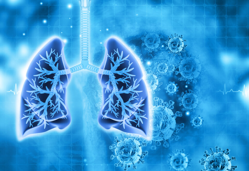
In the human body, the lungs and their vasculature can be likened to a building with an intricate plumbing system. The lungs’ blood vessels are the pipes essential for transporting blood and nutrients for oxygen delivery and carbon dioxide removal. Much like how pipes can get rusty or clogged, disrupting normal water flow, damage from respiratory viruses, like SARS-CoV-2 or influenza, can interfere with this “plumbing system.”

In a recent study, researchers looked at the critical role of vascular endothelial cells in lung repair. Their work, published in Science Translational Medicine, was led by Andrew Vaughan of the University of Pennsylvania’s School of Veterinary Medicine and shows that, by using techniques that deliver vascular endothelial growth factor alpha (VEGFA) via lipid nanoparticles (LNPs), that they were able to greatly enhance modes of repair for these damaged blood vessels, much like how plumbers patch sections of broken pipes and add new ones.

“While our lab and others have previously shown that endothelial cells are among the unsung heroes in repairing the lungs after viral infections like the flu, this tells us more about the story and sheds light on the molecular mechanisms at play,” says Vaughan, assistant professor of biomedical sciences at Penn Vet. “Here we’ve identified and isolated pathways involved in repairing this tissue, delivered mRNA to endothelial cells, and consequently observed enhanced recovery of the damaged tissue. These findings hint at a more efficient way to promote lung recovery after diseases like COVID-19.”

They found VEGFA’s involvement in this recovery, while building on work in which they used single cell RNA sequencing to identify transforming growth factor beta receptor 2 (TGFBR2) as a major signaling pathway. The researchers saw that when TGFBR2 was missing it stopped the activation of VEGFA. This lack of signal made the blood vessel cells less able to multiply and renew themselves, which is vital for the exchange of oxygen and carbon dioxide in the tiny air sacs of the lungs.

“We’d known there was a link between these two pathways, but this motivated us to see if delivering VEGFA mRNA into endothelial cells could improve lung recovery after disease-related injury,” says first author Gan Zhao, a postdoctoral researcher in the Vaughan Lab.

The Vaughan Lab then reached out to Michael Mitchell of the School of Engineering and Applied Science, whose lab specializes in LNPs, to see if delivery of this mRNA cargo would be feasible.

“LNPs have been great for vaccine delivery and have proven incredibly effective delivery vehicles for genetic information. But the challenge here was to get the LNPs into the bloodstream without them heading to the liver, which is where they tend to congregate as its porous structure lends favor to substances passing from the blood into hepatic cells for filtration,” says Mitchell, an associate professor of bioengineering at Penn Engineering and a coauthor of the paper. “So, we had to devise a way to specifically target the endothelial cells in the lungs.”

Lulu Xue, a postdoctoral researcher in the Mitchell Lab and a co-first author of the paper, explains that they engineered the LNP to have an affinity for lung endothelial cells, this is known as extra hepatic delivery, going beyond the liver.

Read the full story in Penn Today.

Leveraging the Body’s Postal System to Understand and Treat Disease

by Nathi Magubane

Microwell device with a solution in the reservoir (Image: Courtesy of David E. Reynolds)

Akin to the packages sent from one person to another via an elaborate postal system, cells send tiny parcels that bear contents and packaging material that serve key purposes: To protect the contents from the outside world and to make sure it gets to the right place via a label with an address. 

These packages are known as extracellular vesicles (EVs)—lipid-bound molecules that serve a variety of regulatory and maintenance functions throughout the body. They assist in the removal of unwanted materials within the cell, and they transport proteins, aid in DNA and RNA transfer, and promote tumorigeneses in cancerous cells. 

Given their myriad roles, EVs have taken center stage for many researchers in the biomedical space as they have the potential to improve current methods of disease detection and treatment. The main challenge, however, is accurately identifying the molecular contents of EVs while also characterizing the EVs, which, unlike other cellular components that are more homogenous, have more heterogeneity.

Now, a team of researchers at the University of Pennsylvania has developed a novel platform, droplet-free double digital assay, for not only profiling individual EVs but also accurately discerning their molecular contents. The researchers took the digital assay, which quantifies the contents of a molecule via binary metric—a 1 corresponds to the presence of a molecule and a zero to the lack thereof—and applies it to the EV. The work is published in Advanced Science.

The team was led by Jina Ko, an assistant professor with appointments in the School of Engineering and Applied Science and Perelman School of Medicine. “Our method allows for highly accurate quantification of the individual molecules inside an EV,” Ko says . “This opens up many doors in the realm of early disease detection and treatment.”

The researchers first compartmentalized individual EVs utilizing a microwell approach to isolate the EVs. Next, they captured individual molecules within the EVs and amplified the signal for clarity. The team then was able to determine the expression levels of pivotal EV biomarkers with remarkable precision via fluorescence.

Read the full story in Penn Today.
Jina Ko is an assistant professor in the Department of Pathology and Laboratory Medicine in the Perelman School of Medicine and an assistant professor in the Department of Bioengineering in the School of Engineering and Applied Science at the University of Pennsylvania.

David Reynolds is a Ph.D. candidate in the Department of Bioengineering in Penn Engineering.

Other authors include, Menghan Pan, George Galanis, Yoon Ho Roh, Renee-Tyler T. Morales, Shailesh Senthil Kumar, and Su-Jin Heo of the Department of Bioengineering at Penn Engineering; Jingbo Yang and Xiaowei Xu of the Department of Pathology and Laboratory Medicine at Penn Medicine; and Wei Guo of the Department of Biology in the School of Arts & Sciences at Penn.

The research was supported by the National Institutes of Health: grants R00CA256353, R35 GM141832, and CA174523 (SPORE).

Senior Design Team “StablEyes” Uses 3D Printing to Simplify Retinal Imaging

A team of Penn Bioengineering Senior Design students was featured as the 3D print of the week by the Penn Biomedical Library’s Biomeditations blog.

The StablEyes team. From left to right, Jake Becker (BE ’23), Ruoming Fan (BE ’23), Ella Atsavapranee (BE ’23), and Savan Patel (M&T ’23).

Fourth-year undergraduate students Ella Atsavapranee, Jake Becker, Ruoming Fan, and Savan Patel created StablEyes, “a stabilization mount that provides fine, motorized control of the handheld OCT to improve ease of use for physicians and machine learning-based software to aid in diagnosis from retinal images.” The team made use of 3D printing services, laboratory space, and expertise across Penn’s campus to create their innovative design, including the Bollinger Digital Fabrication Lab in the Holman Biotech Commons, the Fisher Fine Arts Library, the Children’s Hospital of Philadelphia (CHOP), and the George H. Stephenson Foundation Educational Laboratory & Bio-MakerSpace (aka the Penn BE Labs).

Read “Featured 3D Print: Simplifying Retinal Imaging with StablEyes” by Lexi Voss in Biomeditations.

How HIV Infection Shrinks the Brain’s White Matter

by Katherine Unger Baillie

Researchers from Penn and CHOP detail the mechanism by which HIV infection blocks the maturation process of brain cells that produce myelin, a fatty substance that insulates neurons.

A confocal microscope image shows an oligodendrocyte in cell culture, labeled to show the cell nucleus in blue and myelin proteins in red, green, and yellow. Researchers from Penn and CHOP have shown that HIV infection prevents oligodendrocytes from maturing, leading to a reduction in white matter in the brain. (Image: Raj Putatunda)

It’s long been known that people living with HIV experience a loss of white matter in their brains. As opposed to gray matter, which is composed of the cell bodies of neurons, white matter is made up of a fatty substance called myelin that coats neurons, offering protection and helping them transmit signals quickly and efficiently. A reduction in white matter is associated with motor and cognitive impairment.

Earlier work by a team from the University of Pennsylvania and Children’s Hospital of Philadelphia (CHOP) found that antiretroviral therapy (ART)—the lifesaving suite of drugs that many people with HIV use daily—can reduce white matter, but it wasn’t clear how the virus itself contributed to this loss.

In a new study using both human and rodent cells, the team has hammered out a detailed mechanism, revealing how HIV prevents the myelin-making brain cells called oligodendrocytes from maturing, thus putting a wrench in white matter production. When the researchers applied a compound blocking this process, the cells were once again able to mature.

The work is published in the journal Glia.

“Even when people with HIV have their disease well-controlled by antiretrovirals, they still have the virus present in their bodies, so this study came out of our interest in understanding how HIV infection itself affects white matter,” says Kelly Jordan-Sciutto, a professor in Penn’s School of Dental Medicine and co-senior author on the study. “By understanding those mechanisms, we can take the next step to protect people with HIV infection from these impacts.”

“When people think about the brain, they think of neurons, but they often don’t think about white matter, as important as it is,” says Judith Grinspan, a research scientist at CHOP and the study’s other co-senior author. “But it’s clear that myelination is playing key roles in various stages of life: in infancy, in adolescence, and likely during learning in adulthood too. The more we find out about this biology, the more we can do to prevent white matter loss and the harms that can cause.”

Jordan-Sciutto and Grinspan have been collaborating for several years to elucidate how ART and HIV affect the brain, and specifically oligodendrocytes, a focus of Grinspan’s research. Their previous work on antiretrovirals had shown that commonly used drugs disrupted the function of oligodendrocytes, reducing myelin formation.

In the current study, they aimed to isolate the effect of HIV on this process. Led by Lindsay Roth, who recently earned her doctoral degree within the Biomedical Graduate Studies group at Penn and completed a postdoctoral fellowship working with Jordan-Sciutto and Grinspan, the investigation began by looking at human macrophages, one of the major cell types that HIV infects.

Read the full story in Penn Today.

Kelly Jordan-Sciutto is vice chair and professor in the University of Pennsylvania School of Dental Medicine’s Department of Basic & Translational Sciences and is director of Biomedical Graduate Studies. She is a member of the Penn Bioengineering Graduate Group.

Penn Alumnus Peter Huwe Appointed Assistant Professor at Mercer University

Peter Huwe, Ph.D.

Peter Huwe, a University of Pennsylvania alumnus and graduate of the Radhakrishnan lab, was appointed Assistant Professor of Biomedical Sciences at the Mercer University School of Medicine beginning this summer 2020 semester.

Huwe earned dual B.S. degrees in Biology and Chemistry in 2009 from Mississippi College, where he was inducted into the Hall of Fame. At Mississippi College, Huwe had his first exposure to computational research in the laboratory of David Magers, Professor of Chemistry and Biochemistry. He went on to earn his Ph.D. in Biochemistry and Molecular Biophysics in 2014 in the laboratory of Ravi Radhakrishnan, Chair of the Bioengineering Department at Penn. As an NSF Graduate Research Fellow in Radhakrishnan’s lab, Huwe focused his research on using computational molecular modeling and simulations to elucidate the functional consequences of protein mutations associated with human diseases. Dr. Huwe then joined the structural bioinformatics laboratory Roland Dunbrack, Jr., Professor at the Fox Chase Cancer Center as a T32 post-doctoral trainee. During his post-doctoral training, Huwe held adjunct teaching appointments at Thomas Jefferson University and at the University of Pennsylvania. In 2017, Huwe became an Assistant Professor of Biology at Temple University, where he taught medical biochemistry, medical genetics, cancer biology, and several other subjects.

During each of his appointments, Huwe became increasingly more passionate about teaching, and he decided to dedicate his career to medical education. Huwe is very excited to be joining Mercer University School of Medicine as an Assistant Professor of Biomedical Sciences this summer. There, he will serve in a medical educator track, primarily teaching first and second year medical students.

“Without Ravi Radhakrishnan and Philip Rea, Professor of Biology in Penn’s School of Arts & Sciences, giving me my first teaching opportunities as a graduate guest lecturer at Penn, I may never have discovered how much I love teaching,” says Huwe. “And without the support and guidance of each of my P.I.’s [Dr.’s Magers, Radhakrishnan, and Dunbrack], I certainly would not be where I am, doing what I love.  I am incredibly thankful for all of the people who helped me in my journey to find my dream job.”

Congratulations and best of luck from everyone in Penn Bioengineering, Dr. Huwe!