Penn Bioengineering and COVID-19

A message from Penn Bioengineering Professor and Chair Ravi Radhakrishnan:
In response to the unprecedented challenges presented by the global outbreak of the novel coronavirus SARS-CoV-2, Penn Bioengineering’s faculty, students, and staff are finding innovative ways of pivoting their research and academic projects to contribute to the fight against COVID-19. Though these projects are all works in progress, I think it is vitally important to keep those in our broader communities informed of the critical contributions our people are making. Whether adapting current research to focus on COVID-19, investing time, technology, and equipment to help health care infrastructure, or creating new outreach and educational programs for students, I am incredibly proud of the way Penn Bioengineering is making a difference. I invite you to read more about our ongoing projects below.

RESEARCH

Novel Chest X-Ray Contrast

David Cormode, Associate Professor of Radiology and Bioengineering

Nanomedicine and Molecular Imaging Lab

Peter Noel, Assistant Professor of Radiology and BE Graduate Group Member

Laboratory for Advanced Computed Tomography Imaging

The Cormode and Noel labs are working to develop dark-field X-ray imaging, which may prove very helpful for COVID patients. It involves fabricating diffusers that incorporate gold nanoparticles to modify the X-ray beam. This method gives excellent images of lung structure. Chest X-ray is being used on the front lines for COVID patients, and this could potentially be an easy to implement modification of existing X-ray systems. The additional data give insight into the health state of the microstructures (alveoli) in the lung. This new contrast mechanics could be an early insight into the disease status of COVID-19 patients. For more on this research, see Cormode and Noel’s chapter in the forthcoming volume Spectral, Photon Counting Computed Tomography: Technology and Applications, edited by Katsuyuki Taguchi, Ira Blevis, and Krzysztof Iniewski (Routledge 2020).

Immunotherapy

Michael J. Mitchell, Skirkanich Assistant Professor of Innovation in Bioengineering

Mitchell Lab

Mike Mitchell is working with Saar Gill (Penn Medicine) on engineering drug delivery technologies for COVID-19 mRNA vaccination. He is also developing inhalable drug delivery technologies to block COVID-19 internalization into the lungs. These new technologies are adaptations of prior research published Volume 20 of Nano Letters (“Ionizable Lipid Nanoparticle-Mediated mRNA Delivery for Human CAR T Cell Engineering” January 2020) and discussed in Volume 18 of Nature Reviews Drug Discovery (“Delivery Technologies for Cancer Immunotherapy” January 2019).

Respiratory Distress Therapy Modeling

Ravi Radhakrishnan, Professor, and Chair of Bioengineering and Professor of Chemical and Biomolecular Engineering

Radhakrishnan Lab

Computational Models for Targeting Acute Respiratory Distress Syndrome (ARDS). The severe forms of COVID-19 infections resulting in death proceeds by the propagation of the acute respiratory distress syndrome or ARDS. In ARDS, the lungs fill up with fluid preventing oxygenation and effective delivery of therapeutics through the inhalation route. To overcome this major limitation, delivery of antiinflammatory drugs through the vasculature (IV injection) is a better approach; however, the high injected dose required can lead to toxicity. A group of undergraduate and postdoctoral researchers in the Radhakrishnan Lab (Emma Glass, Christina Eng, Samaneh Farokhirad, and Sreeja Kandy) are developing a computational model that can design drug-filled nanoparticles and target them to the inflamed lung regions. The model combines different length-scales, (namely, pharmacodynamic factors at the organ scale, hydrodynamic and transport factors in the tissue scale, and nanoparticle-cell interaction at the subcellular scale), into one integrated framework. This targeted approach can significantly decrease the required dose for combating ARDS. This project is done in collaboration with Clinical Scientist Dr. Jacob Brenner, who is an attending ER Physician in Penn Medicine. This research is adapted from prior findings published in Volume 13, Issue 4 of Nanomedicine: Nanotechnology, Biology and Medicine: “Mechanisms that determine nanocarrier targeting to healthy versus inflamed lung regions” (May 2017).

Diagnostics

Sydney Shaffer, Assistant Professor of Bioengineering and Pathology and Laboratory Medicine

Syd Shaffer Lab

Arjun Raj, Professor of Bioengineering

Raj Lab for Systems Biology

David Issadore, Associate Professor of Bioengineering and Electrical and Systems Engineering

Issadore Lab

Arjun Raj, David Issadore, and Sydney Shaffer are working on developing an integrated, rapid point-of-care diagnostic for SARS-CoV-2 using single molecule RNA FISH. The platform currently in development uses sequence specific fluorescent probes that bind to the viral RNA when it is present. The fluorescent probes are detected using a iPhone compatible point-of-care reader device that determines whether the specimen is infected or uninfected. As the entire assay takes less than 10 minutes and can be performed with minimal equipment, we envision that this platform could ultimately be used for screening for active COVID19 at doctors’ offices and testing sites. Support for this project will come from a recently-announced IRM Collaborative Research Grant from the Institute of Regenerative Medicine with matching funding provided by the Departments of Bioengineering and Pathology and Laboratory Medicine in the Perelman School of Medicine (PSOM) (PI’s: Sydney Shaffer, Sara Cherry, Ophir Shalem, Arjun Raj). This research is adapted from findings published in the journal Lab on a Chip: “Multiplexed detection of viral infections using rapid in situ RNA analysis on a chip” (Issue 15, 2015). See also United States Provisional Patent Application Serial No. 14/900,494 (2014): “Methods for rapid ribonucleic acid fluorescence in situ hybridization” (Inventors: Raj A., Shaffer S.M., Issadore D.).

HEALTH CARE INFRASTRUCTURE

Penn Health-Tech Coronavirus COVID-19 Collaborations

Brian Litt, Professor of Bioengineering, Neurology, and Neurosurgery

Litt Lab

In his role as one of the faculty directors for Penn Health-Tech, Professor Brian Litt is working closely with me to facilitate all the rapid response team initiatives, and in helping to garner support the center and remove obstacles. These projects include ramping up ventilator capacity and fabrication of ventilator parts, the creation of point-of-care ultrasounds and diagnostic testing, evaluating processes of PPE decontamination, and more. Visit the Penn Health-Tech coronavirus website to learn more, get involved with an existing team, or submit a new idea.

BE Labs COVID-19 Efforts

BE Educational Labs Director Sevile Mannickarottu & Staff
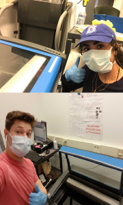
BE Educational Labs staff members Dana Abulez (BE ’19, Master’s BE ’20) and Matthew Zwimpfer (MSE ’18, Master’s MSE ’19) take shifts to laser-cut face shields.

The George H. Stephenson Foundation Educational Laboratory & Bio-MakerSpace staff have donated their PPE to Penn Medicine. Two staff members (Dana Abulez, BE ’19, Master’s BE ’20 and Matthew Zwimpfer, MSE ’18, Master’s MSE ’19) took shifts to laser-cut face shields in collaboration with Penn Health-Tech. Dana and Matthew are also working with Dr. Matthew Maltese on his low-cost ventilator project (details below).

Low-Cost Ventilator

Matthew Maltese, Adjunct Professor of Medical Devices and BE Graduate Group Member

Children’s Hospital of Philadelphia Center for Injury Research and Prevention (CIRP)

Dr. Maltese is rapidly developing a low-cost ventilator that could be deployed in Penn Medicine for the expected surge, and any surge in subsequent waves. This design is currently under consideration by the FDA for Emergency Use Authorization (EUA). This example is one of several designs considered by Penn Medicine in dealing with the patient surge.

Face Shields

David F. Meaney, Solomon R. Pollack Professor of Bioengineering and Senior Associate Dean

Molecular Neuroengineering Lab

Led by David Meaney, Kevin Turner, Peter Bruno and Mark Yim, the face shield team at Penn Health-Tech is working on developing thousands of rapidly producible shields to protect and prolong the usage of Personal Protective Equipment (PPE). Learn more about Penn Health-Tech’s initiatives and apply to get involved here.

Update 4/29/20: The Penn Engineering community has sprung into action over the course of the past few weeks in response to COVID-19. Dr. Meaney shared his perspective on those efforts and the ones that will come online as the pandemic continues to unfold. Read the full post on the Penn Engineering blog.

OUTREACH & EDUCATION

Student Community Building

Yale Cohen, Professor of Otorhinolaryngology, Department of Psychology, BE Graduate Group Member, and BE Graduate Chair

Auditory Research Laboratory

Yale Cohen, and Penn Bioengineering’s Graduate Chair, is working with Penn faculty and peer institutions across the country to identify intellectually engaging and/or community-building activities for Bioengineering students. While those ideas are in progress, he has also worked with BE Department Chair Ravi Radhakrishnan and Undergraduate Chair Andrew Tsourkas to set up a dedicated Penn Bioengineering slack channel open to all Penn Bioengineering Undergrads, Master’s and Doctoral Students, and Postdocs as well as faculty and staff. It has already become an enjoyable place for the Penn BE community to connect and share ideas, articles, and funny memes.

Undergraduate Course: Biotechnology, Immunology, Vaccines and COVID-19 (ENGR 35)

Daniel A. Hammer, Alfred G. and Meta A. Ennis Professor of Bioengineering and Chemical and Biomolecular Engineering

The Hammer Lab

This Summer Session II, Professor Dan Hammer and CBE Senior Lecturer Miriam R. Wattenbarger will teach a brand-new course introducing Penn undergraduates to a basic understanding of biological systems, immunology, viruses, and vaccines. This course will start with the fundamentals of biotechnology, and no prior knowledge of biotechnology is necessary. Some chemistry is needed to understand how biological systems work. The course will cover basic concepts in biotechnology, including DNA, RNA, the Central Dogma, proteins, recombinant DNA technology, polymerase chain reaction, DNA sequencing, the functioning of the immune system, acquired vs. innate immunity, viruses (including HIV, influenza, adenovirus, and coronavirus), gene therapy, CRISPR-Cas9 editing, drug discovery, types of pharmaceuticals (including small molecule inhibitors and monoclonal antibodies), vaccines, clinical trials. Some quantitative principles will be used to quantifying the strength of binding, calculate the dynamics of enzymes, writing and solving simple epidemiological models, methods for making and purifying drugs and vaccines. The course will end with specific case study of coronavirus pandemic, types of drugs proposed and their mechanism of action, and vaccine development.
Update 4/29/20: Read the Penn Engineering blog post on this course published April 27, 2020.

Neuromatch Conference

Konrad Kording, Penn Integrates Knowledge University Professor of Bioengineering, Neuroscience, and Computer and Information Science

Kording Lab

Dr. Kording facilitated Neuromatch 2020, a large virtual neurosciences conferences consisting of over 3,000 registrants. All of the conference talk videos are archived on the conference website and Dr. Kording has blogged about what he learned in the course of running a large  conference entirely online. Based on the success of Neuromatch 1.0, the team are now working on planning Neuromatch 2.0, which will take place in May 2020. Dr. Kording is also working on facilitating the transition of neuroscience communication into the online space, including a weekly social (#neurodrinking) with both US and EU versions.

Neuromatch Academy

Konrad Kording, Penn Integrates Knowledge University Professor of Bioengineering, Neuroscience, and Computer and Information Science

Kording Lab

Dr. Kording is working to launch the Neuromatch Academy, an open, online, 3-week intensive tutorial-based computational neuroscience training event (July 13-31, 2020). Participants from undergraduate to professors as well as industry are welcome. The Neuromatch Academy will introduce traditional and emerging computational neuroscience tools, their complementarity, and what they can tell us about the brain. A main focus is not just on using the techniques, but on understanding how they relate to biological questions. The school will be Python-based making use of Google Colab. The Academy will also include professional development / meta-science, model interpretation, and networking sessions. The goal is to give participants the computational background needed to do research in neuroscience. Interested participants can learn more and apply here.

Journal of Biomedical Engineering Call for Review Articles

Beth Winkelstein, Vice Provost for Education and Eduardo D. Glandt President’s Distinguished Professor of Bioengineering

Spine Pain Research Lab

The American Society of Medical Engineers’ (ASME) Journal of Biomechanical Engineering (JBME), of which Dr. Winkelstein is an Editor, has put out a call for review articles by trainees for a special issue of the journal. The call was made in March 2020 when many labs were ramping down, and trainees began refocusing on review articles and remote work. This call continues the JBME’s long history of supporting junior faculty and trainees and promoting their intellectual contributions during challenging times.
Update 4/29/20: CFP for the special 2021 issue here.

Are you a Penn Bioengineering community member involved in a coronavirus-related project? Let us know! Please reach out to ksas@seas.upenn.edu.



Congratulations to Danielle Bassett and Arjun Raj on Their Promotions to Full Professors

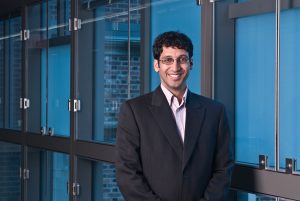
We would like to congratulate Penn Bioengineering faculty members Arjun Raj, Ph.D., and Danielle Bassett, Ph.D., on their promotions from associate to full professors.

Arjun Raj, Ph.D.

The Raj lab studies how biological processes work at the level of individual cells. Their work combines quantitative tools from genomics, imaging, biology, math, and computer science to develop models for how individual cells function, and in particular, how these individual cells can behave differently from each other. One major interest is in cancer, in which the lab is studying how individual cells can drive resistance to anti-cancer drugs. He and his lab also have a regularly updated blog discussing general topics related to scientific academia.

Danielle Bassett, Ph.D.

The Bassett lab takes an in-depth look at the use of network science and complex systems theory to study computational neuroscience in projects that involve the architecture of knowledge networks, the controllability of brain networks, and the dynamic networks in neuroscience. This fall, she will teach an elective course in network neuroscience open to graduate and undergraduate students that covers the use of network science in understanding overall brain circuitry. Bassett was recently profiled in Science Magazine.

Week in BioE (June 14, 2019)

by Sophie Burkholder

Bio-inspiration Informs New Football Helmet Design from IUPUI Students

Art, design, biology, and engineering all interact with each other in a recent design for a football helmet from two students one of media arts and the other of engineering at the Indiana University – Purdue University Indianapolis. Directed by Lecturer in Media Arts and Science Zebulun Wood, M.S., and Associate Professor of Mechanical and Energy Engineering and Assistant Professor of Biomedical Engineering Andres Tovar, Ph.D., the students found inspiration in biological structures like a pomelo peel, nautilus shell, and woodpecker skull to create energy-absorbing helmet liners. The resulting design took these natural concussion-reducing structures and created compliant mechanism lattice-based liners the replace the foam traditionally placed in between two harder shells of a typical helmet. Their work not only exemplifies the benefits of bio-inspiration, but demonstrates the way that several different domains of study can overlap in the innovation of a new product.

Study of Mechanical Properties of Hyaluronic Acid Could Help Inform Current Debates Over Treatment Regulation for Osteoarthritis

Arthritis is an extremely common condition, especially in older patients, in which inflammation of the joints can cause high amounts of stiffness and pain. Osteoarthritis in particular is the result of the degradation of flexible tissue between the bones of a joint, which increases friction in joint motion. A common treatment of this form of arthritis is the injection of hyaluronic acid, which is meant to provide joint lubrication, and decreases this friction between bones. Recently, however, there has been a debate over hyaluronic acid’s classification by the FDA and whether it should remain based on the knowledge of the mechanical actions of the acid in treatment for osteoarthritis or if potential chemical action of the acid should be considered as well.

Because of limited ways of testing the mechanical properties of the acid, many researchers felt that there could be more to hyaluronic acid’s role in pain relief for arthritic patients. But Lawrence Bonassar, Ph.D., the Daljit S. and Elaine Sarkaria Professor in Biomedical Engineering at the Meinig School of Bioengineering of Cornell University, had another idea. With his lab, he created a custom-made tribometer to measure the coefficient of friction of a given lubricant by rubbing a piece of cartilage back and forth across a smooth glass plate. The research demonstrated that hyaluronic acid’s ability to reduce the coefficient of friction aligned with patients’ pain relief. Bonassar and his team hope that these results will demonstrate the heavy contribution of mechanical action that hyaluronic acid has in osteoarthritis treatment, and help bring an end to the debate over its FDA classification.

A New Way of Mapping the Heart Could Lead to Better Understanding of Contractile Activity

Though reduced contractions in certain regions of the heart can be an indicator of a certain condition, there is currently no way to directly measure contractile activity. This is why Cristian Linte, Ph.D., an Associate Professor of Biomedical Engineering in the Kate Gleason College of Engineering at the Rochester Institute of Technology (RIT), hopes to create a map of the heart that can quantify contraction power. In collaboration with Niels Otani, Ph.D., an Associate Professor in the School of Mathematics at RIT, Linte plans to use an $850,000 grant from the National Science Foundation to achieve a more comprehensive understanding of the heart through both medical imaging and mechanical modeling. The group hopes that their approach will lead to not only a better way to diagnose certain heart conditions and diseases, but also open up understanding of active contraction, passive motion, and the stresses within the heart walls that underlie each.

Celebrity Cat Lil Bub Helps Penn and German Researchers Draw Public Attention to Genetics

Lil Bub’s unique appearance has garnered millions of online fans, and now, an avenue for researchers to talk about genetics. (Photo Courtesy of Mike Bridavsky)

In 2015, a group of curious researchers set out to sequence the genome of a celebrity cat named Lil Bub. They were hoping to understand the genetics behind Lil Bub’s extra toes and unique skeletal structure, which contribute to her heart-warming, kitten-like appearance. However, an equally important goal of their “LilBUBome” project was to invite the general public into the world of genetics.

Orsolya “Uschi” Symmons, a postdoctoral researcher at Penn in Associate Professor of Bioengineering Arjun Raj’s lab, led the research team along with Darío Lupiáñez at the Max-Delbrück Center for Molecular Medicine in Berlin, and Daniel Ibrahim at the Max Planck Institute for Molecular Geneticsin Berlin. Lil Bub’s owner, Mike Bridavsky, also contributed to the project.

Because of Lil Bub’s online fame, the project garnered attention from her fans and the media, all hoping to discover the secret to Lil Bub’s charm. As early as 2015, Gizmodo’s Kiona Smith-Strickland reported on the team’s intentions to sequence Lil Bub’s genome, and, since then, many have been awaiting the results of the LilBUBome.

To read more of this story, visit Penn Engineering’s Medium Blog.

People and Places

The Alfred P. Sloan Foundation awarded a six-year grant to Barnard College and Columbia University’s School of Engineering and Applied Science to support graduate education for women in engineering. The funding will go towards a new five-year program that enables Barnard students to attain both a B.A. and M.S. in one year after their traditional four years of undergraduate education. The program will offer M.S. degrees in chemical engineering, biomedical engineering, and industrial engineering and operations research, and is one of the first of its kind for women’s colleges.

We would like to congratulate Jean Paul Allain, Ph.D., on being named the first head of the new Ken and Mary Alice Lindquist Department of Nuclear Engineering at Penn State. Allain, who is currently a Professor and head of graduate programs in the University of Illinois at Urbana-Champaign’s Department of Nuclear, Plasma, and Radiological Engineering, conducts research in models of particle-surface interactions. In addition to being head of the new department at Penn State, Allain will also hold a position as a Professor of Biomedical Engineering at the university.

We would also like to congratulate Andrew Douglas, Ph.D., on his appointment as the Vice Provost for Faculty Affairs at Johns Hopkins University. Douglas currently holds the position of Vice Dean for Faculty at the Whiting School of Engineering, and has joint appointments in Mechanical and Biomedical Engineering. Douglas’s research at Hopkins focuses on mechanical properties and responses of compliant biological tissue and on the nonlinear mechanics of solids, with a focus on soft tissues and organs like the heart and tongue.

Week in BioE (July 31, 2018)

New Data Analysis Methods

Like many other fields, biomedical research is experiencing a data explosion. Some estimates suggest that the amount of data generated from the health sciences is now doubling every eighteen months, and experts expect it to double every seventy-three days by 2020.  One challenge that researchers face is how to meaningfully analyze this data tsunami.

The challenge of interpreting data occurs at all scales, and a recent collaboration shows how new approaches can allow us to handle the volumes of data emerging at the level of individual cells to infer more about how biology “works” at this level.  Wharton Statistics Department researchers Mo Huang and Jingshu Wang (PhD Student and Postdoctoral Researcher, respectively) collaborated with Arjun Raj’s lab in Bioengineering and published their findings in recent issues of Nature Methods and Proceedings of the National Academy of Sciences.  One study focused on a de-noising technique called SAVER to provide more precise data from single cell experiments and significantly improves the ability to detect trends in a dataset, similar to how increasing sample size helps improve the ability to determine differences between experimental groups.  The second method, termed DESCEND, creates more accurate characterization of gene expression that occur in individual cells. Together these two methods will improve data collection for biologists and medical professionals working  to diagnose, monitor, and treat diseased cells.

Dr. Raj’s team contributed data to the cause and acted as consultants on the biological aspects of this research. Further collaboration involved Mingyao Li, PhD, Professor of Biostatistics at the Perelman School of Medicine, and Nancy Zhang, PD, Professor Statistics at the Wharton School. “We are so happy to have had the chance to work with Nancy and Mingyao on analyzing single cell data,” said Dr. Raj. “The things they were able to do with our data are pretty amazing and important for the field.”

Advancements in Biomaterials

This blog features many new biomaterials techniques and substances, and there are several exciting new developments to report this week. First, the journal of Nature Biomedical Engineering published a study announcing a new therapy to treat or even eliminate lung infections, such as those acquired while in hospital or as the result of cystic fibrosis, which are highly common and dangerous. Researchers identified and designed viruses to target and kill the bacteria which causes these infections, but the difficulty of administering them to patients has proven prohibitive. This new therapy, developed by researchers at the Georgia Institute of Technology, is administered as a dry powder directly to the lungs and bypasses many of the delivery problems appearing in past treatments. Further research on the safety of this method is required before clinical trials can begin.

A team at Harvard University published another recent study in Nature Biomedical Engineering announcing their creation of a tissue-engineered scale model of the left human heart ventricle. This model is made from degradable fibers that simulate the natural fibers of heart tissue. Lead investigator Professor Kevin Kit Parker, PhD, and his team eventually hope to build specific models culled from patient stem cells to replicate the features of that patient’s heart, complete with the patient’s unique DNA and any heart defects or diseases. This replica would allow researchers and clinicians to study and test various treatments before applying them to a specific patient.

Lastly, researchers at the Tufts University School of Engineering published in the Proceedings of the National Academy of Sciences on their creation of flexible magnetic composites that respond to light. This material is capable of macroscale motion and is extremely flexible, allowing its adaptation into a variety of substances such as sponges, film, and hydrogels. Author and graduate student Meg Li explained that this material differs from similar substances in its complexity; for example, in the ability for engineers to dictate specific movements, such as toward or away from the light source. Co-author Fiorenzo Omenetto, PhD, suggests that with further research, these movements could be controlled at even more specific and detailed levels.

CFPS: Getting Closer to “On Demand” Medicine

A recent and growing trend in medicine is the move towards personalized or “on demand” medicine, allowing for treatment customized to specific patients’ needs and situations. One leading method is Cell-Free Protein Synthesis (CFPS), a way of engineering cellular biology without using actual cells. CFPS is used to make substances such as medicine, vaccines, and chemicals in a sustainable and portable way. One instance if the rapid manufacture of insulin to treat diabetic patients. Given that many clinics most in need of such substances are found in remote and under-served locations far away from well-equipped hospitals and urban infrastructure, the ability to safely and quickly create and transport these vital substances to patients is vitally important.

The biggest limiting factor to CFPS is difficulty of replicating Glycosylation, a complex modification that most proteins undergo. Glycosylation is important for proteins to exert their biological function, and is very difficult to synthetically duplicate. Previously, achieving successful Glycosylation was a key barrier in CFPS. Fortunately, Matthew DeLisa, PhD, the Williams L. Lewis Professor of Engineering at Cornell University and Michael Jewett, PD, Associate Professor of Chemical and Biological Engineering at Northwestern University, have created a “single-pot” glycoprotein biosynthesis that allows them to make these critical molecules very quickly. The full study was recently published in Nature Communications. With this new method, medicine is one step closer to being fully “on demand.”

People and Places

The Institute of Electrical and Electronics Engineers (IEEE) interviewed our own Penn faculty member Danielle Bassett, PhD, the Edwardo D. Glandt Faculty Fellow and Associate Professor in Bioengineering, for their website. Dr. Bassett, who shares a joint appointment with Electrical Systems Engineering (ESE) at Penn, has published groundbreaking research in Network Neuroscience, Complex Systems, and more. In the video interview (below), Dr. Bassett discusses current research trends in neuroscience and their applications in medicine.

Finally, a new partnership between Case Western Reserve University and Cleveland Clinic seeks to promote education and research in biomedical engineering in the Cleveland area. Cleveland Clinic Lerner Research Institute‘s Chair of Biomedical Engineering, Geoff Vince, PhD, sees this as an opportunity to capitalize on the renown of both institutions, building on the region’s already stellar reputation in the field of BME. Dozens of researchers from both institutions will have the opportunity to collaborate in a variety of disciplines and projects. In addition to professional academics and medical doctors, the leaders of this new partnership hope to create an atmosphere that can benefit all levels of education, all the way down to high school students.

Researchers Visualize Resistant Cancer Cells

by Meagan Ita, Ph.D. Student in Bioengineering and GABE Co-President

Cancer is a disease that affects millions, and over the last several decades, researchers have delved deeply into the biological underpinnings of the disease in the hopes of finding a cure. One major discovery is that mistakes in your DNA “instructions” can lead to cancer by crossing the wires in your cellular circuitry, and researchers have developed amazing new drugs that can cause tumors to melt away by targeting these broken components. The problem though is that, most of the time, the tumors come back, and this is a huge barrier to cures.

Shaffer ASCO
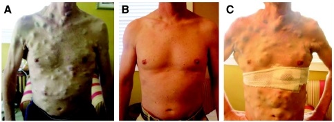
Picture of patient treated with vemurafenib and then developing resistance. Courtesy of the American Society of Clinical Oncology

For a long time, everyone assumed that the reason the tumors came back was  DNA mistakes on top of the original mistakes, with these new mistakes blocking the activity of the anti-cancer drug. However, new work led by Sydney Shaffer from the Arjun Raj Lab at Penn Bioengineering, published this week in Nature, challenges this view by looking all the way down at individual cancer cells and seeing how they respond to these drugs on a cell-by-cell basis.

Sydney found that in melanoma, contrary to what researchers thought, it need not be a DNA mistake that leads a cell to become resistant to the drug, but rather a change in cellular identity. Just like your body has cells of all different types, like skin cells and brain cells, cancer cells appear to change between different types, but unlike in the body, cancer cells do it in a seemingly random and uncontrolled way, and the cells exploit this variability to allow those rare cells that have changed their type to survive the drug.

Here, we talk with Sydney about the inspiration, triumphs, and challenges she faced in her research.

What was the initial inspiration for looking at drug resistance in melanoma?

For the first two years of working on this project, we actually didn’t have a clear question in mind. I was just trying a bunch of different experiments with melanoma cells, and I noticed something that we found thought-provoking. Whenever we gave the melanoma cells a particular drug, they would become resistant at exactly the same point in time. At first, this may not seem unusual, but for example, if everyone showed up at a restaurant to eat lunch at exactly noon, you would guess this was not happening purely by chance. Maybe classes let out right beforehand? Or a big meeting? For the melanoma cells, we would similarly expect there to be a range of different times for the cells to become resistant, but instead it all happened at once.

This observation helped us figure out that the drug-resistant cells probably already exist before we treat them. It also got us curious about the particular processes that make the cells resistant, and we spent many lab meetings discussing this observation until one postdoc, Gautham Nair, suggested trying some experiments based upon the classical molecular biology experiments of Luria and Delbrück.

Who were Luria and Delbrück, and how did they influence your work?

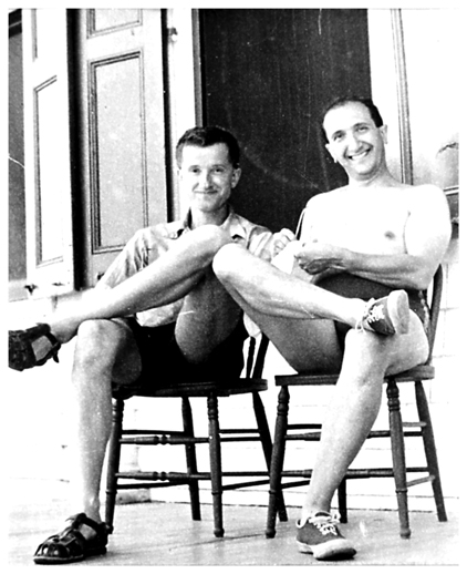
Max Delbrück and Salvador Luria (below) were scientists who, in the 1940s, performed a clever experiment that demonstrated that bacteria become resistant to viruses through random DNA mutations. According to Wikipedia, Luria actually had the idea for these experiments while watching slot machines!

Shaffer D&L
Delbrück (left) and Luria. Courtesy of the Genetics Society of America.

Their experiment was super simple: it was basically a statistical way to see whether cells “sense and respond” to a challenge, or whether they just passively get a mutation that lets some fraction of them survive the challenge, basically like Darwinian evolution. The idea is that, in the first scenario, there is no history: every cell has an equal chance to respond when challenged.

But in the second scenario, history matters in that if your great-grandparent was a survivor, then all your relatives would be too. If you could redo history over and over, then sometimes maybe your great-great-great-great-grandparent would be a survivor, and so you would get a whole bunch of survivors when the challenge came. Luria and Delbrück’s results showed that this second scenario was what happened with bacteria, providing the first evidence for genetic mutations in bacteria occurring in the absence of selection, and they both went on to win a Nobel prize in 1969.

Arjun actually had just lectured about these experiments in our graduate course on modeling biological systems. We adapted the same strategy and theory as Luria and Delbrück’s experiments for our work but applied it to melanoma and actually found a different result. Our experiments showed that resistance in melanoma does not arise through a heritable DNA mutation.

Shaffer colony
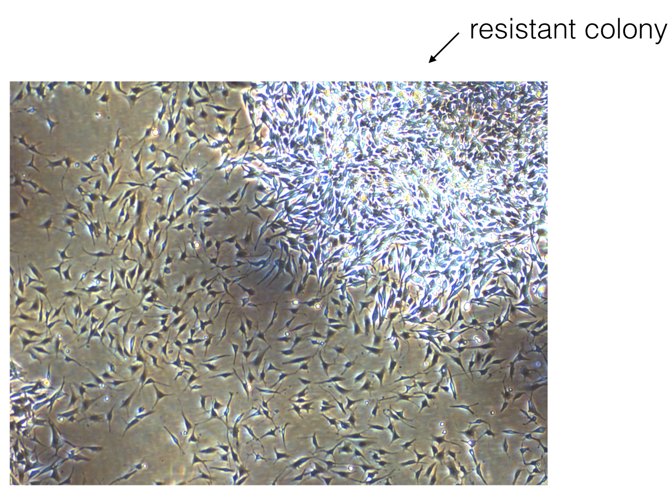
Picture of a resistant colony growing in the Raj lab.

Based upon this work, do you have any ideas for how we might prevent resistance in patients?

Yes. The recommended dosing for many of these drugs is daily. Our work would suggest that something like interval therapy might be more effective, for instance, if you gave the drug for a few days, killed many of the tumor cells, and then stopped the drug. During the time that the drug is stopped, the cells that initially survived the drug (we call these cells pre-resistant) could then transition out of this cell state and back to a sensitive state. Then, when the patient takes the drug again, it would be more effective at killing the remaining tumor cells. Another idea would be to find drugs that are specific to the pre-resistant cells and give these drugs in combination with other targeted therapies.

Were there any “Aha!” moments while working on this project?

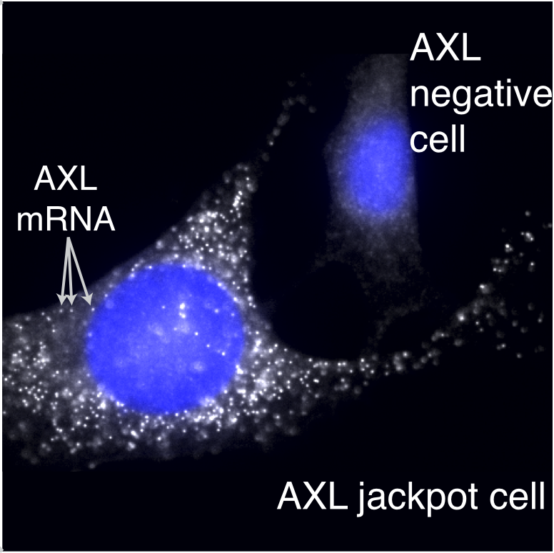
One of the most exciting moments of this research was when we first found the pre-resistant cells. Hidden among thousands of pictures of empty cells, we were shocked to actually see the rare cells full of brightly tagged resistance genes (below).

Shaffer cells
Resistant cells growing in the Raj lab.

What were some low points in working on the project? Do you recall any specific moments that you just felt intellectually and/or emotionally stumped? How did you get through them?

Oh yes, there were definitely low points during this project. One that stands out to me specifically was this one Friday afternoon where I presented at lab meeting. At the time, I only had a little bit of preliminary data. One of the members of the lab asked me a series of questions about resistance: How many different drug doses had I tried? Could I just give a lot and kill them all? What dose of drug is relevant for patients? What about drug resistance? Was I really interested in? All reasonable questions to ask. However, this was really overwhelming to a first-year graduate student because it made me realize that I didn’t have a clearly defined project that I was working on yet. There were just so many different questions that I didn’t know where to start.

Ultimately, with Arjun’s guidance, I came to realize that this was part of the process of figuring out what my thesis project would be, and the vagueness of our ideas at this time was a great thing because it left me open to find a problem that I found really interesting.

At another point in working on this: I remember that we were clearly conceptually stuck. We had identified the rare cells, but it wasn’t clear how to find out if these were the same cells that become resistant to drug. I had an entire lab meeting where we discussed this concern and came to the conclusion that, without some connection between the cells in this state and resistance, the work would be very speculative, which felt unsatisfying to me. Unfortunately, there wasn’t a quick fix to this problem. We just ended up trying a whole bunch of different ideas and eventually one of our strategies worked out.

Were there any funny moments that stand out to you?

Yeah! I was 40 weeks pregnant as we were finishing off our first submission of the paper! As my due date passed, I was really feeling the pressure to finish everything. Each day, I was coming into lab and just hoping I wouldn’t go into labor yet! Actually, the members of our lab had placed bets on when the baby would be born. Fortunately, those who bet on a late arrival ended up winning, and we submitted the paper the day before my daughter, Julien, was born. I was actually still at the hospital when I got the e-mail that the paper went to review.

So even though it might seem like this project is checked off the list with a kick-ass publication, there are probably a bunch of unfinished ideas you have. So,what are you working on next? Will this project ever be “done?”

For sure. The list of unfinished ideas is very long, and some of the questions that came from this work are now being pursued by other people in the lab. Right now, I’m working on ways of measuring the length of time that individual cells remain in these different cell states.

Interested in sharing your research in Penn BE? Contact penngabe@gmail.com for an interview by GABE (Graduate Association of Bioengineers) and let us know!