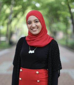
Lamis Elsawah graduated with a B.S.E. in Bioengineering with a concentration in Medical Devices in 2019. She is currently a Design Engineer at Johnson & Johnson’s DePuy Synthes. We caught up with Lamis to hear about why she chose Penn Bioengineering and what she enjoyed about the curriculum.
“Penn had been my dream school for years prior to even applying to college, so their having a top notch bioengineering program was icing on the cake when it was time for me to apply. Prior to applying, I actually had the opportunity to meet with Dr. Meaney (who was the Bioengineering Department Chair up until I graduated) the summer before my senior year in high school and he was always a constant support throughout my bioengineering education up until graduation. Since Bioengineering had less than 100 students per class, it really allowed us to develop that familial feel with our core Bioengineering professors and lab staff. I honestly don’t think I would have survived junior and senior year without the help of Sevile and the entire lab staff, so I will be forever grateful.
I always like to say that junior year labs are really what made me an engineer. Those were some of the most challenging classes I took, but it was really rewarding once I reached the end. Between those lab courses and Biomechatronics taught by Professor Dourte, it prepared me to become a design engineer and apply all that I had learned. I also had the opportunity to get my minor in Engineering Entrepreneurship and be taught by Professor Cassel, which increased my interest in the business side of developing medical devices. The combination of my studies ultimately led me to Imperial College, London where I received my Master’s in Medical Device Design and Entrepreneurship.
The bioengineering curriculum at Penn allowed me to have a vast knowledge of the field that I will always be grateful for. It not only provided me with the mechanical experience, but also the electrical and biological background. I plan on staying an active alumna in both the Engineering Alumni Society and the Penn Alumni Board as a result of my wonderful experience at Penn Engineering and Penn as a whole.”
This post is part of BE’s Alumni Spotlight series. Read more testimonies from BE Alumni on the BE website.
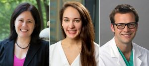
Beth Winkelstein, Megan Sperry, and Eric Granquist
Pain may be a universal experience, but what actually causes that experience within our brains is still poorly understood. Pain often continues long after the relevant receptors in the body have stopped being stimulated and can persist even after those receptors cease to exist, as is the case with “phantom limb” pain.
The exact experience an individual will have after a painful incident comes down to the complex, variable connections formed between several different parts of the brain. The inability to predict how those connections will form and evolve can make pain management a tricky, frustrating endeavor for both healthcare providers and patients.
Now, a team of Penn researchers has shown a way to make such predictions from the pattern of neural connections that begin to take shape soon after the first onset of pain. Though their study was conducted in rats, it suggests that similar brain imaging techniques could be used to guide treatment decisions in humans, such as which individuals are most likely to benefit from different drugs or therapies.
The study, published in the journal Pain, was led by Beth Winkelstein, Eduardo D. Glandt President’s Distinguished Professor in Penn Engineering’s Department of Bioengineering and Deputy Provost of the University of Pennsylvania, along with Megan Sperry, then a graduate student in her lab. Eric Granquist, Director of the Center for Temporomandibular Joint Disease at the Hospital of the University of Pennsylvania in the Department of Oral & Maxillofacial Surgery, and assistant professor of Oral & Maxillofacial Surgery in Penn’s School of Dental Medicine, also contributed to the research.
“Our findings provide the first evidence that brain networks differ between acute and persistent pain states, even before those different groups of rats actually show different pain symptoms,” says Winkelstein.
The Department of Bioengineering is proud to congratulate alumna Brianne Connizzo, PhD on her appointment as a tenure-track Assistant Professor in the Department of Biomedical Engineering in the College of Engineering at Boston University. Connizzo’s appointment will begin in January 2021, after completing her work as a postdoctoral researcher in Biological Engineering at MIT under the supervision of Alan J. Grodzinsky, ScD, Professor of Biological, Electrical, and Mechanical Engineering.
Connizzo got her BS in Engineering Science from Smith College (the first all women’s engineering program in the country) where she graduated in 2010 with highest honors. During her time there, she worked in the laboratory of Borjana Mikic, Rosemary Bradford Hewlett 1940 Professor of Engineering. While working in the lab, she explored the role of myostatin deficiency on Achilles tendon biomechanics and built mechanical testing fixtures for submerged testing of biological tissues. Connizzo continued along this path during her graduate studies in Bioengineering at Penn while working with Louis J. Soslowsky, Fairhill Professor in Orthopaedic Surgery and Professor in Bioengineering, at the McKay Orthopaedic Research Laboratory. Her thesis work focused on the dynamic re-organizations of collagen during tendon loading in the rotator cuff, developing a novel AFM-based method for measuring collagen fibril sliding along the way. During her time at Penn, Connizzo also served as the Social Chair for the Graduate Association of Bioengineers (GABE) and the Graduate Student Engineering Group (GSEG), both of which play a vital role in representing graduate students across the School of Engineering and Applied Sciences. She completed her PhD in Bioengineering in 2015 and then pursued her postdoctoral studies at MIT, focusing on fluid flow during compressive loading and developing novel explant culture models to explore real-time extracellular matrix turnover. For her work she was awarded both an NIH F32 postdoctoral fellowship and the NIH K99/R00 Pathway Independence Award, which are just a few of her long list of impressive accomplishments.
Although Connizzo’s interests in soft tissue mechanobiology span development, injury, and disease, her more recent work has targeted how aging influences tendon function and biology. With a fast-growing active and aging population, she believes that identifying the cause and contributors of age-related changes is critical to finding treatments and therapies that could prevent tendon disease, and thus improve overall population healthspan and quality of life. The primary objectives of the Connizzo Lab at Boston University will be to harness novel in vitro and in vivo models to study cell-controlled extracellular matrix remodeling and tissue biomechanics and to better understand normal tendon maintenance and the initiation of tendon damage in the context of aging.
“I am so grateful to have had the guidance of my mentors and peers at Penn during my doctoral studies, and even more thankful that many of those relationships remain a significant part of my support system to this day,” Connizzo says. “I’m really looking forward to this next chapter — to all the successes and failures in pursuing the science, to building a community at BU and in my own laboratory, and to supporting the next generation of brilliant young scientists.”
Congratulations Dr. Connizzo from everyone at Penn Bioengineering!
To combat the COVID-19 pandemic caused by the SARS-CoV2 virus, Dr. Andrew Tsourkas’s Targeted Imaging Therapeutics and Nanomedicine (Titan) Lab in Penn Bioengineering, in collaboration with the Penn-based startup, AlphaThera, was recently awarded a $667,000 SBIR Phase II Grant Extension to support its efforts in commercializing COVID-19 detection technology. The grant supports work to address the growing need for anti-viral antibody testing. Specifically, the Tsourkas Lab and AlphaThera hope to leverage their expertise with antibody conjugation technologies to reduce the steps and complexity of existing detection assays to enable greater production and higher sensitivity tests. AlphaThera was founded in 2016 by Andrew Tsourkas, PhD, Professor of Bioengineering and James Hui, MD, PhD, a graduate of the Perelman School of Medicine and Penn Bioengineering’s doctoral program.
During this pandemic it is crucial to characterize disease prevalence among populations, understand immunity, test vaccine efficacy and monitor disease resurgence. Projections have indicated that millions of daily tests will be needed to effectively control the virus spread. One important testing method is the serological assay: These tests detect the presence of SARS-CoV2 antibodies in a person’s blood produced by the body’s immune system responding to infection. Serological tests not only diagnose active infections, but also establish prior infection in an individual, which can greatly aid in forecasting disease spread and contact tracing. To perform the serological assays for antibody detection, well-established immunoassay methods are used such as ELISA.
A variety of issues have slowed the distribution of these serological assays for antibody testing. The surge in demand for testing has caused shortages in materials and reagents that are crucial for the assays. Furthermore, complexity in some of the assay formats can slow both production and affect the sensitivity of test results. Recognizing these problems, AlphaThera is leveraging its novel conjugation technology to greatly improve upon traditional assay formats.
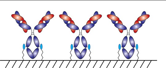
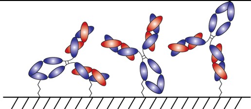
With AlphaThera’s conjugation technology, the orientation of antibodies can be precisely controlled so that they are aligned and uniformly immobilized on assay detection plates. This is crucial as traditional serological assays often bind antibodies to plates in a non-uniform manner, which increases variability of results and reduces sensitivity. See Fig 1 below. With AlphaThera’s uniform antibody immobilization, assay specificity could increase by as much as 1000- fold for detection of a patient’s SaRS-CoV2 antibodies.
Fig 1: Uniform vs Non-Uniform Immobilized Antibodies on Surface: Top is AlphaThera improvement, showing how antibodies would be uniformly immobilized and oriented on a plate for detection. Bottom is how many traditional serological assays immobilize antibodies, resulting in variability of results and lower specificity.
Furthermore, AlphaThera is addressing the shortage of assay reagents, specifically secondary antibody reagents, by removing certain steps from traditional serological assays. Rather than relying on secondary antibodies for detection of the patient antibodies, AlphaThera’s technology can label the patient SaRS-CoV2 primary antibodies directly in serum with a detection reagent. This eliminates several processing steps, reducing the time of the assay by as much as 50%, as well as the costs.
The Tsourkas Lab and AlphaThera have initiated their COVID-19 project, expanding into the Pennovation Center and onboarding new lab staff. Other antibody labeling products have also become available and are currently being prepared for commercialization. Check out the AlphaThera website to learn more about their technology at https://www.alphathera.com.
NIH SBIR Phase II Grant Extension— 5-R44-EB023750-03 (PI: Yu) — 10/07/2020 – 10/07/2021
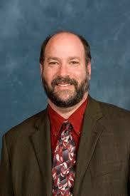
Huwe earned dual B.S. degrees in Biology and Chemistry in 2009 from Mississippi College, where he was inducted into the Hall of Fame. At Mississippi College, Huwe had his first exposure to computational research in the laboratory of David Magers, Professor of Chemistry and Biochemistry. He went on to earn his Ph.D. in Biochemistry and Molecular Biophysics in 2014 in the laboratory of Ravi Radhakrishnan, Chair of the Bioengineering Department at Penn. As an NSF Graduate Research Fellow in Radhakrishnan’s lab, Huwe focused his research on using computational molecular modeling and simulations to elucidate the functional consequences of protein mutations associated with human diseases. Dr. Huwe then joined the structural bioinformatics laboratory Roland Dunbrack, Jr., Professor at the Fox Chase Cancer Center as a T32 post-doctoral trainee. During his post-doctoral training, Huwe held adjunct teaching appointments at Thomas Jefferson University and at the University of Pennsylvania. In 2017, Huwe became an Assistant Professor of Biology at Temple University, where he taught medical biochemistry, medical genetics, cancer biology, and several other subjects.
During each of his appointments, Huwe became increasingly more passionate about teaching, and he decided to dedicate his career to medical education. Huwe is very excited to be joining Mercer University School of Medicine as an Assistant Professor of Biomedical Sciences this summer. There, he will serve in a medical educator track, primarily teaching first and second year medical students.
“Without Ravi Radhakrishnan and Philip Rea, Professor of Biology in Penn’s School of Arts & Sciences, giving me my first teaching opportunities as a graduate guest lecturer at Penn, I may never have discovered how much I love teaching,” says Huwe. “And without the support and guidance of each of my P.I.’s [Dr.’s Magers, Radhakrishnan, and Dunbrack], I certainly would not be where I am, doing what I love. I am incredibly thankful for all of the people who helped me in my journey to find my dream job.”
Congratulations and best of luck from everyone in Penn Bioengineering, Dr. Huwe!
Clear-fronted face masks, better and more frequent interpreters, and amped up involvement from local organizations have made a big difference during the COVID-19 pandemic.
By Michele Berger
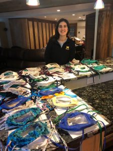
Since April 23, when bioengineering alum Kate Panzer (above) and her partners at the Deaf-Hearing Communication Centre started taking orders for masks with clear fronts, they’ve shipped about 450, with a backlog of requests for hundreds more. (Image: Courtesy Kate Panzer)
Because COVID-19 spreads via respiratory droplets that disperse through sneezes and coughs, shielding the mouth and nose is an important weapon against the virus. But it can also hinder conversations for people who rely on reading lips. “Communication barriers are already difficult sometimes, and this makes it more difficult,” says linguist Jami Fisher, director of Penn’s American Sign Language (ASL)/Deaf Studies program.
It’s one of the trickiest aspects of this pandemic for those in the Deaf and hard-of-hearing communities, Fisher says. The challenge doesn’t stem just from misunderstandings due to wearing masks. It’s also about the dissemination of accurate and timely information, knowing who to rely on and how to assess what’s being said.
Trusted sources like the Swarthmore, Pennsylvania–based nonprofit Deaf-Hearing Communication Centre (DHCC), a Penn community partner, have filled that gap, frequently updating information on its social media channels and websites. Governors and mayors are more frequently using Certified Deaf Interpreters (CDI) during press briefings, and Penn alum Kate Panzer, who graduated in 2018, started a project with DHCC to sew masks with clear fronts to offer both lip-reading access and protection.
Innovative masks
Like much of the country, Panzer has stayed inside for the past several months. When the pandemic started to worsen, she temporarily left a research position in Michigan and returned to her childhood home in Media, Pennsylvania. And like many people, she wanted to give back.
At Penn, she’d taken several American Sign Language classes through the program Fisher runs, so when she read an article about a student in Kentucky making clear-fronted masks, it piqued her interest. She reached out to Fisher, who connected her with Kyle Rosenberg, DHCC’s community development and outreach coordinator.
As a volunteer, she shared her mask idea with Rosenberg. “Even in normal times, the Deaf community really struggles with clear communication,” says Rosenberg, who is himself deaf. “ASL is very visual. It relies on body language. Covering up the mouth with a mask makes communication 10 times harder.”
Rosenberg helped Panzer tweak a design and create a process to reach the community, and they took their first order on April 23. Since then, they’ve shipped about 450 masks, with a backlog of requests for hundreds more.
Though the response has been overwhelmingly positive, when constructive feedback comes in, they do take it to heart, Panzer says. For example, when mask-wearers told them that the elastic bands they’d been using rubbed uncomfortably against hearing aids, they switched to fabric ties that go around the back of the head. The masks are not medical grade, so they can’t be used in a hospital setting, but Panzer says her goal was to improve everyday interactions.
“When you can only see the eyes, it takes a lot out of expressive communication for Deaf people,” says Fisher, whose parents and one brother are deaf. “It’s really important that they be able to more fully convey facial expressions and mouth movements that influence meaning.” Masks with clear fronts help.
NB: Kate has done prior work with ASL during her time at Penn Bioengineering. Kate’s 2018 Senior Design team created a two-way interface to help communication between deaf patients and hearing medical professionals called MEDISIGN. Fellow team members included fellow BE alumni Jackie Valeri, Nick Stiansen, and Karol Szymula. Watch their presentation on the Penn Engineering youtube channel.
Spencer Glantz (left) examines a scheme for light-activated protein cleavage with Dr. Brian Chow (middle) and 2014 iGEM team member Daniel Cabrera (right).
Spencer Glantz, a graduate of the Penn Bioengineering doctoral program and former member of the Brian Chow Lab, was mentioned in a recent WHYY piece highlighting the efforts of Penn labs to develop rapid, at-home testing for COVID-19. Glantz is currently a co-leader of the molecular biology team for 4Catalyzer, a medical device incubator founded by National Medal of Technology and Innovation recipient, and sponsor of the annual Rothberg Catalyzer Makerthon competition, Jonathan Rothberg. 4Catalyzer is developing the testing technology while Penn researchers are working to evaluate its effectiveness.
DNA Microscopy Gives a Better Look at Cell and Tissue Organization
A new technique that researchers from the Broad Institute of MIT and Harvard University are calling DNA microscopy could help map cells for better understanding of genetic and molecular complexities. Joshua Weinstein, Ph.D., a postdoctoral associate at the Broad Institute, who is also an alumnus of Penn’s Physics and Biophysics department and former student in Penn Bioengineering Professor Ravi Radhakrishnan’s lab, is the first author of this paper on optics-free imaging published in Cell.
The primary goal of the study was to find a way of improving analysis of the spatial organization of cells and tissues in terms of their molecules like DNA and RNA. The DNA microscopy method that Weinstein and his team designed involves first tagging DNA, and allowing the DNA to replicate with those tags, which eventually creates a cloud of sorts that diffuses throughout the cell. The DNA tags subsequent interactions with molecules throughout the cell allowed Weinstein and his team to calculate the locations of those molecules within the cell using basic lab equipment. While the researchers on this project focused their application of DNA microscopy on tracking human cancer cells through RNA tags, this new method opens the door to future study of any condition in which the organization of cells is important.
If you’ve ever pressed a picture-hanging strip onto the wall only to realize it’s slightly off-center, you know the disappointment behind adhesion as we typically experience it: it may be strong, but it’s mostly irreversible. While you can un-stick the used strip from the wall, you can’t turn its stickiness back on to adjust its placement; you have to start over with a new strip or tolerate your mistake. Beyond its relevance to interior decorating, durable, reversible adhesion could allow for reusable envelopes, gravity-defying boots, and more heavy-duty industrial applications like car assembly.
Such adhesion has eluded scientists for years but is naturally found in snail slime. A snail’s epiphragm — a slimy layer of moisture that can harden to protect its body from dryness — allows the snail to cement itself in place for long periods of time, making it the ultimate model in adhesion that can be switched on and off as needed. In a new study, Penn Engineers demonstrate a strong, reversible adhesive that uses the same mechanisms that snails do.
Low-Dose Radiation CT Scans Could Be Improved by Machine Learning
Machine learning is a type of artificial intelligence growing more and more popular for applications in bioengineering and therapeutics. Based on learning from patterns in a way similar to the way we do as humans, machine learning is the study of statistical models that can perform specific tasks without explicit instructions. Now, researchers at Rensselaer Polytechnic Institute (RPI) want to use these kinds of models in computerized tomography (CT) scanning by lowering radiation dosage and improving imaging techniques.
A recent paper published in Nature Machine Intelligence details the use of modularized neural networks in low-dose CT scans by RPI bioengineering faculty member Ge Wang, Ph.D., and his lab. Since decreasing the amount of radiation used in a scan will also decrease the quality of the final image, Wang and his team focused on a more optimized approach of image reconstruction with machine learning, so that as little data as possible would be altered or lost in the reconstruction. When tested on CT scans from Massachusetts General Hospital and compared to current image reconstruction methods for the scans, Wang and his team’s method performed just as well if not better than scans performed without the use of machine learning, giving promise to future improvements in low-dose CT scans.
A Mind-Controlled Robotic Arm That Requires No Implants
A new mind-controlled robotic arm designed by researchers at Carnegie Mellon University is the first successful noninvasive brain-computer interface (BCI) of its kind. While BCIs have been around for a while now, this new design from the lab of Bin He, Ph.D., a Trustee Professor and the Department Head of Biomedical Engineering at CMU, hopes to eliminate the brain implant that most interfaces currently use. The key to doing this isn’t in trying to replace the implants with noninvasive sensors, but in improving noisy EEG signals through machine learning, neural decoding, and neural imaging. Paired with increased user engagement and training for the new device, He and his team demonstrated that their design enhanced continuous tracking of a target on a computer screen by 500% when compared to typical noninvasive BCIs. He and his team hope that their innovation will help make BCIs more accessible to the patients that need them by reducing the cost and risk of a surgical implant while also improving interface performance.
KIChE is an organization that aims “to promote constructive and mutually beneficial interactions among Korean Chemical Engineers in the U.S. and facilitate international collaboration between engineers in U.S. and Korea.”
We would also like to congratulate Natalia Trayanova, Ph.D., of the Department of Biomedical Engineering at Johns Hopkins University on being inducted into the Women in Tech International (WITI) Hall of Fame. Beginning in 1996, the Hall of Fame recognizes significant contributions to science and technology from women. Trayanova’s research specializes in computational cardiology with a focus on virtual heart models for the study of individualized heart irregularities in patients. Her research helps to improve treatment plans for patients with cardiac problems by creating virtual simulations that help reduce uncertainty in either diagnosis or courses of therapy.
Finally, we would like to congratulate Andre Churchwell, M.D., on being named Vanderbilt University’s Chief Diversity Officer and Interim Vice Chancellor for Equity, Diversity, and Inclusion. Churchwell is also a professor of medicine, biomedical engineering, and radiology and radiological sciences at Vanderbilt, with a long career focused in cardiology.
Innovations in Vascularization Could Lead to a New Future in Bioprinting
We may be one step closer to 3D-printing organs for transplants thanks to innovations in vascularization from researchers at Rice University and Washington University. Jordan Miller, Ph.D., a Penn Bioengineering alumnus, now an assistant professor of bioengineering at Rice, worked with his colleague Kelly Stevens, Ph.D., an assistant professor of the bioengineering department at Washington, to develop 3D-printed networks that mimicked the vascularized pathways for the transport of blood, lymph, and other fluids in the body. Their work appeared on a recent cover of Science, featuring a visual representation of the 3D-printed vessels in vasculature meant to mirror that of the human lung.
Relying heavily on open source 3D-printing, Miller and Stevens, along with collaborators from a handful of other institutions and start-ups, found ways to model dynamic vasculature systems similar to heart valves, airways systems, and bile ducts to keep 3D-printed tissue viable. The video below demonstrates the way the team successfully modeled vasculature in a small portion of the lung by designing a net-like structure around a sack of air. But Miller, a long-time supporter of open source printing and bioprinting, hopes that this is merely one step closer to what he sees as the ultimate goal of allowing for all organs to be bioprinted. Having that sort of power would reduce the complex issues that come with organ transplants, from organ availability to compatibility, and bring an end to a health issue that affects the over 100,000 people on the organ transplant waiting list.
A Combination of Protein Synthesis and Spectrometry Improve Cell Engineering
One goal of modern medicine is to create individualized therapeutics by figuring out a way to control cell function to perform specific tasks for the body without disrupting normal cell function. Balancing these two goals often proves to be one of the greatest difficulties of this endeavor in the lab, but researchers at Northwestern University found a way to combine the two functions at once in methods they’re calling cell-free protein synthesis and self-assembled monolayer desorption ionization (SAMDI) mass spectrometry. This innovation in the combination of the two methods accelerates the trial and error process that comes with engineering cells for a specific need, allowing researchers to cover a lot more ground in determining what works best in a smaller amount of time.
Leading the study are Milan Mrksich, Ph.D., a Henry Wade Rogers Professor of Biomedical Engineering at Northwestern, and Michael Jewett, Ph.D., a Charles Deering McCormick Professor of Teaching Excellence and co-director of the Center for Synthetic Biology at Northwestern. Together, they hope to continue to take advantage of the factory-like qualities of cell operations in order to use cells from any organisms to our advantage as needed. By helping to reduce the amount of time spent on trial and error, this study brings us one step closer to a world of efficient and individualized medicine.
Non-Invasive Sensory Stimulation as New Way of Treating Alzheimer’s
What if we could reduce the effects of Alzheimer’s disease with a non-invasive therapy comprised of only sensory inputs of light and sound? A recent study between Georgia Tech and MIT tries to make that possible. Alzheimer’s patients often have a larger than normal number of amyloid plaques in their brains, which is a naturally occurring protein that in excess can disrupt neurological function. The treatment — designed in part by Abigail Paulson, a graduate student in the lab of Annabelle Singer, Ph.D., assistant professor of Biomedical Engineering at Georgia Tech and Emory University — uses a combination of light and sound to induce gamma oscillations in brain waves of mice with high amounts of these amyloid plaques. Another lead author of the study is Anthony Martorell, a graduate student in the Tsai Lab at MIT, where Singer was a postdoctoral researcher.
This new approach is different from other non-invasive brain therapies for memory improvement, as tests demonstrated that it had the power to not only reach the visual cortex, but that it also had an effect on the memory centers in the hippocampus. An innovation like this could bring about a more widespread form of treatment for Alzheimer’s patients, as the lack of a need for surgery makes it far more accessible. Singer hopes to continue the project in the future by looking at how these sensory stimulations affect the brain throughout a variety of processes, and more importantly, if the therapy can be successfully applied to human patients.
NIH Grant Awarded to Marquette Biomedical Engineering Professor for Metal Artifact Reduction Techniques in CT Scans
Taly Gilat-Shmidt, Ph.D., an associate professor of biomedical engineering at Marquette University, recently received a $1.4 million grant from the National Institute of Health to improve methods for radiation treatment through metal artifact reduction techniques. When patients have some sort of metal that can’t be removed, such as an orthopaedic implant like a hip or knee replacement, it can interfere with the imaging process for CT scans and lead to inaccuracies by obscuring some tissue in the final images. These inaccuracies can lead to difficulty in devising treatment plans for patients who require radiation, as CT scans are often used to assess patients and determine which line of treatment is most appropriate. Gilat-Schmidt hopes to use the grant to implement tested algorithms to help reduce this variability in imaging that comes from metal implants.
People and Places
Activities for Community Education in Science (ACES), founded by Penn chemistry graduate students in 2014, aims to inspire interest and provide a positive outlook in STEM for kids and their families. The biannual event provides students grades 3–8 with an afternoon of demonstrations, experiments, and hands-on activities focused on physics and chemistry.
After an explosive opening demonstration, more than 70 students made their way between experiments in small groups, each participating in different experiments based on their age.
The Society of Women Engineers (SWE) is a non-profit organization serving as one of the world’s largest advocates for women in engineering and technology over the past six decades. With a mission to empower women to become the next leading engineers of the world, SWE is just one of many agents hoping to bring more diversity to the field. Our chapter of SWE at Penn focuses particularly on professional development, local educational outreach, and social activities across all general body members. In a new article from SWE Magazine, the organization collected social media responses from the public on the women engineers we should all know. With a diverse list of engineers from both the past and present, the article helps bring to light just how much even a handful of women contributed to the field of engineering already.
Penn has one of the most distinctive graduate programs in the country, and is proud to graduate the first Ph.D. in Bioengineering in the United States. With such a history, our alumni have succeeded as professors, entrepreneurs, policy leaders, and industry pioneers. One recent example of this Penn tradition is leadership in national organizations.
Withing the field of biomaterials, the preeminent international organization is the Society for Biomaterials (SfB). Dedicated to the advancement of biomaterials science, the SfB was created more than four decades at nearly the same time the Bioengineering department was established at Penn. Many of our alumni are now part of the senior leadership in the SfB, including the following.
President: David Kohn
President-elect: Andrés García
Member-at-large: Helen Lu
In fact, of the three officers elected this year, two were from Penn (Andrés and Helen). We also have strong alumni representation across the various committees within the SfB. We extend our congratulations — with great pride — to our Penn family.


 A new technique that researchers from the Broad Institute of MIT and Harvard University are calling DNA microscopy could help map cells for better understanding of genetic and molecular complexities.
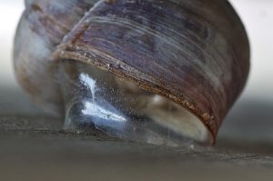

