Penn Bioengineers Recommend Improvements to Science Communication

Three graduate students in Bioengineering have collaborated to craft a list of recommendations to improve science communication during national health emergencies.

Doctoral students Miles J. Arnett, Dimitris Boufidis, and Melanie Hilman are part of the Penn Science Policy and Diplomacy Group (PSPDG), student organization which creates opportunities for students to get hands-on experience in Science Policy, Diplomacy, and Communication.

Their brief reviews the public health response to the COVID-19 pandemic and recommends specific improvements to science policy and communication by national scientific institutions:

The public health response to the pandemic was dramatically weakened by an uncoordinated communication strategy, inconsistent messaging, and fractured media environments. These shortcomings had a real human cost, with an estimated hundreds of thousands of Americans dying as a consequence of high rates of vaccine hesitancy. Now, in the aftermath of the pandemic, we have a chance to learn from this crisis and develop a more robust science communication infrastructure for future health emergencies.

Read “From Chaos to Clarity: Reinventing Science Communication After COVID-19” at Medium.

For a New Generation of Antibiotics, Scientists are Bringing Extinct Molecules Back to Life – and Discovering the Hidden Genetics of Immunity Along the Way

by Devorah Fischler

Marrying artificial intelligence with advanced experimental methods, the Machine Biology Group has mined the ancient past for future medical breakthroughs, bringing extinct molecules back to life. (Image credit: Ella Marushchenko)

“We need to think big in antibiotics research,” says Cesar de la Fuente. “Over one million people die every year from drug-resistant infections, and this is predicted to reach 10 million by 2050. There hasn’t been a truly new class of antibiotics in decades, and there are so few of us tackling this issue that we need to be thinking about more than just new drugs. We need new frameworks.”

De la Fuente is Presidential Assistant Professor in the Department of Bioengineering and the Department of Chemical and Biomolecular Engineering at the University of Pennsylvania School of Engineering and Applied Science. He holds additional primary appointments in Psychiatry and Microbiology in the Perelman School of Medicine.

De la Fuente’s lab, the Machine Biology Group, creates these new frameworks using potent partnerships in engineering and the health sciences, drawing on the “power of machines to accelerate discoveries in biology and medicine.”

Marrying artificial intelligence with advanced experimental methods, the group has mined the ancient past for future medical breakthroughs. In a recent study published in Cell Host and Microbe, the team has launched the field of “molecular de-extinction.”

Our genomes – our genetic material – and the genomes of our ancient ancestors, express proteins with natural antimicrobial properties. “Molecular de-extinction” hypothesizes that these molecules could be prime candidates for safe new drugs. Naturally produced and selected through evolution, these molecules offer promising advantages over molecular discovery using AI alone.

In this paper, the team explored the proteomic expressions of two extinct organisms –Neanderthals and Denisovans, archaic precursors to the human species – and found dozens of small protein sequences with antibiotic qualities. Their lab then worked to synthesize these molecules, bringing these long-since-vanished chemistries back to life.

“The computer gives us a sequence of amino acids,” says de la Fuente. “These are the building blocks of a peptide, a small protein. Then we can make these molecules using a method called ‘solid-phase chemical synthesis.’ We translate the recipe of amino acids into an actual molecule and then build it.”

The team next applied these molecules to pathogens in a dish and in mice to test the veracity and efficacy of their computational predictions.

“The ones that worked, worked quite well,” continues de la Fuente. “In two cases, the peptides were comparable – if not better – than the standard of care. The ones that didn’t work helped us learn what needed to be improved in our AI tools. We think this research opens the door to new ways of thinking about antibiotics and drug discovery, and this first step will allow scientists to explore it with increasing creativity and precision.”

Read the full story in Penn Engineering Today.

Student Spotlight: Cosette Tomita

Cosette TomitaCosette Tomita, a master’s student in Bioengineering, spoke with Penn Engineering Graduate Admissions about her research in cellular therapy and her path to Penn Engineering.

“What were you doing before you came to Penn Engineering? 

After college I wanted to get some industry experience before going to graduate school, so I spent a year working for a pharmaceutical company in New Jersey. I learned a lot—but mostly I learned that I wanted to go back into academia. So I was looking for a more research-oriented position to boost my graduate school applications, and I found a position at Penn’s cyclotron facility. Shortly after that, I applied to the master’s program. I’m still working at the cyclotron, so I’m doing the program part time. 

How has your experience in the program been so far? 

I love the research I’m doing here. I love the collaboration we have and the fact that I’m able to work with whoever I want to. And I can only say good things about my PI, Robert Mach. He’s a very busy man, but he makes time for his people. And he recognizes when somebody has a lot on their plate and he will go to bat for that person.

What’s your research all about? 

The focus of my PI’s lab is on neurodegenerative diseases and opiate use, so we’re looking to make imaging agents and antagonists that can help with the opioid crisis. 

For my project, I wanted to look at treating neurodegenerative disease from the perspective of cellular therapy. My PI doesn’t have that expertise, so when I came to him with this idea, he said I should talk to Mark Sellmyer in the bioengineering department. He does a lot of cellular therapies, cell engineering, protein engineering and things of that nature. So his lab is more biological. 

I don’t have a grant for my research, so my advisors are supporting it out of their own pockets. They could have said, no, you need to work on this project that’s already going on in the lab. But they gave me the intellectual freedom to do what I wanted to do.”

Read the full Q&A at the Penn Engineering Graduate Admissions website.

Mark Sellmeyer is Assistant Professor of Radiology in the Perelman School of Medicine and member of the Penn Bioengineering Graduate Group.

Could Psychedelics Simultaneously Treat Chronic Pain and Depression?

Ahmad Hammo

Ongoing clinical trials have demonstrated that psychedelics like psilocybin and LSD can have rapid and long-lived antidepressant and anti-anxiety effects. A related clinical problem is chronic pain, which is notoriously difficult to treat and often associated with depression and anxiety.

This summer, Ahmad Hammo, a rising third-year student in bioengineering in the School of Engineering and Applied Science, is conducting a pilot study to explore psilocybin’s potential as a therapy for chronic pain and the depression that often accompanies it.

“There’s a strong correlation between chronic pain and depression, so I’m looking at how a psychedelic might be used for treating both of these things simultaneously,” says Hammo, who is originally from Amman, Jordan.

Hammo is working under the guidance of neuroanesthesiologist and neuroscientist Joseph Cichon, an assistant professor in the Perelman School of Medicine. The effort is supported by the Penn Undergraduate Research Mentoring (PURM) program, administered by the Center for Undergraduate Research and Fellowships, which awards undergraduate students $5,000 to spend 10 weeks conducting research alongside Penn faculty.

Hammo’s project focuses on neuropathic pain, pain associated with nerve damage. Like other forms of chronic pain, most experts believe that chronic neuropathic pain is stored in the brain.

“Neuropathic pain can lead to a centralized pain syndrome where the pain is still being processed in the brain,” Cichon says. “It’s as if there’s a loop that keeps playing over and over again, and this chronic form is completely divorced from that initial injury.”

Read the full story in Penn Today.

Could the Age of the Universe Be Twice as Old as Current Estimates Suggest?

by Nathi Magubane

NASA’s James Webb Space Telescope has produced the deepest and sharpest infrared image of the distant universe to date. Known as Webb’s First Deep Field, this image of galaxy cluster SMACS 0723 is rich with detail. Thousands of galaxies—including the faintest objects ever observed in the infrared—have appeared in Webb’s view for the first time. The image shows the galaxy cluster SMACS 0723 as it appeared 4.6 billion years ago. The combined mass of this galaxy cluster acts as a gravitational lens, magnifying much more distant galaxies behind it. Webb’s Near-Infra Red Cam has brought those distant galaxies into sharp focus—they have tiny, faint structures that have never been seen before, including star clusters and diffuse features. (Image: NASA, ESA, CSA, and STScI)

Could the universe be twice as old as current estimates put forward? Rajendra Gupta of the University of Ottawa recently published a paper suggesting just that. Gupta claims the universe may be around 26.7 billion years rather than the commonly accepted 13.8 billion. The news has generated many headlines as well as criticism from astronomers and the larger scientific community.

Penn Today met with professors Vijay Balasubramanian and Mark Devlin to discuss Gupta’s findings and better understand the rationale of these claims and how they fit in the broader context of problems astronomers are attempting to solve.

How do we know how old the universe actually is?

Balasubramanian: The universe is often reported to be 13.8 billion years old, but, truth be told, this is an amalgamation of various measurements that factor in different kinds of data involving the apparent ages of ‘stuff’ in the universe.

This stuff includes observable or ordinary matter like you, me, galaxies far and near, stars, radiation, and the planets, then dark matter—the sort of matter that doesn’t interact with light and which makes up about 27% of the universe—and finally, dark energy, which makes up a massive chunk of the universe, around 68%, and is what we believe is causing the universe to expand.

And so, we take as much information as we can about the stuff and build what we call a consensus model of the universe, essentially a line of best fit. We call the model the Lambda Cold Dark Matter (ΛCDM).

Lambda represents the cosmological constant, which is linked to dark energy, namely how it drives the expansion of the universe according to Einstein’s theory of general relativity. In this framework, how matter and energy behave in the universe determines the geometry of spacetime, which in turn influences how matter and energy move throughout the cosmos. Including this cosmological constant, Lambda, allows for an explanation of a universe that expands at an accelerating rate, which is consistent with our observations.

Now, the Cold Dark Matter part represents a hypothetical form of dark matter. ‘Dark’ here means that it neither interacts with nor emits light, so it’s very hard to detect. ‘Cold’ refers to the fact that its particles move slowly because when things cool down their components move less, whereas when they heat up the components get excited and move around more relative to the speed of light.

So, when you consider the early formation of the universe, this ‘slowness’ influences the formation of structures in the universe like galaxies and clusters of galaxies, in that smaller structures like the galaxies form before the larger ones, the clusters.

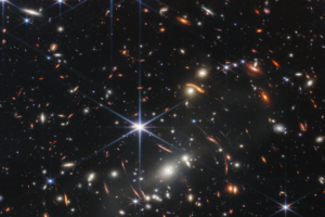
Devlin: And then taking a step back, the way cosmology works and pieces how old things are is that we look at the way the universe looks today, how all the structures are arranged within it, and we compare it to how it used to be with a set of cosmological parameters like Cosmic Microwave Background (CMB) radiation, the afterglow of the Big Bang, and the oldest known source of electromagnetic radiation, or light. We also refer to it as the baby picture of the universe because it offers us a glimpse of what it looked like at 380,000 years old, long before stars and galaxies were formed.

And what we know about the physical nature of the universe from the CMB is that it was something really smooth, dense, and hot. And as it continued to expand and cool, the density started to vary, and these variations became the seeds for the formation of cosmic structures.
The denser regions of the universe began to collapse under their own gravity, forming the first stars, galaxies, and clusters of galaxies. So, this is why, when we look at the universe today, we see this massive cosmic web of galaxies and clusters separated by vast voids. This process of structure formation is still ongoing.

And, so, the ΛCDM model suggests that the primary driver of this structure formation was dark matter, which exerts gravity and which began to clump together soon after the Big Bang. These clumps of dark matter attracted the ordinary matter, forming the seeds of galaxies and larger cosmic structures.

So, with models like the ΛCDM and the knowledge of how fast light travels, we can add bits of information, or parameters, and we have from things like the CMB and other sources of light in our universe, like the ones we get from other distant galaxies, and we see this roadmap for the universe that gives us it’s likely age. Which we think is somewhere in the ballpark of 13.8 billion years.

Read the full Q&A in Penn Today.

Vijay Balasubramanian is the Cathy and Marc Lasry Professor in the Department of Physics and Astronomy in the School of Arts & Sciences at the University of Pennsylvania. He is a member of the Penn Bioengineering Graduate Group.

Mark Devlin is the Reese W. Flower Professor of Astronomy and Astrophysics in the Department of Physics and Astronomy in the School of Arts & Sciences at Penn.

Riccardo Gottardi Recognized for Airway Research

Matthew Aronson (left), Ph.D. student in Bioengineering, and Riccardo Gottardi, Assistant Proessor in Bioengineering and Pediatrics.

Riccardo Gottardi, Assistant Professor in Pediatrics in the Perelman School of Medicine and in Bioengineering in the School of Engineering and Applied Science, has been named a “Young Innovator of Cellular and Molecular Bioengineering” by Cellular and Molecular Bioengineering, the official journal of the Biomedical Engineering Society (BMES). Gottardi is Chief Scientist in the Pediatric Airway Frontier Program at the Children’s Hospital of Philadelphia (CHOP). He leads the Bioengineering and Biomaterials (Bio2) Lab, and was recognized here for his research to prevent subglottic stenosis in children.

Gottardi’s work in subglottic stensosis, a severe narrowing of the airway in response to intubation, was recently profiled in CHOP’s Cornerstone Blog. CHOP’s award press release describes Gottardi’s innovative treatment:

“Prior studies by Dr. Gottardi’s lab used in vitro models to demonstrate that incorporating AMPs into polymer-coated tubes can inhibit bacterial growth and modulate the upper-airway microbiome. In a recent study in Cellular and Molecular Engineering, led by [Bioengineering] PhD student Matthew Aronson of the Gottardi Lab, the researchers went a step further and used both ex vivo and in vivo models to show how their patent-pending antimicrobial peptide-eluting endotracheal tube (AMP-ET) effectively targeted the local airway microbiota, reducing inflammation and resolving stenosis.

‘I am honored to be recognized by Cellular and Molecular Engineering for this exciting and notable award,” Dr. Gottardi said. “We are hopeful that our airway innovation will show similar success in human trials, so that we can improve outcomes for intubated pediatric patients.’”

Read CHOP’s full announcement of the award here.

“QR Code for Cancer Cells” – Uncovering Why Some Cells Become Resistant to Anti-Cancer Therapies

by Win Reynolds

QR codeA research team led by engineers at the University of Pennsylvania and Northwestern University scientists has created a new synthetic biology approach, or a “QR code for cancer cells,” to follow tumor cells over time, finding there are meaningful differences in why a cancer cell dies or survives in response to anti-cancer therapies.

Remarkably, what fate cancer cells choose after months of therapy is “entirely predictable” based on seemingly small, yet important, differences that appear even before treatment begins. The researchers also discovered the reason is not genetics, contrary to beliefs held in the field.

The findings were recently published in Nature.

The study outlined the team’s new technology platform that developed a QR code for each of the millions of cells for scientists to find and use later — much like tagging swans in a pond. The QR code directs researchers to a genome-wide molecular makeup of these cells and provides information about how they’ve reacted to cancer treatment.

“We think this work stands to really change how we think about therapy resistance,” said Arjun Raj, co-senior author and Professor in Bioengineering in the School of Engineering and Applied Science at the University of Pennsylvania. “Rather than drug-resistant cells coming in just one flavor, we show that even in highly controlled conditions, different ‘flavors’ can emerge, raising the possibility that each of these flavors may need to be treated individually.”

In the study, the lab and collaborators sought to apply synthetic biology tools to answer a key question in cancer research: What makes certain tumors come back a few months or years after therapy? In other words, could the lab understand what causes some rare cells to develop therapeutic resistance to a drug?

“There are many ways cells become different from each other,” said Yogesh Goyal, the co-senior author at Northwestern University. “Our lab asks, how do individual cells make decisions? Understanding this in the context of cancer is all the more exciting because there’s a clinically relevant dichotomy: A cell dies or becomes resistant when faced with therapies.”

Using the interdisciplinary team, the scientists put the before-and-after cloned cells through a whole genome sequencing pipeline to compare the populations and found no systematic underlying genetic mutations to investigate the hypothesis. Raj and Goyal  helped develop the QR code framework, FateMap, that could identify each unique cell that seemed to develop resistance to drug therapy. “Fate” refers to whether a cell dies or survives (and if so, how), and the scientists “map” the cells across their lifespan, prior to and following anti-cancer therapy. FateMap is the result of work from several research institutions, and it applies an amalgamation of concepts spanning several disciplines, including synthetic biology, genome engineering, bioinformatics, machine learning and thermodynamics.

“Some are different by chance — just as not all leaves on a tree look the same — but we wanted to determine if that matters,” Goyal said. “The cell biology field has a hard time defining if differences have meaning.”

Read the full story in Penn Engineering Today.

AI-guided Brain Stimulation Aids Memory in Traumatic Brain Injury

by Erica Moser

Illustration of a human brain
Image: iStock/Ogzu Arslan

Traumatic brain injury (TBI) has disabled 1 to 2% of the population, and one of their most common disabilities is problems with short-term memory. Electrical stimulation has emerged as a viable tool to improve brain function in people with other neurological disorders.

Now, a new study in the journal Brain Stimulation shows that targeted electrical stimulation in patients with traumatic brain injury led to an average 19% boost in recalling words.

Led by University of Pennsylvania psychology professor Michael Jacob Kahana, a team of neuroscientists studied TBI patients with implanted electrodes, analyzed neural data as patients studied words, and used a machine learning algorithm to predict momentary memory lapses. Other lead authors included Wesleyan University psychology professor Youssef Ezzyat and Penn research scientist Paul Wanda.

“The last decade has seen tremendous advances in the use of brain stimulation as a therapy for several neurological and psychiatric disorders including epilepsy, Parkinson’s disease, and depression,” Kahana says. “Memory loss, however, represents a huge burden on society. We lack effective therapies for the 27 million Americans suffering.”

Read the full story in Penn Today.

Michael Kahana is the Edmund J. and Louise W. Kahn Term Professor of Psychology at the University of Pennsylvania. He is a member of the Penn Bioengineering Graduate Group.

Artificial Intelligence is Leveling Up the Fight Against Infectious Diseases

by

Image credit: NIAID

Artificial intelligence is a new addition to the infectious disease researcher’s toolbox. Yet in merely half a decade, AI has accelerated progress on some of the most urgent issues in medical science and public health. Researchers in this field blend knowledge of life sciences with skill in computation, chemistry and design, satisfying decades-long appeals for interdisciplinary tactics to treat these disorders and stop their spread.

Diseases are “infectious” when they are caused by organisms, including parasites, viruses, bacteria and fungi. People and animals can contract infectious diseases from their environments or food, or through interactions with one another. Some, but not all, are contagious.

Infectious diseases are an intractable global challenge, posing problems that continue to grow in severity even as science has offered a steady pace of solutions. The world continues to become more interconnected, bringing people into new kinds and levels of relation, and the climate crisis is throwing environmental and ecological networks out of balance. Diseases that were once treatable by drugs have become resistant, and new drug discovery is more costly than ever. Uneven resource distribution means that certain parts of the world are perennial hotspots for diseases that others never fear.

Cesar de la Fuente brings an expert eye to how AI has transformed infectious disease research in a recently published piece in Science with co-authors Felix Wong and James J. Collins from MIT.

Presidential Assistant Professor in the Department of Bioengineering and the Department of Chemical and Biomolecular Engineering at the University of Pennsylvania School of Engineering and Applied Science, with additional primary appointments in Psychiatry and Microbiology within the Perelman School of Medicine, de la Fuente brings a multifaceted perspective to his survey of the field.

In the paper, de la Fuente and co-authors assess the progress, limitations and promise of research in AI and infectious diseases in three major areas of inquiry: anti-infective drug discovery, infection biology, and diagnostics for infectious diseases.

Read more in Penn Engineering Today.

Penn Bioengineers Create Non-invasive Cartilage Implants for Pediatric Subglottic Stenosis

by Emily Shafer

Paul Gehret and Riccardo Gottardi accept the International Society for Biofabrication New Investigator Award onstage at the international conference.
Paul Gehret (left) and Riccardo Gottardi, PhD, at Biofabrication 2022, the International Conference on Biofabrication.

Bioengineering researchers at Children’s Hospital of Philadelphia are developing a less invasive and quicker method to create cartilage implants as an alternative to the current treatment for severe subglottic stenosis, which occurs in 10 percent of premature infants in the U.S.

Subglottic stenosis is a narrowing of the airway, in response to intubation. Severe cases require laryngotracheal reconstruction that involves grafting cartilage from the rib cage with an invasive surgery. With grant support from the National Institutes of Health, Riccardo Gottardi, PhD, who leads the Bioengineering and Biomaterials (Bio2) Lab at CHOP, is refining a technology called Meniscal Decellularized scaffold (MEND). Working with a porcine model meniscus, the researchers remove blood vessels and elastin fibers to create pathways that allow for recellularization. Dr. Gottardi and his team then harvest ear cartilage progenitor cells (CPCs) with a minimally invasive biopsy, combine them with MEND, and create cartilage implants that could be a substitute for the standard laryngotracheal reconstruction.

This work and similar work on the tympanic membrane earned Paul Gehret, a doctoral student in the Gottardi Lab, the International Society for Biofabrication New Investigator Award and the Wake Forest Institute for Regenerative Medicine Young Investigator Award.  Gehret and Dr. Gottardi accepted the awards at Biofabrication 2022, the International Conference on Biofabrication, in Pisa Italy.

While laryngotracheal reconstruction in the adult population has a success rate of up to 96%, success rates in children range from 75% to 85%, and children often require revision surgery due to a high incidence of restenosis. The procedure also involves major surgery to remove cartilage from the rib cage, which is more difficult for childrens’ smaller bodies.

“Luckily not many children suffer from severe subglottic stenosis, but for those who do, it is really serious,” said Dr. Gottardi, who also is assistant professor in the Department of Pediatrics and Department of Bioengineering at CHOP and the University of Pennsylvania. “With our procedure, we have an easily accessible source for the cartilage and the cells, providing a straightforward and noninvasive treatment option with much potential.”

Read the full story in CHOP’s Cornerstone Blog.

Riccardo Gottardi is an Assistant Professor in the Department of Pediatrics, Division of Pulmonary Medicine in the Perelman School of Medicine and in the Department of Bioengineering in the School of Engineering and Applied Science. He also holds an appointment in the Children’s Hospital of Philadelphia (CHOP).

Paul Gehret is a Ph.D. student in Bioengineering, an Ashton Fellow and a NSF Fellow. His research focuses on leveraging decellularized cartilage scaffolds and novel cell sources to reconstruct the pediatric airway.