Speaker: Sumita Pennathur, Ph.D.
Professor of Mechanical Engineering
University of California, Santa Barbara
Date: Thursday, November 21, 2019
Time: 12:00-1:00 pm
Location: Room 337, Towne Building
Title: “Nanofluidic Technologies for Biomolecule Manipulation”
Abstract:
In the last 20 years, microfabrication techniques have allowed researchers to miniaturize tools for a plethora of bioanalytical applications. In addition to better sensitivity, accuracy and precision, scaling down the size of bioanalytical tools has led to the exploitation of new technologies to further manipulate biomolecules in ways that has never before been achieved. For example, when microfluidic channels are on the same order of magnitude of the electric double layers that form due to localized charge at the surfaces, there exists unique physics that create different flow phenomenon, such as analyte concentration and/or separation, mainly due to the couples physics of electrostatics and fluid dynamics. This talk will outline the basis of such interesting phenomena, such as nanofluidic separation and concentration, and well as probe the applications of such coupled systems, for example, handheld DNA detection. Most importantly, we will focus on the most recent work in the Pennathur lab in this field — biopolar electrode (BPE)-based phenomenon. Bipolar electrodes (BPE) have been studied in microfluidic systems over the past few decades, and through rigorous experimentally-validated modeling of the rich combined physics of fluid dynamics, electrokinetics, and electrochemistry at BPEs, I will show the potential of utilizing microfluidic-based BPEs for the design and development of low power, accurate, low volume fluid pumping mechanisms, with the ultimate goal of integration into wearable drug delivery and µTAS systems.
Bio:
Professor Pennathur has been a Professor of Mechanical Engineering at University of California, Santa Barbara in 2007, specializing in the fields of MEMS, nanofludics, and electrokinetics. Her most significant contributions include: 1) unearthing a novel mechanism for separation and concentration of analytes for bioanalytical applications, 2) developing a label-free detection mechanism for nucleic acids (that has since spun off into a point-of-care diagnostic company), 3) developing commercial medical diagnostic products, 4) building optical and acoustic biosensors and 5) developing revolutionized methods for measuring blood glucose for patients with diabetes. She received her B.S. and M.S. from MIT and PhD. From Stanford University.
Growing up in Sri Lanka and being surrounded by relatives who were doctors, I have been fascinated by both modern and traditional medicine. However, during physician shadowing in high school, I came to the realization that I was far more fascinated with the technology doctors use rather than practicing medicine. Therefore, I made the decision to turn down studying medicine in the U.K. and come to Penn to study Bioengineering in the hopes of being more hands-on with medical technology.
Have you done research with a professor on campus? What did you like, and what didn’t you like about it?
I currently work in the Interventional Radiology Lab at the Hospital of the University of Pennsylvania (HUP) under Assistant Professor of Radiology Chamith Rajapakse. The best thing about research here is that I get to be hands-on with some of the most cutting edge technology in the world and help pioneer medical diagnostic techniques that aren’t traditionally being used anywhere else. The only downside is that the learning curve can be a little too steep.
What have been some of your favorite courses and/or projects in Bioengineering so far?
Without a doubt, my favorite BE class has to be BE 309 (Bioengineering Modeling, Analysis and Design Laboratory I) and especially the Computer-Cockroach Interface we have to develop for this lab.
What advice would you give to your freshman self?
There are way too many things happening at a given time at Penn. Take it easy and plan it out so you can do everything you want to! It’s totally possible. Who says you can’t work hard and play hard?!
What do you hope to pursue after obtaining your undergraduate degree?
My hope is to head my own health-tech startup and create technologies that will aid developing countries, starting out with my humble island of Sri Lanka first.
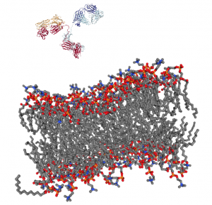
Getting a complex protein like an antibody through the membrane of a cell without damaging either is a long-standing challenge in the life sciences. Penn Engineers have found a plug-and-play solution that makes antibodies compatible with the delivery vehicles commonly used to ferry nucleic acids across that barrier.
For almost any conceivable protein, corresponding antibodies can be developed to block it from binding or changing shape, which ultimately prevents it from carrying out its normal function. As such, scientists have looked to antibodies as a way of shutting down proteins inside cells for decades, but there is still no consistent way to get them past the cell membrane in meaningful numbers.
Now, Penn Engineering researchers have figured out a way for antibodies to hitch a ride with transfection agents, positively charged bubbles of fat that biologists routinely use to transport DNA and RNA into cells. These delivery vehicles only accept cargo with a highly negative charge, a quality that nucleic acids have but antibodies lack. By designing a negatively charged amino acid chain that can be attached to any antibody without disrupting its function, they have made antibodies broadly compatible with common transfection agents.
Beyond the technique’s usefulness towards studying intracellular dynamics, the researchers conducted functional experiments with antibodies that highlight the technique’s potential for therapeutic applications. One antibody blocked a protein that decreases the efficacy of certain drugs by prematurely ejecting them from cells. Another blocked a protein involved in the transcription process, which could be an even more fundamental way of knocking out proteins with unwanted effects.
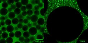
The researchers’ experiments involved making synthetic tissues with artificial “cells.” The fibrin network that surrounds these beads pull on them when compressed; by changing the number of beads in their experimental tissues, the researchers could suss out how cell-fiber interplay contributes to the tissue’s overall properties.
Tissue gets stiffer when it’s compressed. That property can become even more pronounced with injury or disease, which is why doctors palpate tissue as part of a diagnosis, such as when they check for lumps in a cancer screening. That stiffening response is a long-standing biomedical paradox, however: tissue consists of cells within a complex network of fibers, and common sense dictates that when you push the ends of a string together, it loosens tension, rather than increasing it.
Now, in a study published in Nature, University of Pennsylvania’s School of Engineering and Applied Science researchers have solved this mystery by better understanding the mechanical interplay between that fiber network and the cells it contains.
The researchers found that when tissue is compressed, the cells inside expand laterally, pulling on attached fibers and putting more overall tension on the network. Targeting the proteins that connect cells to the surrounding fiber network might therefore be the optimal way of reducing overall tissue stiffness, a goal in medical treatments for everything from cancer to obesity.
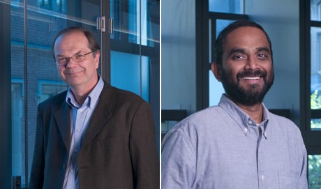
Paul Janmey and Vivek Shenoy
The study was led by Paul Janmey, Professor in the Perelman School of Medicine’s Department of Physiology and in Penn Engineering’s Department of Bioengineering, and Vivek Shenoy, Eduardo D. Glandt President’s Distinguished Professor in Penn Engineering’s Department of Materials Science and Engineering, Mechanical Engineering and Applied Mechanics, and Bioengineering, along with Anne van Oosten and Xingyu Chen, graduate students in Janmey’s and Shenoy’s labs. Van Oosten is now a postdoctoral fellow at Leiden University in The Netherlands.
Shenoy is Director of Penn’s Center for Engineering Mechanobiology, which studies how physical forces influence the behavior of biological systems; Janmey is the co-director of one of the Center’s working groups, organized around the question, “How do cells adapt to and change their mechanical environment?”
Together, they have been interested in solving the paradox surrounding tissue stiffness.
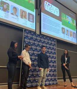
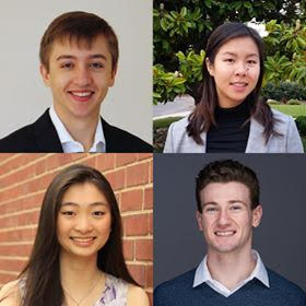
We would like to congratulate Penn Bioengineering Senior Design team MeVR on winning a Berkman Prize. MeVR consists of current BE seniors Nicole Chiou, Gabriel DeSantis, Ben Habermeyer, and Vera Lee. Awarded by the Penn Engineering Entrepreneurship Program, the Berkman Opportunity Fund provides grants to support students with innovative ideas that might turn into products and companies.
Bioengineering Seniors Ben Habermeyer (top left), Nicole Chiou (top right), Gabriel DeSantis (bottom right), and Vera Lee (bottom left)
MeVR is a bioresponsive virtual reality platform for administering biofeedback therapy. Biofeedback is the process of gaining greater awareness of involuntary physiological functions using sensors that provide information on the activity of those bodily systems, with the goal of gaining voluntary control over functions such as heart rate, muscle tension, and pain perception. This therapy is used to treat a variety of conditions such as chronic pain, stress, anxiety, and PTSD. These treatments cost on the order of hundreds to thousands of dollars, require the presence of a therapist to set up and deliver the therapy session, and are generally not interactive or immersive. MeVR is a platform to reduce these limitations of biofeedback therapy through an individualized, immersive, and portable device which guides users through biofeedback therapy using wearable sensors and a virtual reality environment which responds in real-time to biological feedback from the user’s body.
The blue circle is the global symbol for diabetes. Wikimedia Commons.
Diabetes is one of the more common diseases among Americans today, with the American Diabetes Association estimating that approximately 9.5 percent of the population battles the condition today. Though symptoms and causes may vary across types and patients, diabetes generally results from the body’s inability to produce enough insulin to keep blood sugar levels in check. A new experimental treatment from the lab of Sha Jin, Ph.D., a biomedical engineering professor at Binghamton University, aims to use about $1.2 million in recent federal grants to develop a method for pancreatic islet cell transplantation, as those are the cells responsible for producing insulin.
But the catch to this new approach is that relying on healthy donors of these islet cells won’t easily meet the vast need for them in diabetic patients. Sha Jin wants to use her grants to consider the molecular mechanisms that can lead pluripotent stem cells to become islet-like organoids. Because pluripotent stem cells have the capability to evolve into nearly any kind of cell in the human body, the key to Jin’s research is learning how to control their mechanisms and signaling pathways so that they only become islet cells. Jin also wants to improve the eventual culture of these islet cells into three-dimensional scaffolds by finding ways of circulating appropriate levels of oxygen to all parts of the scaffold, particularly those at the center, which are notoriously difficult to accommodate. If successful in her tissue engineering efforts, Jin will not only be able to help diabetic patients, but also open the door to new methods of evolving pluripotent stem cells into mini-organ models for clinical testing of other diseases as well.
A Treatment to Help Others See Better
Permanently crossed eyes, a medical condition called strabismus, affects almost 18 million people in the United States, and is particularly common among children. For a person with strabismus, the eyes don’t line up to look at the same place at the same time, which can cause blurriness, double vision, and eye strain, among other symptoms. Associate professor of bioengineering at George Mason University, Qi Wei, Ph.D., hopes to use almost $2 million in recent funding from the National Institute of Health to treat and diagnose strabismus with a data-driven computer model of the condition. Currently, the most common method of treating strabismus is through surgery on one of the extraocular muscles that contribute to it, but Wei wants her model to eventually offer a noninvasive approach. Using data from patient MRIs, current surgical procedures, and the outcomes of those procedures, Wei hopes to advance and innovate knowledge on treating strabismus.
A Newly Analyzed Brain Mechanism Could be the Key to Stopping Seizures
Among neurological disorders, epilepsy is one of the most common. An umbrella term for a lot of different seizure-inducing conditions, many versions of epilepsy can be treated pharmaceutically. Some, however, are resistant to the drugs used for treatment, and require surgical intervention. Bin He, Ph. D., the Head of the Department of Biomedical Engineering at Carnegie Mellon University, recently published a paper in collaboration with researchers at Mayo Clinic that describes the way that seizures originating at a single point in the brain can be regulated by what he calls “push-pull” dynamics within the brain. This means that the propagation of a seizure through the brain relies on the impact of surrounding tissue. The “pull” he refers to is of the surrounding tissue towards the seizure onset zone, while the “push” is what propagates from the seizure onset zone. Thus, the strength of the “pull” largely dictates whether or not a seizure will spread. He and his lab looked at different speeds of brain rhythms to perform analysis of functional networks for each rhythm band. They found that this “push-pull” mechanism dictated the propagation of seizures in the brain, and suggest future pathways of treatment options for epilepsy focused on this mechanism.
Hyperspectral Imaging Might Provide New Ways of Finding Cancer
A new method of imaging called hyperspectral imaging could help improve the prediction of cancerous cells in tissue specimens. A recent study from a University of Texas Dallas team of researchers led by professor of bioengineering Baowei Fei, Ph.D., found that a combination of hyperspectral imaging and artificial intelligence led to an 80% to 90% level of accuracy in identifying the presence of cancer cells in a sample of 293 tissue specimens from 102 patients. With a $1.6 million grant from the Cancer Prevention and Research Institute of Texas, Fei wants to develop a smart surgical microscope that will help surgeons better detect cancer during surgery.
Fei’s use of hyperspectral imaging allows him to see the unique cellular reflections and absorptions of light across the electromagnetic spectrum, giving each cell its own specific marker and mode of identification. When paired with artificial intelligence algorithms, the microscope Fei has in mind can be trained to specifically recognize cancerous cells based on their hyperspectral imaging patterns. If successful, Fei’s innovations will speed the process of diagnosis, and potentially improve cancer treatments.
People and Places
A group of Penn engineering seniors won the Pioneer Award at the Rothberg Catalyzer Makerthon led be Penn Health-Tech that took place from October 19-20, 2019. SchistoSpot is a senior design project created by students Vishal Tien (BE ‘20), Justin Swirbul (CIS ‘20), Alec Bayliff (BE ‘20), and Bram Bruno (CIS ‘20) in which the group will design a low-cost microscopy dianostic tool that uses computer vision capabilities to automate the diagnosis of schistosomiasis, which is a common parasitic disease. Read about all the winners here.
Virginia Tech University will launch a new Cancer Research Initiative with the hope of creating an intellectual community across engineers, veterinarians, biomedical researchers, and other relevant scientists. The initiative will focus not only on building better connections throughout departments at the university, but also in working with local hospitals like the Carilion Clinic and the Children’s National Hospital in Washington, D.C. Through these new connections, people from all different areas of science and engineering and come together to share ideas.
Associate Professor of Penn Bioengineering Dani Bassett, Ph.D., recently sat down with the Penn Integrates Knowledge University Professor Duncan Watts, Ph.D., for an interview published in Penn Engineering. Bassett discusses the origins of network science, her research in small-world brain networks, academic teamwork, and the pedagogy of science and engineering. You can read the full interview here.
An all-female group of researchers from Northern Illinois University developed a device for use by occupational therapists that can capture three-dimensional images of a patient’s hand, helping to more accurately measure the hand or wrist’s range of motion. The group presented the abstract for their design at this year’s meeting of the Biomedical Engineering Society here in Philadelphia, where Penn students and researchers presented as well.
We would like to congratulate Brit Shields, Ph.D., of the Penn Department of Bioengineering, on her recent promotion to Senior Lecturer. Shields got her start at Penn by completing her Ph.D. here in 2015 in History and Sociology of Science, with a dissertation on scientific diplomacy through the example of Richard Courant and New York University, where Shields completed an M.A. in Humanities and Social Thought: Science Studies. Following the conclusion of her doctorate, Shields immediately joined Penn as a lecturer in the Department of Bioengineering, teaching core undergraduate classes like the Senior Thesis course for B.A.S. degree candidates, and Engineering Ethics, one of the courses that fulfills the ethics requirement for all Penn engineering students. Furthermore, Shields has served as an advisor for undergraduate students on senior thesis in the History and Sociology of Science as well as Bioengineering.
In her new position, Shields will have the chance to further develop the engineering ethics curriculum for SEAS students. She will also take on a direct role with freshman bioengineering students as one of two bioengineering faculty members in charge of advising the incoming classes. Through these opportunities to better connect with students, Shields will be able to continue improving the ethics curriculum for all engineering majors, and increase its efficacy in imparting lessons that all engineers should take to the workforce with them. Beyond her roles in the classroom and as an advisor, Shields will also continue her research in the history and sociology of science and technology focusing on both scientific diplomacy and educational programs for engineers. She says that she “look[s] forward to collaborating with the school’s administration, faculty and students to further develop the engineering ethics curriculum. Being able to innovate in this field with such talented students is incredibly rewarding.”
We hope you will join us for the Fall 2019 Herman P. Schwan Distinguished Lecture by Dr. Gordana Vunjak-Novakovic, hosted by the Department of Bioengineering.
Date: Wednesday, November 6th, 2019
Time: 3:30-4:30 PM
Location: Glandt Forum, Singh Center, 3205 Walnut Street
Gordana Vunjak-Novakovic, PhD, Columbia University
Speaker:Gordana Vunjak-Novakovic, PhD, University Professor, The Mikati Foundation Professor of Biomedical Engineering and Medical Sciences, Columbia University in the City of New York
Abstract:
The classical paradigm of tissue engineering involves the integrated use of human stem cells, biomaterial scaffolds (providing a structural and logistic template for tissue formation) and bioreactors (providing environmental control, dynamic sequences of molecular and physical signaling, and insights into the structure and function of the forming tissues). This “biomimetic” approach results in an increasingly successful representation of the environmental milieu of tissue development, regeneration and disease. Living human tissues are now being engineered from various types of human stem cells, and tailored to the patient and the condition being treated. A reverse paradigm is now emerging with the development of the “organs on a chip” platforms for modeling of integrated human physiology, using micro-tissues that are derived from human iPS cells and functionally connected by vascular perfusion. In all cases, the critical questions relate to our ability to recapitulate the cell niches, using bioengineering tools. To illustrate the state of the art in the field and reflect on the current challenges and opportunities, this talk will discuss: (i) anatomically correct bone regeneration, (ii) bioengineering of the lung, (iii) heart repair by a cell-free therapy, and (iv) the use of “organs on a chip” for patient-specific studies of human physiology, injury, healing and disease.
Funding: NIH, NSF, New York State, Mikati Foundation, Schwartz Foundation
Bio:
Gordana Vunjak-Novakovic is a University Professor, the highest academic rank at Columbia University that is reserved for only 16 professors out of 4,000, and the first engineer in the history of Columbia to receive this highest distinction. She is also the Mikati Foundation Professor of Biomedical Engineering and Medical Sciences, and on faculty in the Irving Comprehensive Cancer Center, College of Dental Medicine, Center for Human Development, and Mortimer B Zuckerman Mind Brain Behavior Institute. She directs the Laboratory for Stem Cells and Tissue Engineering that is a bioengineering lead of the Columbia Stem Cell Initiative and a home of the NIH Tissue Engineering Resource Center. She also serves on the Columbia President’s Task Force for Precision Medicine and the Executive Leadership of the Columbia University Medical Center. She received her Ph.D. in Chemical Engineering from the University of Belgrade in Serbia where she was on faculty until 1993, holds a doctorate honoris causa from the University of Novi Sad, and was a Fulbright Fellow at MIT.
The focus of her research is on engineering functional human tissues for regenerative medicine and studies of development and disease. Gordana published 3 books, 60 book chapters, 400 articles (including those in Nature, Cell, Nature Biotechnology, Nature Biomedical Engineering, Nature Communications, Nature Protocols, PNAS, Cell Stem Cell, Science Advances, Science Translational Medicine). With over 44,000 citations and impact factor h=121, she is one of the most highly cited individuals. She gave 420 invited talks, and has 101 licensed, issued or pending patents. With her students, she co-founded four biotech companies: epiBone (epibone.com), Tara Biosystems (tarabiosystems.com), Xylyx Bio (xylyxbio.com), and Immplacate (immplacatehealth.com).
She is a member of the Academia Europaea, Serbian Academy of Arts and Sciences, National Academy of Engineering, National Academy Medicine, National Academy of Inventors, and the American Academy of Arts and Sciences.
NB: Penn Bioengineering would like to congratulate one of its current Senior Design teams (Alec Bayliff, Bram Bruno, Justin Swirbul, and Vishal Then) which took home the $500 Pioneer Award at this year’s Rothberg Catalyzer competition this past weekend! Keep reading for more information on the competition, awards, and winners.
Penn Health-Tech’s Rothberg Catalyzer is a two-day makerthon that challenges interdisciplinary student teams to prototype and pitch medical devices that aim to address an unmet clinical need.
The Catalyzer’s third competition was held last weekend and was won by MAR Designs, a team of Mechanical Engineering and Applied Mechanics graduate students: Rebecca Li, Ariella Mansfield and Michael Sobrepera.
MAR Designs took home the top prize of $10,000 for their project, an orthotic device that children with cerebral palsy can more comfortably wear as they sleep.
According to the team’s presentation, existing wrist orthoses “improve function and treat/prevent spasticity. However, patients report that these devices are uncomfortable which leads to lack of compliance and may also prevent patient’s eligibility for surgeries.” MAR Designs’ device initially allows full range of motion, but gradually straightens the wrist as the child is falling asleep.
In second place was Splash Throne. Team members Greg Chen, Nik Evitt, Jake Crawford and Meghan Lockwood proposed a toilet safety frame intended for elderly users. Embedded sensors track basic health information, like weight and heart-rate, as part of a preventative health routine.
Integrated Product Design students Jonah Arheim, Laura Ceccacci, Julia Lin and Alex Wan took third place with ONESCOPE, an untethered, hands-free laproscope designed to make minimally-invasive surgeries faster and safer.
Finally, SchistoSpot took home the Catalyzer’s Pioneer Award. Bioengineering and Computer and Information Science seniors Alec Bayliff, Bram Bruno, Justin Swirbul and Vishal Then designed a low-cost microscopy system that can aid in the diagnosis of the parasitic disease schistosomiasis by detecting eggs in urine samples, eliminating the need for a hospital visit.
The event was made possible by a three-year donation by scientist and entrepreneur Jonathan Rothberg, with the intent of inspiring the next generation of healthcare innovators.
A Q&A with neuroscientist Konrad Kording on how connections between minds and machines are portrayed in popular culture, and what the future holds for this reality-defying technology.
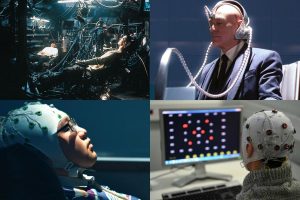
Science fiction and superhero films portray brain-machine interfaces as malevolent robots that plug into human brains for fuel in The Matrix (top left) or as power-enhancing devices in X-Men (top right). In reality, they can help patients use artificial limbs or directly connect to computers. (Image credits, from top left to bottom right: Warner Brothers, 20th Century Fox, Intelligent Films, AFP Photo/Jean-Pierre Clatot)
For the many superheroes that use high-powered gadgets to save the day, there’s an equal number of villains who use technology nefariously. From robots that plug into human brains for fuel in “The Matrix” to the memory-warping devices seen in “Men in Black,” “Captain Marvel,” and “Total Recall,” technology that can control people’s minds is one of the most terrifying examples of technology gone wrong in science fiction and superhero films.
Now, progress made on brain-machine interfaces, technology that provides a direct communication link between a brain and an external device, is bringing us closer to a world that feels like science fiction. Elon Musk’s company NeuraLink is working on a device to let people control computers with their minds, while Facebook’s “mind-reading initiative” can decode speech from brain activity. Is this progress a glimpse into a dark future, or are there more empowering ways in which brain-machine interfaces could become a force for good?
Q: What are the main challenges in connecting brains to devices?
The key problem is that you need to get a lot of information out of brains. Today’s prosthetic devices are very slow, and if we want to go faster it’s a tradeoff: I can go slower and then I am more precise, or I can go faster and be more noisy. We need to get more data out of brains, and we want to do it electrically, meaning we need to get more electrodes into brains.
So what do you need? You need a way of getting electrodes into the brain without making your brain into a pulp, you want the electrodes to be flexible so they can stay in longer, and then you want the system to be wireless. You don’t want to have a big connector on the top of your head.
It’s primarily a hardware problem. We can get electrodes into brains, but they deteriorate quickly because they are too thick. We can have plugs on people’s heads, but it’s ruling out any real-world usage. All these factors hold us back at the moment.
That’s why the Neuralink announcement was very interesting. They get a rather large number of electrodes into brains using well-engineered approaches that make that possible. What makes the difference is that Neuralink takes the best ideas in all the different domains and puts them together.
Q: Most examples in pop culture of connecting brains to machines have villainous or nefarious ends. Does that match up with how brain-machine interfaces are currently being developed?
Let’s say you’ve had a stroke, you can’t talk, but there’s a prosthetic device that allows you to talk again. Or if you lost your arm, and you get a new one that’s as good as the original—that’s absolutely a force for good.
It’s not a dark, ugly future thing, it’s a beautiful step forward for medicine. I want to make massive progress in these diseases. I want patients who had a stroke to talk again; I want vets to have prosthetic devices that are as good as the real thing. I think short-term this is what’s going to happen, but we are starting to worry about the dark sides.


 In the last 20 years, microfabrication techniques have allowed researchers to miniaturize tools for a plethora of bioanalytical applications. In addition to better sensitivity, accuracy and precision, scaling down the size of bioanalytical tools has led to the exploitation of new technologies to further manipulate biomolecules in ways that has never before been achieved. For example, when microfluidic channels are on the same order of magnitude of the electric double layers that form due to localized charge at the surfaces, there exists unique physics that create different flow phenomenon, such as analyte concentration and/or separation, mainly due to the couples physics of electrostatics and fluid dynamics. This talk will outline the basis of such interesting phenomena, such as nanofluidic separation and concentration, and well as probe the applications of such coupled systems, for example, handheld DNA detection. Most importantly, we will focus on the most recent work in the Pennathur lab in this field — biopolar electrode (BPE)-based phenomenon. Bipolar electrodes (BPE) have been studied in microfluidic systems over the past few decades, and through rigorous experimentally-validated modeling of the rich combined physics of fluid dynamics, electrokinetics, and electrochemistry at BPEs, I will show the potential of utilizing microfluidic-based BPEs for the design and development of low power, accurate, low volume fluid pumping mechanisms, with the ultimate goal of integration into wearable drug delivery and µTAS systems.