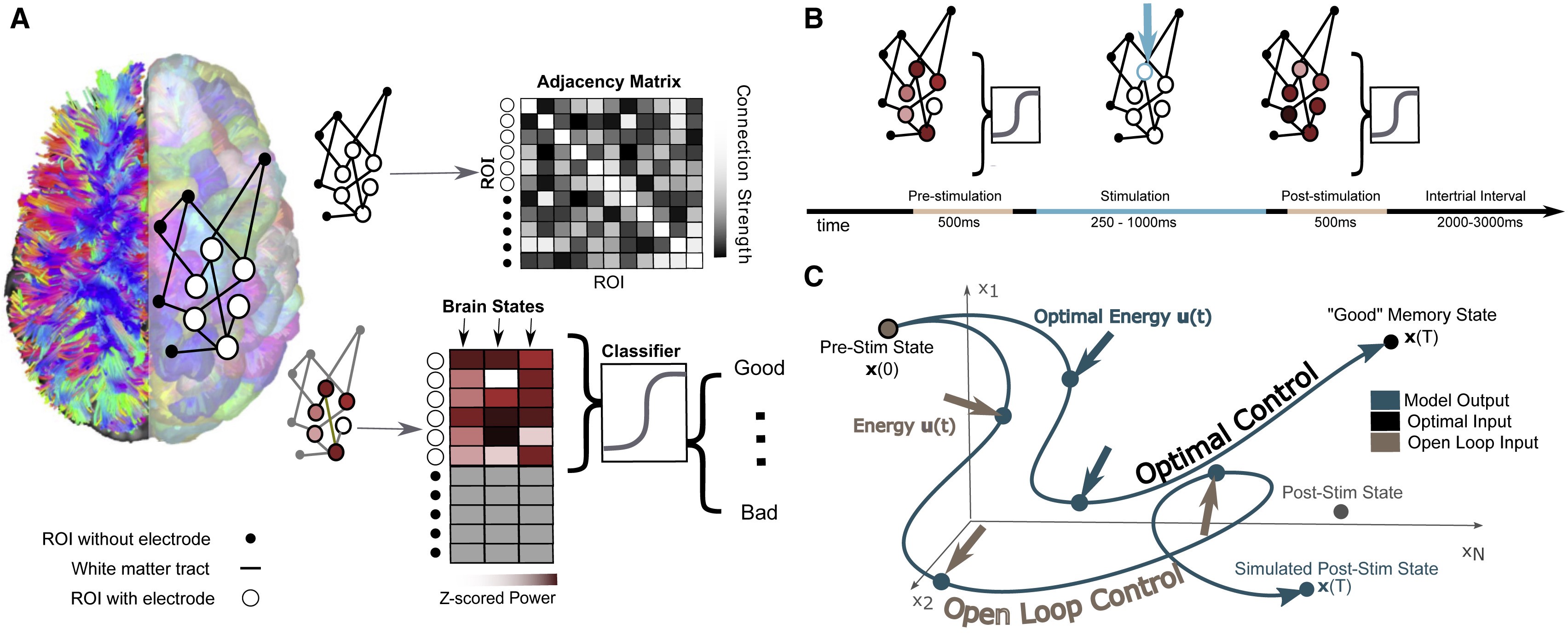
The researchers’ model involves mapping the connections between different regions of an individual’s brain while they performed a basic memory task, then using that data to predict how electrical stimulation in one region would affect activity throughout the network. Individuals’ improved performance on the same memory task after stimulation suggests the model could eventually be generalized toward a variety of stimulation therapies.
Brain stimulation, where targeted electrical impulses are directly applied to a patient’s brain, is already an effective therapy for depression, epilepsy, Parkinson’s and other neurological disorders, but many more applications are on the horizon. Clinicians and researchers believe the technique could be used to restore or improve memory and motor function after an injury, for example, but progress is hampered by how difficult it is to predict how the entire brain will respond to stimulation at a given region.
In an effort to better personalize and optimize this type of therapy, researchers from the University of Pennsylvania’s School of Engineering and Applied Science and Perelman School of Medicine, as well as Thomas Jefferson University Hospital and the University of California, Riverside, have developed a way to model how a given patient’s brain activity will change in response to targeted stimulation.
To test the accuracy of their model, they recruited a group of study participants who were undergoing an unrelated treatment for severe epilepsy, and thus had a series of electrodes already implanted in their brains. Using each individual’s brain activity data as inputs for their model, the researchers made predictions about how to best stimulate that participant’s brain to improve their performance on a basic memory test.
The participants’ brain activity before and after stimulation suggest the researchers’ models have meaningful predictive power and offer a first step towards a more generalizable approach to specific stimulation therapies.
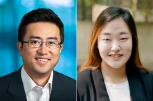
Danielle Bassett and Jennifer Stiso.
The study, published in the journal Cell Reports, was led by Danielle Bassett, J. Peter Skirkanich Professor in Penn Engineering’s Department of Bioengineering, and Jennifer Stiso, a neuroscience graduate student in Penn Medicine and a member of Bassett’s Complex Systems Lab.
A New Sprayable Gel Can Help Prevent Surgical Adhesions
Adhesions are a common kind of scar tissue that can occur after surgery, and though usually not painful, they have the potential to result in complications like chronic pain or decreased heart efficiency, depending on where the scar tissue forms. Now, a sprayable gel developed by researchers at Stanford University will help to prevent adhesions from forming during surgical procedures. The gel, called PNP 1:10 in reference to its polymer-nanoparticle structure, has a similar stiffness to mayonnaise and achieves an ideal balance of slipperiness and stickiness that allows it to adhere easily to tissue of irregular shapes and surfaces. The flexible gel will actually dissolve in the body after two weeks, which is about how long most adhesions take to heal. Though lead author Lyndsay Stapleton, M.S., and senior authors Joseph Woo, M.D., and Eric Appel, Ph.D., have only tested the gel in rats and sheep so far, they hope that human applications are not too far in the future.
Learning to Understand Blood Clots in a New Model
Blood clots are the source of some of the deadliest human conditions and diseases. When a clot forms, blood flow can be interrupted, cutting off supply to the brain, heart, or other vital organs, resulting in potentially serious damage to the mind and body. For patients with certain bleeding disorders, clotting or the lack thereof can hold tremendous importance in surgery, and affect some of the typical procedures of a given operation. To help plan for such situations, researchers at the University of Buffalo created an in vitro model to help better illustrate the complex fluid mechanics of blood flow and clotting. Most importantly, this new model better demonstrates the role of shear stress in blood flow, and the way that it can affect the formation or destruction of blood clots – an aspect that current clinical devices often overlook. Led by Ruogang Zhao, Ph.D., the model can allow surgeons and hematologists to consider the way that certain chemical or physical treatments can affect clot formation on a patient-to-patient basis. The two key factors of the model are its incorporation of blood flow, and its relationship to shear stress, with clot stiffness through microfabrication technology using micropillars as force sensors for the stiffness. Going forward, Zhao and his research team hope to test the model on more patients, to help diversify the different bleeding disorders it can exhibit.
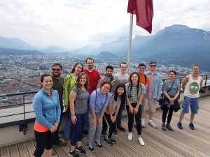
Training the Next Generation of Researchers
REACT 2019 students and Grenoble summer program interns, including undergraduate Rebecca Zappala (third from left, front), pose in front of the Chartreuse Mountains after completing a challenging ropes course. (Photo: Hermine Vincent)
Rebecca Zappala, a junior from Miami, Florida who is majoring in bioengineering, worked in Grenoble this summer on new ways to harvest water from fog. She describes her research project as a “futuristic” way to collect water and says that she’s thankful for the opportunity to work on her first independent research project through the Research and Education in Active Coatings Technology (REACT) program.
After learning the technical skills she needed for her project, Zappala spent her summer independently working on new ways to modify her material’s properties while working closely with her French PI and a post-doc in the lab. She was surprised to see how diverse the lab was, with researchers working on everything from biomolecular research to energy in the same space.
“I learned a lot,” she says about being in such an interdisciplinary setting. “I hadn’t been part of a research team before, and I got a lot of exposure to things that I wouldn’t have been exposed to otherwise.”
Virginia Tech Course Addresses the Needs of Wounded Veterans
A new course at Virginia Tech encourages students to apply engineering skills to real-life problems in the biomedical world by designing medical devices or other applications to assist veterans suffering from serious injuries or illnesses. Funded by the National Institute of Health, faculty from the Department of Biomedical Engineering and Mechanics hope that the course will allow students to see how theoretical knowledge from the classroom actually works in a clinical setting, and to understand how different stakeholder interests factor into designing a real device. What makes this new class unique from other traditional design-focused courses at other universities is its level of patient interaction. Students at Virginia Tech who choose to take this class will have the chance to gain input from field professionals like clinicians and engineers from the Salem Veterans Affairs Medical Center, while also being able to get direct feedback from the patients that the devices will actually help. Beginning in the spring of 2020, students can take the new course, and volunteer in the veterans clinics to gain even more experience with patients.
People and Places
Sevile Mannickarottu, the Director of the Educational Laboratories in Penn’s Department of Bioengineering and recent recipient of the Staff Recognition Award from the School of Engineering and Applied Sciences, presented a paper to highlight the Stephenson Foundation Bioengineering Educational Lab and Bio-Makerspace at the 126th annual conference of the American Society for Engineering Education. Thanks to the dedication of Mannickarottu and the lab staff to creating a space for simultaneous education and innovation, the Bioengineering Lab continues to be a hub for student community and projects of all kinds.
A week-long program for high school girls interested in STEM allows students to explore ideas and opportunities in the field through lab tours, guest speakers, and hands-on challenges. A collaboration across the University of Virginia Department of Biomedical Engineering, Charlottesville Women in Tech, and St. Anne’s Belfield School, the program gave this year’s students a chance to design therapies for children with disorders like hemiplegia and cerebral palsy, in the hopes that these interactive design challenges will inspire the girls to pursue future endeavors in engineering.
We would like to congratulate Nancy Albritton, Ph.D., on her appointment as the next Frank & Julie Jungers Dean of the College of Engineering at the University of Washington. Albritton brings both a deep knowledge of the research-to-marketplace pipeline and experience in the development of biomedical microdevices and pharmacoengineering to the new position.
We would also like to congratulate Jeffrey Brock, Ph.D., on his appointment as the dean of the Yale School of Engineering and Applied Science. Already both a professor of mathematics and a dean of science in the Faculty of Arts and Sciences at Yale, Brock’s new position will help him to foster collaborations across different departments of academia and research in science and engineering.
Rachel Young, a graduate student in Huh’s lab, holds up the new eye-on-a-chip device. The latest iteration of the lab’s eye-on-a-chip has a mechanical eyelid to simulate blinking, and was used to test an experimental drug for dry eye disease. By incorporating human cells into an engineered scaffolding, the eye-on-a-chip has many of the benefits of testing on living subjects, while minimizing risks and ethical concerns.
People who spend eight or more hours a day staring at a computer screen may notice their eyes becoming tired or dry, and, if those conditions are severe enough, they may eventually develop dry eye disease (DED). DED is a common disease with shockingly few FDA-approved drug options, partially because of the difficulties of modeling the complex pathophysiology in human eyes. Enter the blinking eye-on-a-chip: an artificial human eye replica constructed in the laboratory of Penn Engineering researchers.
This eye-on-a-chip, complete with a blinking eyelid, is helping scientists and drug developers to improve their understanding and treatment of DED, among other potential uses. The research, published in Nature Medicine, outlines the accuracy of the eye-on-a-chip as an organ stand-in and demonstrates its utility as a drug testing platform.
They collaborated with Vivian Lee, Vatinee Bunya and Mina Massaro-Giordano from the Department of Ophthalmology in Penn’s Perelman School of Medicine, as well as with Vivek Shenoy, Eduardo D. Glandt President’s Distinguished Professor in Penn Engineering’s Department of Materials Science and Engineering. Other collaborators included Woo Byun, Andrei Georgescu and Yoon-suk Yi, members of Huh’s lab, and Farid Alisafaei, a member of Shenoy’s lab.
Huh’s lab specializes in creating organs-on-a-chip that provide microengineered in vitro platforms to mimic their in vivo counterparts, including lung and bone marrow proxies launched into space this May to study astronaut illness. The lab has spent years fine-tuning its eye-on-a-chip, which earned them the 2018 Lush Prize for its promise in animal-free testing of drugs, chemicals, and cosmetics.
In this study, Huh and Seo focused on engineering an eye model that could imitate a healthy eye and an eye with DED, allowing them to test an experimental drug without risk of human harm.
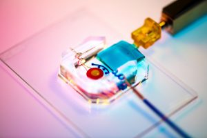
The Huh lab’s eye-on-a-chip attached to a motorized, gelatin-based eyelid. Blinking spreads tears over the corneal surface, and so was a critical aspect to replicate in the researchers’ model of dry eye disease. cells. The cells of the cornea grow on the inner circle of scaffolding, dyed yellow, and the cells of the conjunctiva grow on the surrounding red circle. Artificial tears are supplied by a tear duct, dyed blue.
To construct their eye-on-a-chip, Huh’s team starts with a porous scaffold engineered with 3D printing, about the size of a dime and the shape of a contact lens, on which they grow human eye cells. The cells of the cornea grow on the inner circle of scaffolding, dyed yellow, and the cells of the conjunctiva, the specialized tissue covering the white part of human eyes, grow on the surrounding red circle. A slab of gelatin acts as the eyelid, mechanically sliding over the eye at the same rate as human blinking. Fed by a tear duct, dyed blue, the eyelid spreads artificial tear secretions over the eye to form what is called a tear film.
“From an engineering standpoint, we found it interesting to think about the possibility of mimicking the dynamic environment of a blinking human eye. Blinking serves to spread tears and generate a thin film that keeps the ocular surface hydrated. It also helps form a smooth refractive surface for light transmission. This was a key feature of the ocular surface that we wanted to recapitulate in our device,” says Huh.
For people with DED, that tear film evaporates faster than it’s replenished, resulting in inflammation and irritation. A common cause of DED is the reduced blinking that occurs during excessive computer usage, but people can develop the disease for other reasons as well. DED affects about 14 percent of the world’s population but has been notably difficult to develop new treatments for, with 200 failed clinical drug trials since 2010 and only two currently available FDA-approved drugs for treatment.
Huh’s lab has been considering the drug-testing potential of organs-on-a-chip since their initial conceptualization, and, because of its surface-level area of impact, DED seemed the perfect place to start putting their eye model to the test. But before they started a drug trial, the team had to ensure their model could really imitate the conditions of DED.
“Initially, we thought modeling DED would be as simple as just keeping the culture environment dry. But as it turns out, it’s an incredibly complex multifactorial disease with a variety of sub-types,” Huh says. “Regardless of type, however, there are two core mechanisms that underlie the development and progression of DED. First, as water evaporates from the tear film, salt concentration increases dramatically, resulting in hyperosmolarity of tears. And second, with increased tear evaporation, the tear film becomes thinner more rapidly and often ruptures prematurely, which is referred to as tear film instability. The question was: Is our model capable of modeling these core mechanisms of dry eye?”
The answer, after much experimentation, was yes. The team evoked DED conditions in their eye-on-a-chip by cutting their device’s artificial blinking in half and carefully creating an enclosed environment that simulated the humidity of real-life conditions. When put to the test against real human eyes, both healthy and with DED, the corresponding eye-on-a-chip models proved their similarity to the actual organ on multiple clinical measures. The eyes-on-a-chip mimicked actual eyes’ performance in a Schirmer strip, which tests liquid production; in an osmolarity test, which looks at tear film salt content; and in a keratography test, which evaluates the time it takes for a tear film to break up.
Having confirmed their eye-on-a-chip’s ability to mirror the performance of a human eye in normal and DED-inducing settings, Huh’s team turned to the pharmaceutical industry to find a promising DED drug candidate to test-drive their model. They landed on an upcoming drug based on lubricin, a protein primarily found in the lubricating fluid that protects joints.
“When people think of DED, they normally treat it as a chronic disease driven by inflammation,” says Huh, “but there’s now increasing evidence suggesting that mechanical forces are important for understanding the pathophysiology of DED. As the tear film becomes thinner and more unstable, friction between the eyelids and the ocular surface increases, and this can damage the epithelial surface and also trigger adverse biological responses such as inflammation. Based on these observations, there is emerging interest in developing ophthalmic lubricants as a topical treatment for dry eye. In our study, we used an lubricin-based drug that is currently undergoing clinical trials. When we tested this drug in our device, we were able to demonstrate its friction-lowering effects, but, more importantly, using this model we discovered its previously unknown capacity to suppress inflammation of the ocular surface.”
By comparing the testing results of their models of a healthy eye, an eye with DED, and an eye with DED plus lubricin, Huh and Seo were able to further scientists’ understanding of how lubricin works and show the drug’s promise as a DED treatment.
Similarly, the process of building a blinking eye-on-a-chip pushed forward scientists’ understanding of the eye itself, providing insights into the role of mechanics in biology. Collaborating with Shenoy, director of the Center for Engineering MechanoBiology, the team’s attention was drawn to how the physical blinking action was affecting the cells they cultivated to engineer an artificial eye on top of their scaffolding.
“Initially, the corneal cells start off as a single layer, but they become stratified and form multiple layers as a result of differentiation, which happens when these cells are cultured at the air-liquid interface. They also form tight cell-cell junctions and express a set of markers during differentiation,” Huh says. “Interestingly, we found out that mechanical forces due to blinking actually help the cells differentiate more rapidly and more efficiently. When the corneal cells were cultured under air in the presence of blinking, the rate and extent of differentiation increased significantly in comparison to static models without blinking. Based on this result, we speculate that blink-induced physiological forces may contribute to differentiation and maintenance of the cornea.”
In other words, human cornea cells growing on the scientists’ scaffold more quickly became specialized and efficient at their particular jobs when the artificial eyelid was blinking on top of them, suggesting that mechanical forces like blinking contribute significantly to how cells function. These types of conceptual advances, coupled with drug discovery applications, highlight the multifaceted value that engineered organs-on-a-chip can contribute to science.
Huh and Seo’s eye-on-a-chip is still just dipping its toes into the field of drug testing, but this first step is a victory that represents years of work refining their artificial eye to reach this level of accuracy and utility.
“Although we have just demonstrated proof-of-concept,” says Seo, “I hope our eye-on-a-chip platform is further advanced and used for a variety of applications besides drug screening, such as testing of contact lenses and eye surgeries in the future.”
“We are particularly proud of the fact that our work offers a great and rare example of interdisciplinary efforts encompassing a broad spectrum of research activities from design and fabrication of novel bioengineering systems to in vitro modeling of complex human disease to drug testing,” says Huh. “I think this is what makes our study unique and representative of innovation that can be brought about by organ-on-a-chip technology.”
This work was supported by the National Institutes of Health through grants 1DP2HL127720–0, R01EY026972 and K08EY025742–01, the National Science Foundation through grants CMMI:15–48571, and Research to Prevent Blindness.
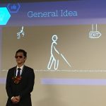
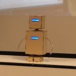
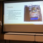
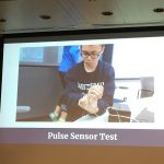
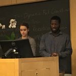
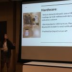
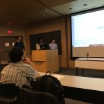
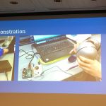
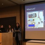
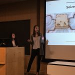
On May 8, 2019, first year Bioengineering students at the University of Pennsylvania gathered together for a marathon two-hour session in which no fewer than twenty-one groups presented the results of their final projects. These projects were the culmination of two semesters’ work in the courses BE 100 and 101, the department’s year-long introduction to Bioengineering. The topics were as diverse and creative as the students, ranging from medical devices and pediatric monitors to plant-care and diagnostic apps. They covered a variety of issues and needs, including tools to help the blind; lockboxes that incorporate breathalyzers (to stop you getting to your keys when intoxicated); mechanisms to sense epileptic seizures and monitor heart rate; and more. Each group had only four minutes to present the research, concept, and results of their project and give a brief demonstration. In the end, the entire class voted and two clear winners emerged. In first place was Group R7 with Heart Guide, a heart-shaped ultrasonic collision device for the blind. Group R3 came in second place with Pulsar the Robot, an adorable pediatric heart rate monitor. The course’s instructor, Dr. Michael Rizk, ended by saying that all of the students should be very proud of their work and that these final projects and the skills learned in year one are the foundation on which the rest of their BE curriculum will be based.
Congratulations to all of our first years on their amazing work. Check out some photos of their impressive work below! For more information on the Penn Bioengineering Undergraduate Curriculum, visit the department website. Most BE student projects are created in the George H. Stephenson Foundation Education Laboratory and “Bio-MakerSpace”, the department’s primary teaching lab.
In this series of posts, University of Pennsylvania students who took the spring 2019 APOC (Appropriate Point of Care Diagnostics) course write about their experience traveling to Ghana in May-June 2019.
by Allaire Morgan (Electrical Engineering, ’22)
Today, the team took various field trips related to water supply and public health. After being picked up on the bus, we drove for about an hour through the busy (and bumpy) streets of Kumasi to the Barakese Headworks of Ghana Water Company Limited. We spent the majority of the morning and early afternoon on a tour of the plant, following in chronological order the retrieval and treatment of water from the reservoir.
Upon walking up to the dam, the roar of the water was powerful. We went into a garage-like building which housed the four large pumps from the dam, pumping water uphill for further processing. After climbing approximately five stories on a shaky ladder, we reached the top of the dam to observe the reservoir from which the Ghana Water Company extracts its water. From there, our team ventured up the hill to observe the water treatment plant.
With the sun beating down, we slowly made our way through the facility, observing each stage of the treatment process in which millions of gallons of water were being treated at a time. The first stage, aeration, takes about six hours to complete. From there, water is pumped into giant drums for sedimentation, where water is stirred and polymers are added to force harmful chemicals to settle at the bottom. Clean water then slowly rises up in the million-gallon drum, with the clean water spilling over the edges to be collected and further processed in large sand filters. The sludge at the bottom of the drum is currently pumped back into the river, which may propose a serious public health problem in the future. The team then followed our guide into the labs, where we observed the various tests which are performed daily on the water after treatment to ensure proper sanitation. Lab technicians perform chemical experiments and culture the water to ensure that water-borne diseases cannot be carried by the filtered water.
After learning so much at the treatment plant, the team jumped on the bus to escape the heat and then traveled, after lunch, to the Komfo Akoye Teaching Hospital to observe the patient intake process in the hypertension clinic. We watched carefully in small groups from the corner of each doctor’s office to see how patients are treated and diagnosed. The doctors see around twenty to thirty patients per day, but on worse days they can see up to forty, with around two being new referrals from peripheral clinics. After speaking with the patient, the doctor makes a prescription recommendation on the patient’s paper file and gives it to the nurse for further processing. Each patient has a paper book which contains all of their medical data and history since coming to the hospital, and they retrieve it from a records room every time they visit. When asked about digitizing the process, the nurses were surprisingly resistant, arguing that they already were used to the paper filing system and they do not have the proper training to efficiently use a computer to file records.
After a long day of observations, the team traveled back to the guest house to eat dinner. Over our meal of pizza , spring rolls, and ambiguous but delicious juice, we discussed the events of the day and refocused our project, ironing out a specific plan for how we want to design our program and creating a vision for its implementation. We went to bed exhausted from a long day’s work but motivated for the project developments to come.
In this series of posts, University of Pennsylvania students who took the spring 2019 APOC (Appropriate Point of Care Diagnostics) course write about their experience traveling to Ghana in May-June 2019.
by Aime Bienfait Igiraneza (Computer Science, ’20)
Operation: Relaxation…and laundry
Itinerary:
Breakfast: Eggs, bread and tea. 8:30am
Laundry
Lunch at the Magnificent Foods
Running (or more of walking in my case) and swimming at the KNUST university
After what was a fun, informative, but busy week filled with hospital and clinic visits and walks through communities (and not forgetting the activity-filled day we had before, of course), this Sunday was supposed to be the time to relax…and do laundry.
Team meeting to talk about project.
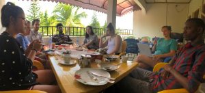
Our breakfast started a little later than usual. Though breakfast was ready at 8:30 am, most of us had a lazy morning in and came out to eat at 9:00 am. When all the team members were assembled before the usual omelet and tea breakfast, we decided to do an impromptu recap of the week and brainstormed on how we could adapt our initial project to fit the clinics and hospitals we had visited during the week. This session, as spontaneous as it was, became a good way for us to build on our observations from the week not just for the applications of our project, but for identifying certain problems that can become future projects for future APOC teams.
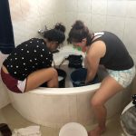
El and Laura doing laundry in the bathtub
Keeping the theme of lazy Sunday in mind, we did not start laundry until late morning, around 10:30 am. This didn’t prove to be a very wise idea, especially since we were supposed to do laundry the old-fashioned way with water, a bar of soap, and our good old hands. Furthermore, there proved to be a shortage of buckets for all of us to do laundry at the same time, which didn’t seem to leave enough time before the bus was supposed to pick us up at 1:00 pm for lunch. Nonetheless, we bonded as we shared our buckets (who knew that manual laundry-washing was such a social activity) and we made it in time for lunch; also nothing a little adrenaline and team work couldn’t fix.
Kyler and Bienfait doing laundry.
After laundry, we had lunch at the same restaurant we visited last Sunday: Magnificent Foods. The food was just as fantastic as we know it is in Ghana (the serving sizes are still too large for any of us to finish), but the most eventful thing was that the tailor came to take our measurements for the traditional clothes we are to wear to the King’s palace next Sunday. Everyone had their designs on their phones, their cloths bought from the market, and a bit of excitement on their faces as we saw our plans on the clothes being born. We anticipate receiving the clothes sometime this week. (Emotion check: beaming with excitement!!)
Once our long lunch meal was over, we decided to do a very un-lazy thing and went running on the KNUST campus before the sun set. This created another team bonding moment and we went swimming afterwards. This was the most memorable part for me because, though I can’t swim, I had my teammates with me and they taught me some basic swimming skills.
The rest of the night was very lazy. We played cards until late and each prepared for the week to come. (Emotion check: tired but excited to start the week.)
In this series of posts, University of Pennsylvania students who took the spring 2019 APOC (Appropriate Point of Care Diagnostics) course write about their experience traveling to Ghana in May-June 2019.
by Aime Bienfait Igiraneza (Computer Science, ’20)
The morning was fantastic! Several group members were having a slightly enhanced digestion, which turned out not so great. It did not help that we also saw at least three guys peeing on the street, a serious public health issue that should be looked into. Nonetheless, we were excited for the day and we were not disappointed. We were about to visit one of the regional hospitals. If I were to summarize the day, there were two main takeaways: First, the hospital has an amazing system that works. Second, the potential of the system in place is not fully explored.
We started by meeting the medical director of the hospital along with another doctor specializing in tuberculosis. The main methods used to diagnose TB are microscopy and GeneXpert when the patient can produce sputum. Otherwise, the use of X-rays test is the remaining option. Although patients with TB can be diagnosed within two hours, it is still challenging to catch all cases due to stigma and the fact that testing is only ever done in the morning. Even the detected cases are more likely to be very advanced as people tend to first try other medicines when they encounter symptoms such as coughing, thinking that it will simply go away. However, an interesting program to collect sputum from pharmacies has been initiated which allows professionals to catch those who simply buy cough medicines from drug stores. After being diagnosed, patients are prescribed TB pills and these patients are re-checked after six months, which unfortunately is not always followed up on. When possible, patients are also followed up on at home by special health workers who ensure that pills are taken as prescribed. Even better, injectables are administered once in four months, thus reducing the burden of taking pills every day.
A few technical challenges were mentioned. The first one is that medical records for every patient are still saved in books, which pile up over time making it difficult to manage. A software called Health Administration Management System (HAMS) is used to save patients’ identifications and registration book numbers. When this book is updated, so is the HAMS account corresponding to the particular patient. All this is done in real time. Then, at the end of the day, staff from the Health and Information office use a software called District Health Management System (DHMS) to collect the information about the types of patients seen that day. This information is drawn from HAMS as well as record books. Thus, the challenge is how to automate the uploading of data from HAMS to DHMS in real time without waiting until the end of the day to manually do it. This has not been possible in part because HAMS does not contain all the necessary details.
Overall, it’s impressive how well everything works, given the current structure and workflow. At least, we learned about a few challenges, which by the way, were not always revealed to us. As usual, a ridiculously delicious lunch was provided. We also passed by the mall and bought a few fabrics. When we came back from the hospital, we picked up some KFC chickens for dinner and kept enjoying the incredible Ghanaian hospitality until we called it a day.
In this series of posts, University of Pennsylvania students who took the spring 2019 APOC (Appropriate Point of Care Diagnostics) course write about their experience traveling to Ghana in May-June 2019.
We started the day very early at 7:00 am with a drive to the Suntreso General Hospital where we were to visit Dr. Agyarko-Poku, a venerologist who would help us understand more about mother-to-child transmission of HIV/AIDS to aid in our study of noncommunicable diseases.
We arrived at the hospital, where the in-charge for the STI-OPD unit welcomed us and gave us a general overview of how events are run at the hospital’s HIV clinic which occurs on Wednesdays for adults and on Fridays for children. She gave us also an overview of how viral testing, data collection, and drug dispensary and treatment occur for the confirmed patients in the hospital. We got to see the relevant data points that were collected on each patient visit to help us better understand how well to modify our machine learning system.
We then went to the Disease Control Unit where the professionals there have the job of collating all hospital cases at the end of the week to identify diseases that are on the rise and have the potential to become public health concerns. We also got to understand more about how tuberculosis is diagnosed in the clinics and how treatment occurs for the disease. We found out also that drug-resistant tuberculosis has not been much of a problem for that particular hospital, contrary to what we thought. After this, we split into teams and went to visit the ART center, the counseling center, the dispensary, the data management room, as well as the consulting room where we got to interact more with the health professionals (i.e. nurses and pharmacists) in order to understand the processes that the patients have to go through from entrance into the hospital facility to diagnosis and the reception of their medication.
We left the Suntreso hospital and came back to KNUST where we had lunch before meeting with the civil engineers to discuss writing a report and making possible recommendations for the communities that we visited in order to improve their sanitation and water supply. We arrived at the Komfo Anokye Teaching Hospital’s Nursing Training School at about 2.30pm where we presented our Tuberculosis Triaging system to the students and received their questions which ranged from the usefulness of our algorithm and project to concerns about data contamination and invalidation. From KATH, we made a final run through KFC to grab dinner before making our way back to the guest house to enjoy our meal and conclude the day.
Vector Flow Imaging Helps Visualize Blood Flow in Pediatric Hearts
A group of biomedical engineers at the University of Arkansas used a new ultrasound-based imaging technique called vector flow imaging to help improve the diagnosis of congenital heart disease in pediatric patients. The study, led by associate professor of biomedical engineering Morten Jensen, Ph.D., collaborated with cardiologists at the local Children’s Hospital in Little Rock to produce images of the heart in infants to help potentially diagnose congenital heart defects. Though the use of vector flow imaging has yet to be developed for adult patients, this type of imaging could possibly provide more detail about the direction of blood flow through the heart than traditional techniques like echocardiography do. In the future, the use of both techniques could provide information about both the causes and larger effects of heart defects in patients.
Using Stem Cells to Improve Fertility in Leukemia Survivors
One of the more common side effects of leukemia treatment in female patients is infertility, but researchers at the University of Michigan want to change that. Led by associate professor of biomedical engineering Ariella Shikanov, Ph.D., researchers in her lab found ways of increasing ovarian follicle productivity in mice, which directly relates to the development of mature eggs. The project involves the use of adipose-derived stem cells, that can be found in human fat tissue, to surround the follicles in an ovary-like, three-dimensional scaffold. Because the radiation treatments for leukemia and some other cancers are harmful to follicles, increasing their survival rate with this stem cell method could reduce the rate of infertility in patients undergoing these treatments. Furthermore, this new approach is innovative in its use of a three-dimensional scaffold as opposed to a two-dimensional one, as it stimulates follicle growth in all directions and thus helps to increase the follicle survival rate.
Penn Engineers Look at How Stretching & Alignment of Collagen Fibers Help Cancer Cells Spread
Cancer has such a massive impact on people’s lives that it might be easy to forget that the disease originates at the cellular level. To spread and cause significant damage, individual cancer cells must navigate the fibrous extracellular environment that cells live in, an environment that Penn Engineer Vivek Shenoy has been investigating for years.
Shenoy’s most recent study on cancer’s mechanical environment was led by a postdoctoral researcher in his lab, Ehsan Ban. Paul Janmey, professor in Physiology and Bioengineering, and colleagues at Stanford University also contributed to the study. Shenoy also received the Heilmeier Award this March and delivered the Heilmeier Award Lecture in April.
Controlled Electrical Stimulation Can Prevent Joint Replacement Infections
Joint replacements are one of the most common kinds of surgery today, but they still require intense post-operative therapy and have a risk of infection from the replacement implant. These infections are usually due to the inflammatory response that the body has to any foreign object, and can become serious and life-threatening if left untreated. Researchers at the University of Buffalo Jacobs School of Medicine and Biomedical Sciences hope to offer a solution to preventing infections through the use of controlled electrical stimulation. Led by Mark Ehrensberger, Ph.D., Kenneth A. Krackow, M.D., and Anthony A. Campagnari, Ph.D., the treatment system uses the electrical signal to create an antibacterial environment at the interface of the body and the implant. While the signal does not prevent infections completely, these antibacterial properties will prevent infections from worsening to a more serious level. Patented as the Biofilm Disruption Device TM, the final product uses two electrode skin patches and a minimally invasive probe that delivers the electrical signal directly to the joint-body interface. The researchers behind the design hope that it can help create a more standard way of effectively treating joint replacement infections.
People and Places
TBx: Gabriel Koo, Ethan Zhao, Daphne Cheung, and Shelly Teng
For their senior design project, four bioengineering seniors — Gabriel Koo, Ethan Zhao, Daphne Cheung, and Shelly Teng — created a low-cost tuberculosis diagnostic that they called TBx. Using their knowledge of the photoacoustic effect of certain dyes, the platform the group created can detect the presence of lipoarabinomannan in patient urine. The four seniors presented TBx at the Rice360 Design Competition in Houston, Texas this spring, which annually features student-designed low-cost global health technologies.
In this series of posts, University of Pennsylvania students who took the spring 2019 APOC (Appropriate Point of Care Diagnostics) course write about their experience traveling to Ghana in May-June 2019.
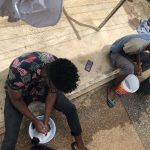
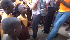
On this beautiful Tuesday, we woke up at our hotel to a lovely breakfast of sausage and pepper omelets. After our meal we loaded up on the bus to drive over to Aprade. On the way to Aprade we picked up our water and sanitation expert of friends: Sophia, Kingsley, and Alfred. We first stopped at Aprade Government School where we met with the entire population of the school to present a demonstration on hand-washing. After the presentation we took a tour of the school to survey their existing water and sanitation systems. We learned about their construction projects and about the school itself, which educates about six hundred students!
Our second stop was to the main village of Aprade. We were taken through the village and observed their rainwater collection systems and boreholes. We discovered the public and private boreholes around the village. Everyone was so hospitable and welcoming. Before continuing we took a quick break to eat lunch on the bus. The food was catered and we had a delicious meal of jollof rice and fried chicken. There was refreshing mango and ginger juice to cool us down from the hot day.
After lunch we drove over to Mmeswam. We were able to make an appointment with the Chief! He brought us into his home with welcoming arms; it was amazing being able to shake the chief’s hand. The chief showed us around Mmeswam and led us to his boreholes and wells. The main borehole pumps water to a large elevated tank. Underneath that tank is a pipe and valve mechanism where villagers come to collect water in large basins. The chief encouraged us to take the basin on our heads and try to carry some of the water. Ellie, Kyler and El were chosen among the group to test their strength.
From the boreholes we walked over to the village’s wells. The oldest well was spoiled and is unusable. The working well in the village was a traditional pulley system well. Several of our students cast the bucket and fished up some water. After our day of water we thanked the chief and hopped back on the bus. We drove back to campus, dropped off the graduate students at campus, and made a stop at the mall. We grabbed dinner at Game and quickly made our way home. It had been a long day so we chowed down on dinner and passed out for the night. A great and very busy day!


 A group of biomedical engineers at the University of Arkansas used a
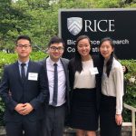
 On this beautiful Tuesday, we woke up at our hotel to a lovely breakfast of sausage and pepper omelets. After our meal we loaded up on the bus to drive over to Aprade. On the way to Aprade we picked up our water and sanitation expert of friends: Sophia, Kingsley, and Alfred. We first stopped at Aprade Government School where we met with the entire population of the school to present a demonstration on hand-washing. After the presentation we took a tour of the school to survey their existing water and sanitation systems. We learned about their construction projects and about the school itself, which educates about six hundred students!
 Our second stop was to the main village of Aprade. We were taken through the village and observed their rainwater collection systems and boreholes. We discovered the public and private boreholes around the village. Everyone was so hospitable and welcoming. Before continuing we took a quick break to eat lunch on the bus. The food was catered and we had a delicious meal of jollof rice and fried chicken. There was refreshing mango and ginger juice to cool us down from the hot day.
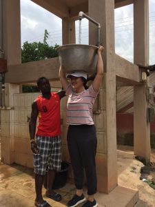 After lunch we drove over to Mmeswam. We were able to make an appointment with the Chief! He brought us into his home with welcoming arms; it was amazing being able to shake the chief’s hand. The chief showed us around Mmeswam and led us to his boreholes and wells. The main borehole pumps water to a large elevated tank. Underneath that tank is a pipe and valve mechanism where villagers come to collect water in large basins. The chief encouraged us to take the basin on our heads and try to carry some of the water. Ellie, Kyler and El were chosen among the group to test their strength.
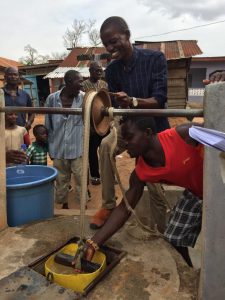 From the boreholes we walked over to the village’s wells. The oldest well was spoiled and is unusable. The working well in the village was a traditional pulley system well. Several of our students cast the bucket and fished up some water. After our day of water we thanked the chief and hopped back on the bus. We drove back to campus, dropped off the graduate students at campus, and made a stop at the mall. We grabbed dinner at Game and quickly made our way home. It had been a long day so we chowed down on dinner and passed out for the night. A great and very busy day!