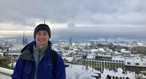
Magnetic Field and Hydrogels Could Be Used to Grow New Cartilage

by Frank Otto

MRI Knee joint or Magnetic resonance imaging sagittal view for detect tear or sprain of the anterior cruciate ligament (ACL).

Using a magnetic field and hydrogels, a team of researchers in the Perelman School of Medicine have demonstrated a new possible way to rebuild complex body tissues, which could result in more lasting fixes to common injuries, such as cartilage degeneration. This research was published in Advanced Materials.

“We found that we were able to arrange objects, such as cells, in ways that could generate new, complex tissues without having to alter the cells themselves,” says the study’s first author, Hannah Zlotnick, a graduate student in bioengineering who works in the McKay Orthopaedic Research Laboratory at Penn Medicine. “Others have had to add magnetic particles to the cells so that they respond to a magnetic field, but that approach can have unwanted long-term effects on cell health. Instead, we manipulated the magnetic character of the environment surrounding the cells, allowing us to arrange the objects with magnets.”

In humans, tissues like cartilage can often break down, causing joint instability or pain. Often, the breakdown isn’t in total, but covers an area, forming a hole. Current fixes are to fill those holes in with synthetic or biologic materials, which can work but often wear away because they are not the same exact material as what was there before. It’s similar to fixing a pothole in a road by filling it with gravel and making a tar patch: The hole will be smoothed out but eventually wear away with use because it’s not the same material and can’t bond the same way.

What complicates fixing cartilage or other similar tissues is that their makeup is complex.

“There is a natural gradient from the top of cartilage to the bottom, where it contacts the bone,” Zlotnick explains. “Superficially, or at the surface, cartilage has a high cellularity, meaning there is a higher number of cells. But where cartilage attaches to the bone, deeper inside, its cellularity is low.”

So the researchers, which included senior author Robert Mauck, PhD, director of the McKay Lab and a professor of Orthopaedic Surgery and Bioengineering, sought to find a way to fix the potholes by repaving them instead of filling them in. With that in mind, the research team found that if they added a magnetic liquid to a three-dimensional hydrogel solution, cells, and other non-magnetic objects including drug delivery microcapsules, could be arranged into specific patterns that mimicked natural tissue through the use of an external magnetic field.

Read more at Penn Medicine News.

Through Brain Imaging Analysis in Rats, Penn Researchers Show Potential to Predict Whether Pain Will be Acute or Persistent

Beth Winkelstein, Megan Sperry, and Eric Granquist

Pain may be a universal experience, but what actually causes that experience within our brains is still poorly understood. Pain often continues long after the relevant receptors in the body have stopped being stimulated and can persist even after those receptors cease to exist, as is the case with “phantom limb” pain.

The exact experience an individual will have after a painful incident comes down to the complex, variable connections formed between several different parts of the brain. The inability to predict how those connections will form and evolve can make pain management a tricky, frustrating endeavor for both healthcare providers and patients.

Now, a team of Penn researchers has shown a way to make such predictions from the pattern of neural connections that begin to take shape soon after the first onset of pain. Though their study was conducted in rats, it suggests that similar brain imaging techniques could be used to guide treatment decisions in humans, such as which individuals are most likely to benefit from different drugs or therapies.

The study, published in the journal Pain, was led by Beth Winkelstein, Eduardo D. Glandt President’s Distinguished Professor in Penn Engineering’s Department of Bioengineering and Deputy Provost of the University of Pennsylvania, along with Megan Sperry, then a graduate student in her lab. Eric Granquist, Director of the Center for Temporomandibular Joint Disease at the Hospital of the University of Pennsylvania in the Department of Oral & Maxillofacial Surgery, and assistant professor of Oral & Maxillofacial Surgery in Penn’s School of Dental Medicine, also contributed to the research.

“Our findings provide the first evidence that brain networks differ between acute and persistent pain states, even before those different groups of rats actually show different pain symptoms,” says Winkelstein.

Read the full story at Penn Engineering Today. Media contact Evan Lerner.

Brianne Connizzo Appointed Assistant Professor at Boston University

by Mahelet Asrat

Brianne Connizzo, PhD

The Department of Bioengineering is proud to congratulate alumna Brianne Connizzo, PhD on her appointment as a tenure-track Assistant Professor in the Department of Biomedical Engineering in the College of Engineering at Boston University. Connizzo’s appointment will begin in January 2021, after completing her work as a postdoctoral researcher in Biological Engineering at MIT under the supervision of Alan J. Grodzinsky, ScD, Professor of Biological, Electrical, and Mechanical Engineering.

Connizzo got her BS in Engineering Science from Smith College (the first all women’s engineering program in the country) where she graduated in 2010 with highest honors. During her time there, she worked in the laboratory of Borjana Mikic, Rosemary Bradford Hewlett 1940 Professor of Engineering. While working in the lab, she explored the role of myostatin deficiency on Achilles tendon biomechanics and built mechanical testing fixtures for submerged testing of biological tissues. Connizzo continued along this path during her graduate studies in Bioengineering at Penn while working with Louis J. Soslowsky, Fairhill Professor in Orthopaedic Surgery and Professor in Bioengineering, at the McKay Orthopaedic Research Laboratory. Her thesis work focused on the dynamic re-organizations of collagen during tendon loading in the rotator cuff, developing a novel AFM-based method for measuring collagen fibril sliding along the way. During her time at Penn, Connizzo also served as the Social Chair for the Graduate Association of Bioengineers (GABE) and the Graduate Student Engineering Group (GSEG), both of which play a vital role in representing graduate students across the School of Engineering and Applied Sciences. She completed her PhD in Bioengineering in 2015 and then pursued her postdoctoral studies at MIT, focusing on fluid flow during compressive loading and developing novel explant culture models to explore real-time extracellular matrix turnover. For her work she was awarded both an NIH F32 postdoctoral fellowship and the NIH K99/R00 Pathway Independence Award, which are just a few of her long list of impressive accomplishments.

Although Connizzo’s interests in soft tissue mechanobiology span development, injury, and disease, her more recent work has targeted how aging influences tendon function and biology. With a fast-growing active and aging population, she believes that identifying the cause and contributors of age-related changes is critical to finding treatments and therapies that could prevent tendon disease, and thus improve overall population healthspan and quality of life. The primary objectives of the Connizzo Lab at Boston University will be to harness novel in vitro and in vivo models to study cell-controlled extracellular matrix remodeling and tissue biomechanics and to better understand normal tendon maintenance and the initiation of tendon damage in the context of aging.

“I am so grateful to have had the guidance of my mentors and peers at Penn during my doctoral studies, and even more thankful that many of those relationships remain a significant part of my support system to this day,” Connizzo says. “I’m really looking forward to this next chapter to all the successes and failures in pursuing the science, to building a community at BU and in my own laboratory, and to supporting the next generation of brilliant young scientists.”

Congratulations Dr. Connizzo from everyone at Penn Bioengineering!

Gardening in Costa Rica Yields Inspiration During Quarantine

by

María Suarez in her garden.

When the coronavirus pandemic began in March, María Suarez, junior in bioengineering, left Penn’s campus and returned home to Costa Rica. What should have been the final weeks of club activities, social events and end-of-year celebrations shifted to months spent at home, far away from Philadelphia. But Suarez, like many others, wanted to do something productive with her time in quarantine. Drawing on her bucolic roots, she decided to start a garden.

“I was born and raised in a very rural area,” Suarez says. “There is a huge river in my backyard where I learned how to count by throwing pebbles in the river with my mother and sister. Nature is a big part of my life, and it’s really shaped my personality. As a child, I planted herbs, like basil, mint and oregano, with my parents. When you are close to the land like this, gardening was something that grew naturally out of our lifestyle.”

As the spring semester shifted into summer, Suarez returned to her love of planting and embarked on an ambitious project to grow a vegetable garden in her backyard. Unlike the smaller herb gardens she had grown as a child, this vegetable garden required deeper horticultural knowledge as well as intense work under the hot sun.

“To begin the garden, I had to clear the land I wanted to use and remove all the grass and stones from the soil,” Suarez shares. “It was the dry season in Costa Rica and the ground was very difficult to work with.”

After clearing the land, Suarez had to bring nutrients back into the soil of her garden plot. Luckily, her family has been maintaining a natural compost pile for many years.

“Basically, the compost pile is a hole in the ground where we put our natural food waste. There are worms and animals there that help us naturally decompose the waste and they produce a very nutrient rich soil.” Suarez explains. “The compost is a five-minute walk from my garden, and I had to take at least ten trips with a wheelbarrow to bring enough back. It was a great arm workout.”

Once the soil was placed and watered, Suarez was finally able to plant her seeds. After a few days, she saw celery and zucchini plants beginning to sprout. Throughout the summer, Suarez’s crops grew well, and she was able to harvest the vegetables and share them with her family.

“It was very fulfilling to see the products of my efforts,” Suarez says.

Read the full story on the Penn Engineering blog.

Meet Bioengineering Sophomore and SNF Paideia Fellow Catherine Michelutti

Catherine Michelutti (BSE, BS ’23)

Rising Bioengineering Sophomore Catherine Michelluti (BSE 2023) has been featured on Penn’s SNF Paideia Program Instagram which discusses her diverse interests in machine learning in medicine, computer science, playing the violin and more. Catherine is a pre-med student who is pursuing an uncoordinated dual degree between the School of Engineering and Applied Science and the Wharton School of Business (BS in Economics 2023). She is also an incoming fellow in the SNF Paideia Program, which is supported by the Stavros Niarchos Foundation, is an interdisciplinary program which “encourage[s] the free exchange of ideas, civil and robust discussion of divergent views, and the integration of individual and community wellness, service, and citizenship through SNF Paideia designated courses, a fellows program, and campus events” (SNF Paideia website).

Read more about Catherine and other Fellows on the SNF Paideia Instagram.

Why This Bioengineering Ph.D. Student Pursued Impact Investing

In a Q&A, Bioengineering doctoral candidate Ana P. Peredo explains how the idea of “regeneration” motivated her to join WIVA, Wharton Social Impact’s impact investing program.

Why would you — a bioengineering Ph.D. student — seek to join WIVA?

As a bioengineering Ph.D. student, Ana P. Peredo is currently working on the development of regenerative methods and drug-delivery approaches for musculoskeletal tissue

“As a high school student, I was motivated to study bioengineering because of its potential to generate impact through technical innovation. To me, bioengineering was a way to apply engineering principles to create medical technology in the hopes of devising solutions for global health concerns.

Though I have gained significant understanding of the current pressing healthcare needs, I felt that I was missing a key understanding of how investors think about social impact. To better understand how to apply my science background to the impact space, I joined WIVA. I also wanted to venture outside of healthcare and learn about other important social impact sectors such as education, energy, and environment, all of which WIVA explores in its deal-sourcing process.”

What have you learned through WIVA that you have not been exposed to before?

“I learned how to assess early-stage startups for their impact and return-on-investment potential, as well as how to rigorously analyze company financials and projections.

I also had the opportunity to meet leading social impact professionals through WIVA. I attended a Wharton Social Impact Initiative event with Vincent Stanley, the Director of Philosophy at Patagonia. From this discussion, I learned about how the word ‘sustainable’ continues to be misused by companies and how companies should try to ‘regenerate’ the resources they consume to be truly deemed sustainable.

This conversation brought to mind my research experience with regeneration — could I use my WIVA deal-sourcing techniques to find impactful startups that use this concept?”

Continue reading at Wharton Stories.

Penn Alumnus Peter Huwe Appointed Assistant Professor at Mercer University

Peter Huwe, Ph.D.

Peter Huwe, a University of Pennsylvania alumnus and graduate of the Radhakrishnan lab, was appointed Assistant Professor of Biomedical Sciences at the Mercer University School of Medicine beginning this summer 2020 semester.

Huwe earned dual B.S. degrees in Biology and Chemistry in 2009 from Mississippi College, where he was inducted into the Hall of Fame. At Mississippi College, Huwe had his first exposure to computational research in the laboratory of David Magers, Professor of Chemistry and Biochemistry. He went on to earn his Ph.D. in Biochemistry and Molecular Biophysics in 2014 in the laboratory of Ravi Radhakrishnan, Chair of the Bioengineering Department at Penn. As an NSF Graduate Research Fellow in Radhakrishnan’s lab, Huwe focused his research on using computational molecular modeling and simulations to elucidate the functional consequences of protein mutations associated with human diseases. Dr. Huwe then joined the structural bioinformatics laboratory Roland Dunbrack, Jr., Professor at the Fox Chase Cancer Center as a T32 post-doctoral trainee. During his post-doctoral training, Huwe held adjunct teaching appointments at Thomas Jefferson University and at the University of Pennsylvania. In 2017, Huwe became an Assistant Professor of Biology at Temple University, where he taught medical biochemistry, medical genetics, cancer biology, and several other subjects.

During each of his appointments, Huwe became increasingly more passionate about teaching, and he decided to dedicate his career to medical education. Huwe is very excited to be joining Mercer University School of Medicine as an Assistant Professor of Biomedical Sciences this summer. There, he will serve in a medical educator track, primarily teaching first and second year medical students.

“Without Ravi Radhakrishnan and Philip Rea, Professor of Biology in Penn’s School of Arts & Sciences, giving me my first teaching opportunities as a graduate guest lecturer at Penn, I may never have discovered how much I love teaching,” says Huwe. “And without the support and guidance of each of my P.I.’s [Dr.’s Magers, Radhakrishnan, and Dunbrack], I certainly would not be where I am, doing what I love.  I am incredibly thankful for all of the people who helped me in my journey to find my dream job.”

Congratulations and best of luck from everyone in Penn Bioengineering, Dr. Huwe!

Video Series Explores Penn Bioengineering’s Unique Bio-MakerSpace

A new series of short videos on the BE Labs Youtube Channel highlights the unique and innovative approach to engineering education found in The George H. Stephenson Foundation Educational Laboratory & Bio-MakerSpace, the primary teaching lab for the Department of Bioengineering at Penn Engineering. This video series explores how “engineering is fundamentally interdisciplinary” and demonstrates the ways in which Penn students from Bioengineering and beyond have combined the fields of biology, chemistry, and electrical, mechanical, and materials engineering into one exciting and dynamic “MakerSpace.”

“Our Bio-MakerSpace” takes viewers on a tour inside BE’s one-of-a-kind educational laboratories.

Produced primarily on smart phones and with equipment borrowed from the Penn Libraries, and software provided by Computing and Educational Technology Services, the videos were made by rising Bioengineering junior Nicole Wojnowski (BAS ‘22). Nicole works on staff as a student employee of the BE Labs and as a student researcher in the Gottardi Lab at the Children’s Hospital of Philadelphia (CHOP), helmed by Assistant Professor of Pediatrics Riccardo Gottardi.

Sevile Mannickarottu, Director of the Educational Labs in Bioengineering, says that the philosophy of the Bio-MakerSpace “encourages a free flow of ideas, creativity, and entrepreneurship between Bioengineering students and students throughout Penn. We are the only open Bio-MakerSpace with biological, chemical, electrical, materials, and mechanical testing and fabrication facilities, all in one place, anywhere.”

Previous stories on the BE blog have gone into detail about how BE’s Bio-MakerSpace has become a hub for start-ups in recent years, how students can build their own makerspace for under $1500, and more. Major award-winning start-ups including Strella Biotechnology and InstaHub got their start in the BE Labs.

To learn more about the Bio-MakerSpace, check out the other videos below.

Katherine Sizov (Biology ‘19), founder of the 2019 President’s Innovation Prize (PIP) award-winning company Strella Biotechnology, discusses how the Bio-MakerSpace is a hub for interdisciplinary innovation.

Bioengineering doctoral student Dayo Adewole co-founded the company Instahub, which also took home a PIP award in 2019. Dayo also graduated from the BE undergraduate program in 2014. In this video, he discusses the helpfulness and expertise of the BE Labs staff.

Senior Associate Dean for Penn Engineering and Solomon R. Pollack Professor in Bioengineering David Meaney discusses how the Bio-MakerSpace is the only educational lab on campus to provide “all of the components that one would need to make the kinds of systems that bioengineers make.”

What do ‘Bohemian Rhapsody,’ ‘Macbeth,’ and a list of Facebook Friends All Have in Common?

New research finds that works of literature, musical pieces, and social networks have a similar underlying structure that allows them to share large amounts of information efficiently.

Examples of statistical network analysis of characters in two of Shakespeare’s tragedies. Two characters are connected by a line, or edge, if they appear in the same scene. The size of the circles that represent these characters, called nodes, indicate how many other characters one is connected to. The network’s density relates to how complete the graph is, with 100% density meaning that it has all of the characters are connected. (Image: Martin Grandjean)


By Erica K. Brockmeier

To an English scholar or avid reader, the Shakespeare Canon represents some of the greatest literary works of the English language. To a network scientist, Shakespeare’s 37 plays and the 884,421 words they contain also represent a massively complex communication network. Network scientists, who employ math, physics, and computer science to study vast and interconnected systems, are tasked with using statistically rigorous approaches to understand how complex networks, like all of Shakespeare, convey information to the human brain.

New research published in Nature Physics uses tools from network science to explain how complex communication networks can efficiently convey large amounts of information to the human brain. Conducted by postdoc Christopher Lynn, graduate students Ari Kahn and Lia Papadopoulos, and professor Danielle S. Bassett, the study found that different types of networks, including those found in works of literature, musical pieces, and social connections, have a similar underlying structure that allows them to share information rapidly and efficiently.

Technically speaking, a network is simply a statistical and graphical representation of connections, known as edges, between different endpoints, called nodes. In pieces of literature, for example, a node can be a word, and an edge can connect words when they appear next to each other (“my” — “kingdom” — “for” — “a” — “horse”) or when they convey similar ideas or concepts (“yellow” — “orange” — “red”).

The advantage of using network science to study things like languages, says Lynn, is that once relationships are defined on a small scale, researchers can use those connections to make inferences about a network’s structure on a much larger scale. “Once you define the nodes and edges, you can zoom out and start to ask about what the structure of this whole object looks like and why it has that specific structure,” says Lynn.

Building on the group’s recent study that models how the brain processes complex information, the researchers developed a new analytical framework for determining how much information a network conveys and how efficient it is in conveying that information. “In order to calculate the efficiency of the communication, you need a model of how humans receive the information,” he says.

Continue reading at Penn Today.

Bridging the Communication Divide for Deaf and Hard-of-hearing Communities

Clear-fronted face masks, better and more frequent interpreters, and amped up involvement from local organizations have made a big difference during the COVID-19 pandemic.

By Michele Berger

Since April 23, when bioengineering alum Kate Panzer (above) and her partners at the Deaf-Hearing Communication Centre started taking orders for masks with clear fronts, they’ve shipped about 450, with a backlog of requests for hundreds more. (Image: Courtesy Kate Panzer)

Because COVID-19 spreads via respiratory droplets that disperse through sneezes and coughs, shielding the mouth and nose is an important weapon against the virus. But it can also hinder conversations for people who rely on reading lips. “Communication barriers are already difficult sometimes, and this makes it more difficult,” says linguist , director of ’s .

It’s one of the trickiest aspects of this pandemic for those in the Deaf and hard-of-hearing communities, Fisher says. The challenge doesn’t stem just from misunderstandings due to wearing masks. It’s also about the dissemination of accurate and timely information, knowing who to rely on and how to assess what’s being said.

Trusted sources like the Swarthmore, Pennsylvania–based nonprofit Deaf-Hearing Communication Centre (DHCC), a Penn community partner, have filled that gap, frequently updating information on its social media channels and websites. Governors and mayors are more frequently using Certified Deaf Interpreters (CDI) during press briefings, and Penn alum Kate Panzer, who graduated in 2018, started a project with DHCC to sew masks with clear fronts to offer both lip-reading access and protection.

Innovative masks

Like much of the country, Panzer has stayed inside for the past several months. When the pandemic started to worsen, she temporarily left a research position in Michigan and returned to her childhood home in Media, Pennsylvania. And like many people, she wanted to give back.

At Penn, she’d taken several American Sign Language classes through the program Fisher runs, so when she read an article about a student in Kentucky making clear-fronted masks, it piqued her interest. She reached out to Fisher, who connected her with Kyle Rosenberg, DHCC’s community development and outreach coordinator.

As a volunteer, she shared her mask idea with Rosenberg. “Even in normal times, the Deaf community really struggles with clear communication,” says Rosenberg, who is himself deaf. “ASL is very visual. It relies on body language. Covering up the mouth with a mask makes communication 10 times harder.”

Rosenberg helped Panzer tweak a design and create a process to reach the community, and they took their first order on April 23. Since then, they’ve shipped about 450 masks, with a backlog of requests for hundreds more.

Though the response has been overwhelmingly positive, when constructive feedback comes in, they do take it to heart, Panzer says. For example, when mask-wearers told them that the elastic bands they’d been using rubbed uncomfortably against hearing aids, they switched to fabric ties that go around the back of the head. The masks are not medical grade, so they can’t be used in a hospital setting, but Panzer says her goal was to improve everyday interactions.

“When you can only see the eyes, it takes a lot out of expressive communication for Deaf people,” says Fisher, whose parents and one brother are deaf. “It’s really important that they be able to more fully convey facial expressions and mouth movements that influence meaning.” Masks with clear fronts help.

Continue reading at .

Kate Panzer earned her bachelor’s degree in 2018 from the Department of Bioengineering in the School of Engineering and Applied Science at the University of Pennsylvania. She is currently a disability health and family medicine research assistant at Michigan Medicine at the University of Michigan. 

NB: Kate has done prior work with ASL during her time at Penn Bioengineering. Kate’s 2018 Senior Design team created a two-way interface to help communication between deaf patients and hearing medical professionals called MEDISIGN. Fellow team members included fellow BE alumni Jackie Valeri, Nick Stiansen, and Karol Szymula. Watch their presentation on the Penn Engineering youtube channel.