BE Seminar Series: February 13th with Jeffrey J. Tabor, Ph.D.

Our next Penn Bioengineering seminar is coming up soon. We hope to see you there!

Jeffrey J. Tabor, Ph.D.

Speaker: Jeffrey J. Tabor, Ph.D.
Associate Professor of Bioengineering and BioSciences
Rice University

Date: Thursday, February 13, 2020
Time: 12:00-1:00 pm
Location: Room 337, Towne Building


Title: “Repurposing bacterial two-component systems as sensors for synthetic biology applications”

Abstract:

Two-component systems (TCSs) are the largest family of signal transduction pathways in biology, and a treasure trove of biosensors for engineering applications. Though present in plants and other eukaryotes, TCSs are ubiquitous in bacteria. Bacteria use TCSs to sense everything from metal ions to carbohydrates and light, and activate responses such as biofilm formation, antibiotic-resistance, and virulence. Despite their importance, the vast majority of TCSs remain uncharacterized. The major challenges are that most bacteria cannot be cultured nor genetically manipulated in the laboratory, and that many TCSs are silenced by poorly-understood gene regulatory networks in laboratory conditions. We have recently developed synthetic biology technologies to address these challenges. In particular, we have developed dual inducible promoter systems that allow us simultaneously express both TCS proteins to optimal levels in the model Gram-negative and Gram-positive bacteria E. coli and B. subtilis. In addition, we have developed a method to modularly interchange the DNA-binding domains of response regulator proteins, enabling unknown or silent TCS output promoters to be replaced with well-characterized alternatives. Finally, we have developed a method to rationally tune the amount of input signal required to activate a TCS over several orders of magnitude by introducing mutations that specifically alter the intrinsic phosphatase activity of the sensor histidine kinase protein. Using these methods, we have repurposed cyanobacterial TCSs to function as optogenetic tools with wavelength specificities from the ultraviolet (380 nm) to the near infrared (770 nm), engineered gut bacteria that diagnose colon inflammation in mice, and discovered a novel pH-sensing TCS in the genome of Yersinia pestis, the causative agent of bubonic plague. Additionally, we have constructed a library of >500 uncharacterized TCSs from the human gut microbiome, which we are screening for novel sensors of gut metabolites and diseases in humans. Finally, we are using our methods to develop new anti-virulence compounds that inhibit TCSs that regulate pathogenesis in major human pathogens. Our work is accelerating fundamental microbiological discoveries and has broad applications in synthetic biology.

Bio:

Since coming to Rice in 2010, Tabor’s work at the interface of synthetic chemistry and molecular/cell biology has led to more than 30 peer-reviewed journal publications and five patent applications. Additional awards he has received include a Collaborative Research Award from the John S. Dunn Foundation (2016), a Michel Systems Biology Innovation Award (2013), a Hamill Innovation Award (2011) by Rice’s Institute of Biosciences and Bioengineering, and a National Academies Keck Futures Initiative (NAKFI) award (2009). Tabor is an affiliated investigator of the NSF Synthetic Biology Engineering Research Center (SynBERC), a member of the editorial board of ACS Synthetic Biology, and has served on an NIH study section and five NSF panels. He also co-organized Synthetic Biology 5.0 – the leading conference in the field.


BE Seminar Series: February 6th with Kara Spiller, Ph.D.

We hope you’ll join us for our next Penn Bioengineering seminar!

Kara L. Spiller, Ph.D.

Speaker: Kara Spiller, Ph.D.
Associate Professor of the School of Biomedical Engineering, Science, and Health Systems
Drexel University

Date: Thursday, February 6, 2020
Time: 12:00-1:00 pm
Location: Room 337, Towne Building


Title: “Immunomodulatory Biomaterials for Limb Salvage”

Abstract:

Diabetes and peripheral arterial disease affect hundreds of millions of people worldwide. Patients with these conditions frequently develop chronic wounds on the lower limbs that lead to amputation, with a 5-year mortality rate as high as 77%. Macrophages, the primary cell of the innate immune system, are critical regulators of angiogenesis and wound healing. Their dysfunction is strongly implicated in arterial dysfunction, limb ischemia, and poorly healing chronic wounds. The goal of the Biomaterials and Regenerative Medicine Laboratory at Drexel University is to understand the mechanisms by which macrophages orchestrate successful angiogenesis and tissue regeneration and to develop novel biomaterial strategies that apply these principles to pathological situations, in order to ultimately prevent limb amputation. This talk will focus on the effects of temporal changes in macrophage phenotype on angiogenesis, the design of biomaterials and drug delivery systems to modulate macrophage phenotype for enhanced angiogenesis, and the development of macrophage phenotype-related biomarkers to assist in clinical decision making for a personalized medicine approach to wound care.

Bio:

Dr. Kara Spiller is an Associate Professor in Drexel University’s School of Biomedical Engineering, Science, and Health Systems. Her research interests include the role of immune cells in tissue regeneration, the design of immunomodulatory biomaterials, and international engineering education. Her research is funded by the NIH, the NSF, and private foundations. Her awards include a Fulbright fellowship, the NSF CAREER award, and the United States nomination for the ASPIRE prize.

BE Seminar Series: January 9th with Ning Jenny Jiang, Ph.D.

Our first seminar in our Penn Bioengineering seminar series will happen shortly after the winter break, so be sure to mark your calendars now!

Jenny Jiang, Ph.D.

Speaker: Ning Jenny Jiang, Ph.D.
Associate Professor of Biomedical Engineering
University of Texas at Austin

Date: Thursday, January 9, 2020
Time: 12:00-1:00 pm
Location: Room 337, Towne Building


Title: “High-throughput T Cell Repertoire Profiling Enabled Systems Immunology and Immune Engineering”


Abstract:

T cells are important to the initiation, prevention, and cure of many diseases. For example, various T cells based cancer immunotherapies have been quite effective in treating several types of cancers. However, a significant fraction of patients do not respond. A comprehensive understanding of the complexity of the T cells repertoire in health and diseases not only provide underlying mechanisms but also new therapeutic targets. In the past several years, we have developed several tools to profile the T cell repertoire from T cell receptor diversity to T cell receptor affinity to multi-dimensional profiling of single T cells in high-throughput. In this talk, I will first introduce these tools and then give examples on how we use them to answer some of the fundamental questions in systems immunology, which in turn help us design new approaches in immune engineering.

Bio:

Dr. Jenny Jiang is an associate professor in the Department of Biomedical Engineering at the University of Texas at Austin. She obtained her Ph.D. from Georgia Institute of Technology and did her postdoc training at Stanford University. Her lab focuses on systems immunology by developing technologies that enable high-throughput, high-content, single cell profiling of T cells in health and disease. Dr. Jiang is a recipient of the prestigious NIH Pathway to Independence Award (K99/R00), Cancer Prevention and Research Institute of Texas, Damon Runyon-Rachleff Innovator Award, NSF CAREER Award, a Chan Zuckerberg Initiative Ben Barres Early Career Acceleration award, and was recently selected as one of National Academy of Medicine 2019 Emerging Leaders in Health and Medicine Scholars.

BE Seminar Series: November 21st with Sumita Pennathur, Ph.D.

Sumita Pennathur, Ph.D.

Speaker: Sumita Pennathur, Ph.D.
Professor of Mechanical Engineering
University of California, Santa Barbara

Date: Thursday, November 21, 2019
Time: 12:00-1:00 pm
Location: Room 337, Towne Building

Title: “Nanofluidic Technologies for Biomolecule Manipulation”

Abstract:

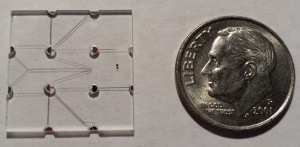
In the last 20 years, microfabrication techniques have allowed researchers to miniaturize tools for a plethora of bioanalytical applications.  In addition to better sensitivity, accuracy and precision, scaling down the size of bioanalytical tools has led to the exploitation of new technologies to further manipulate biomolecules in ways that has never before been achieved. For example, when microfluidic channels are on the same order of magnitude of the electric double layers that form due to localized charge at the surfaces, there exists unique physics that create different flow phenomenon, such as analyte concentration and/or separation, mainly due to the couples physics of electrostatics and fluid dynamics. This talk will outline the basis of such interesting phenomena, such as nanofluidic  separation and concentration, and well as probe the applications of such coupled systems, for example, handheld DNA detection. Most importantly, we will focus on the most recent work in the Pennathur lab in this field —  biopolar electrode (BPE)-based phenomenon. Bipolar electrodes (BPE) have been studied in microfluidic systems over the past few decades, and through rigorous experimentally-validated modeling of the rich combined physics of fluid dynamics, electrokinetics, and electrochemistry at BPEs, I will show the potential of utilizing microfluidic-based BPEs for the design and development of low power, accurate, low volume fluid pumping mechanisms, with the ultimate goal of integration into wearable drug delivery and µTAS systems.

Bio:

Professor Pennathur has been a Professor of Mechanical Engineering at University of California, Santa Barbara in 2007, specializing in the fields of MEMS, nanofludics, and electrokinetics.  Her most significant contributions include: 1) unearthing a novel mechanism for separation and concentration of analytes for bioanalytical applications, 2) developing a label-free detection mechanism for nucleic acids (that has since spun off into a point-of-care diagnostic company), 3) developing commercial medical diagnostic products, 4) building optical and acoustic biosensors and 5) developing revolutionized methods for measuring blood glucose for patients with diabetes. She received her B.S. and M.S. from MIT and PhD. From Stanford University.

Herman P. Schwan Distinguished Lecture: “Engineering human tissues for medical impact”

We hope you will join us for the Fall 2019 Herman P. Schwan Distinguished Lecture by Dr. Gordana Vunjak-Novakovic, hosted by the Department of Bioengineering.

Date: Wednesday, November 6th, 2019
Time: 3:30-4:30 PM
Location: Glandt Forum, Singh Center, 3205 Walnut Street

Gordana Vunjak-Novakovic, PhD, Columbia University

Speaker: Gordana Vunjak-Novakovic, PhD, University Professor, The Mikati Foundation Professor of Biomedical Engineering and Medical Sciences, Columbia University in the City of New York

Abstract:

The classical paradigm of tissue engineering involves the integrated use of human stem cells, biomaterial scaffolds (providing a structural and logistic template for tissue formation) and bioreactors (providing environmental control, dynamic sequences of molecular and physical signaling, and insights into the structure and function of the forming tissues). This “biomimetic” approach results in an increasingly successful representation of the environmental milieu of tissue development, regeneration and disease. Living human tissues are now being engineered from various types of human stem cells, and tailored to the patient and the condition being treated. A reverse paradigm is now emerging with the development of the “organs on a chip” platforms for modeling of integrated human physiology, using micro-tissues that are derived from human iPS cells and functionally connected by vascular perfusion. In all cases, the critical questions relate to our ability to recapitulate the cell niches, using bioengineering tools. To illustrate the state of the art in the field and reflect on the current challenges and opportunities, this talk will discuss: (i) anatomically correct bone regeneration, (ii) bioengineering of the lung, (iii) heart repair by a cell-free therapy, and (iv) the use of “organs on a chip” for patient-specific studies of human physiology, injury, healing and disease.

Funding: NIH, NSF, New York State, Mikati Foundation, Schwartz Foundation

Bio:

Gordana Vunjak-Novakovic is a University Professor, the highest academic rank at Columbia University that is reserved for only 16 professors out of 4,000, and the first engineer in the history of Columbia to receive this highest distinction. She is also the Mikati Foundation Professor of Biomedical Engineering and Medical Sciences, and on faculty in the Irving Comprehensive Cancer Center, College of Dental Medicine, Center for Human Development, and Mortimer B Zuckerman Mind Brain Behavior Institute. She directs the Laboratory for Stem Cells and Tissue Engineering that is a bioengineering lead of the Columbia Stem Cell Initiative and a home of the NIH Tissue Engineering Resource Center. She also serves on the Columbia President’s Task Force for Precision Medicine and the Executive Leadership of the Columbia University Medical Center. She received her Ph.D. in Chemical Engineering from the University of Belgrade in Serbia where she was on faculty until 1993, holds a doctorate honoris causa from the University of Novi Sad, and was a Fulbright Fellow at MIT.

The focus of her research is on engineering functional human tissues for regenerative medicine and studies of development and disease. Gordana published 3 books, 60 book chapters, 400 articles (including those in Nature, Cell, Nature Biotechnology, Nature Biomedical Engineering, Nature Communications, Nature Protocols, PNAS, Cell Stem Cell, Science Advances, Science Translational Medicine). With over 44,000 citations and impact factor h=121, she is one of the most highly cited individuals. She gave 420 invited talks, and has 101 licensed, issued or pending patents. With her students, she co-founded four biotech companies: epiBone (epibone.com), Tara Biosystems (tarabiosystems.com), Xylyx Bio (xylyxbio.com), and Immplacate (immplacatehealth.com).

She is a member of the Academia Europaea, Serbian Academy of Arts and Sciences, National Academy of Engineering, National Academy Medicine, National Academy of Inventors, and the American Academy of Arts and Sciences.

BE Grace Hopper Lecture: Powering Tumor Cell Migration Through Hetergeneous Microenvironments

We hope you will join us for the 2019 Bioengineering Grace Hopper lecture by Dr. Cynthia Reinhart-King.

Date: Thursday, April 4, 2019
Time: 3:30-4:30 PM
Location: Glandt Forum, Singh Center, 3205 Walnut Street

Dr. Cynthia Reinhart-King, Engineering, BME, Photo by Joe Howell

Speaker: Cynthia Reinhart-King, Ph.D.
Cornelius Vanderbilt Professor of Engineering, Director of Graduate Studies, Biomedical Engienering
Vanderbilt University

Title: “Powering Tumor Cell Migration Through Heterogeneous Microenvironments”

Abstract:
To move through tissues, cancer cells must navigate a complex, heterogeneous network of fibers in the extracellular matrix. This network of fibers also provides chemical, structural and mechanical cues to the resident cells. In this talk, I will describe my lab’s efforts to understand the forces driving cell movements in the tumor microenvironment. Combining tissue engineering approaches, mouse models, and patient samples, we create and validate in vitro systems to understand how cells navigate the tumor stroma environment. Microfabrication and native biomaterials are used to build mimics of the paths created and taken by cells during metastasis. Using these platforms, we have described a role for a balance between cellular energetics, cell and matrix stiffness, and confinement in determining migration behavior. Moreover, we have extended this work into investigating the role of the mechanical microenvironment in tumor angiogenesis to show that mechanics guides vessel growth and integrity. I will discuss the mechanical influences at play during tumor progression and the underlying biological mechanisms driving angiogenesis and metastatic cell migration as a function of the ECM with an eye towards potential therapeutic avenues.

Bio:
Cynthia Reinhart-King is the Cornelius Vanderbilt Professor of Engineering and the Director of Graduate Studies in Biomedical Engineering at Vanderbilt University.  Prior to joining the Vanderbilt faculty in 2017, she was on the faculty of Cornell University where she received tenure in the Department of Biomedical Engineering. She obtained undergraduate degrees in chemical engineering and biology at MIT and her PhD at the University of Pennsylvania in the Department of Bioengineering as a Whitaker Fellow working with Daniel Hammer. She then completed postdoctoral training as an Individual NIH NRSA postdoctoral fellow at the University of Rochester.  Her lab’s research interests are in the areas of cell mechanics and cell migration specifically in the context of cancer and atherosclerosis. Her lab has received funding from the American Heart Association, the National Institutes of Health, the National Science Foundation and the American Federation of Aging Research.  She has been awarded the Rita Schaffer Young Investigator Award in 2010 and the Mid-Career Award in 2018 from the Biomedical Engineering Society, an NSF CAREER Award, the 2010 Sonny Yau ‘72 Excellence in Teaching Award, a Cook Award for “contributions towards improving the climate for women at Cornell,” and the Zellman Warhaft Commitment to Diversity Award from the Cornell College of Engineering. She is a fellow of the Biomedical Engineering Society and the American Institute for Medical and Biological Engineering, and she is a New Voices Fellow of the National Academies of Science, Engineering and Medicine. She is currently a standing member of the NIH CMT study section panel and Secretary of the Biomedical Engineering Society.

Information on the Grace Hopper Lecture:
In support of its educational mission of promoting the role of all engineers in society, the School of Engineering and Applied Science presents the Grace Hopper Lecture Series. This series is intended to serve the dual purpose of recognizing successful women in engineering and of inspiring students to achieve at the highest level.
Rear Admiral Grace Hopper was a mathematician, computer scientist, systems designer and the inventor of the compiler. Her outstanding contributions to computer science benefited academia, industry and the military. In 1928 she graduated from Vassar College with a B.A. in mathematics and physics and joined the Vassar faculty. While an instructor, she continued her studies in mathematics at Yale University where she earned an M.A. in 1930 and a Ph.D. in 1934. Grace Hopper is known worldwide for her work with the first large-scale digital computer, the Navy’s Mark I. In 1949 she joined Philadelphia’s Eckert-Mauchly, founded by the builders of ENIAC, which was building UNIVAC I. Her work on compilers and on making machines understand ordinary language instructions lead ultimately to the development of the business language, COBOL. Grace Hopper served on the faculty of the Moore School for 15 years, and in 1974 received an honorary degree from the University. In support of the accomplishments of women in engineering, each department within the School invites a prominent speaker to campus for a one or two-day visit that incorporates a public lecture, various mini-talks and opportunities to interact with undergraduate and graduate students and faculty. The lecture is open to everyone!

BE Seminar Series: March 14th

The BE Seminar Series continues this week. We hope to see you there!

Shuichi Takayama, Ph.D.

Speaker: Shuichi Takayama, Ph.D.
Professor, GRA Eminent Scholar, Price Gilbert, Jr. Chair in Regenerative Engineering and Medicine
Wallace H. Coulter Department of Biomedical Engineering
Georgia Institute of Technology and Emory University

Date: Thursday, March 14th, 2019
Time: 12:00 pm
Location: Room 337, Towne Building

“Microfluidics and Immuno-Materials for Organs-on-a-Chip”

This presentation will describe microfluidic technologies to conveniently produce life-like pulsatile flows along with applications to study of lung injury, enhancement of in vitro fertilization, and analysis of frequency-dependent cellular responses. The microfluidic technologies range from adaptation of piezo-electric actuator arrays from Braille displays to design of microfluidic circuits that can be designed to switch fluid flow on and off periodically on their own. The presentation will also describe engineered materials to mimic an aspect of the innate immune system to combat bacterial infection. More specifically, reconstituted chromatin microwebs inspired by neutrophil extracellular traps. Using a defined composition reconstituted chromatin microweb, we reveal impact of microweb DNA-histone ratio on bacteria capture. Additionally, we found that E. coli, including clinical isolates and resistant strains, are killed more efficiently by the last-resort antibiotic, colistin, when bound to microwebs. Recent efforts towards incorporation of these materials into human cell systems will also be described. Time permitting, topics on organoids, fibrosis, liquid-liquid phase separation, and scaling may be incorporated.

BE Seminar Series: November 29th

The BE Seminar Series continues this week. We hope to see you there!

Speaker: Alison Pouch, Ph.D.
Research Associate, Penn Image Computing & Science Lab, Department of Radiology
Gorman Cardiovascular Research Group, Department of Surgery, University of Pennsylvania

Date: November 29, 2018
Time: 12:00 pm
Location: Room 337, Towne Building

“Shaping Innovation in Heart Valve Surgery with Image-Based Modeling”

A number of challenging questions routinely arise before heart valve surgery. Should a regurgitant valve be repaired or replaced? If repaired, which techniques and maneuvers should be employed? When is the optimal time to perform surgery? This is just the beginning of a series of questions whose answers rely on an individualized assessment of heart valve morphology and function. With advanced imaging technology like 3D echocardiography already in the operating room, we have a window into patient-specific disease characteristics that cannot otherwise be appreciated immediately before surgery. Unfortunately, imaging resources are often not tapped to their full potential, which contributes to delays in surgical innovation. This presentation introduces an image-based modeling approach to creating enhanced visualizations and quantification of heart valves from real-time 3D echocardiography. The goal of this approach is to fundamentally change the way that surgeons interact with and utilize images in the operating room. Pre-operative image-based heart valve models can be used to characterize mechanisms of disease prior to cardiopulmonary bypass and to identify connections between pre-operative image features and clinical outcomes. Image-based modeling provides a means for surgeons to innovate and master new surgical techniques like bicuspid aortic valve repair and to devise new standardized approaches for surgical treatment of ischemic mitral regurgitation. Image analysis and surgical innovation are linked within and beyond the cardiovascular domain, and this work aims to optimize the potential of imaging and shape modeling for pre-surgical planning.

BE Seminar Series: November 15th

The BE Seminar Series continues this week with two lectures delivered by our current PhD Students. We hope to see you there!

Date: November 15, 2018
Location: Room 337, Towne Building

Claim Extraction for Biomedical Publications

Speaker: Titipat Achikulvisut, Ph.D. Student
Advisor: Konrad Kording, Ph.D.
Time: 12:05-12:25 pm

Abstract:

Scientific claims are a foundation of scientific discourse. Extracting claims from scientific articles is primarily done by researchers during literature review and discussions. The enormous growth in scientific articles makes this ever more challenging and time-consuming. Here, we develop a deep neural network architecture to solve the problem. Our model an F1 score of 0.704 on a large corpus of expertly annotated claims within abstracts. Our results suggest that we can use a small dataset of annotated resources to achieve high-accuracy claim detection. We release a tool for discourse and claim detection, and a novel dataset annotated by experts. We discuss further applications beyond Biomedical literature.

Multiple Sclerosis Lesion Segmentation with Joint Label Fusion Evaluated on OASIS and CNN

Speaker: Mengjin Dong, Ph.D. Student
Advisor: Paul Yushkevich, Ph.D.
Time: 12:30-12:50 pm

Abstract:

Scientific claims are a foundation of scientific discourse. Extracting claims from scientific articles is primarily done by researchers during literature review and discussions. The enormous growth in scientific articles makes this ever more challenging and time-consuming. Here, we develop a deep neural network architecture to solve the problem. Our model an F1 score of 0.704 on a large corpus of expertly annotated claims within abstracts. Our results suggest that we can use a small dataset of annotated resources to achieve high-accuracy claim detection. We release a tool for discourse and claim detection, and a novel dataset annotated by experts. We discuss further applications beyond Biomedical literature.


BE Seminar Series: September 20, 2018

The BE Seminar Series continues next week with three lectures delivered by our current PhD Students. We hope to see you there!

“Magnetic Susceptibility of Hemorrhagic Myocardial Infarction”

Brianna Moon, PhD Candidate

Speaker: Brianna Moon, PhD Candidate
Research Advisor: Walter Witschey, PhD

Date: Thursday, September 20, 2018
Time: 12:05pm-12:25pm
Location: Room 337 Towne Building

Abstract: 
Hemorrhagic myocardial infarction (MI) has been reported in 41% and 54% of ST-elevated MI patients after primary percutaneous coronary intervention. These patients are at high risk for adverse left ventricle (LV) remodeling, impaired LV function and increased risk of fatal arrhythmias.  Relaxation time MRI such as T2*-maps are sensitive to hemorrhagic infarct iron content, but are also affected by myocardial edema and fibrosis. Quantitative Susceptibility Mapping (QSM), which uses the MR signal phase to quantify tissue magnetic susceptibility, may be a more specific and sensitive marker of hemorrhagic MI. The objective of this study was to develop and validate cardiac QSM in a large animal model of myocardial infarction, investigate the association of magnetic susceptibility with iron content and infarct pathophysiology, and compare QSM to relaxation time mapping, susceptibility-weighted imaging (SWI), and late-gadolinium enhanced (LGE) MRI.


“Role of ACTG2 Mutations in Visceral Myopathy”

Sohaib Hashmi, MD/PhD Student

Speaker: Sohaib Hashmi, MD/PhD Student
Research Advisor: Robert O. Heuckeroth, MD, PhD

Date: Thursday, September 20, 2018
Time: 12:30-12:50pm
Location: Room 337 Towne Building

Abstract: 

Visceral myopathy is a debilitating chronic medical condition in which smooth muscle of the bowel, bladder, and uterus is weak or dysfunctional. When the bowel muscle is weak and unable to efficiently contract, the bowel becomes distended which causes pain, bilious vomiting, growth failure, and nutritional deficiencies. The abdominal distension can become life-threatening. Patients often become dependent on intravenous nutrition and undergo multiple rounds of abdominal surgery, which only partially alleviates symptoms. Recently, rare mutations in gamma smooth muscle actin (ACTG2) have been shown to be responsible for a large subset of visceral myopathies. ACTG2 is a critical protein in the smooth muscle contractile apparatus. However, we have only limited knowledge of how ACTG2 mutations may cause human disease. To improve our understanding of the pathophysiology of ACTG2 mutations, my work has the following specific aims:

1) Determine how pathogenic ACTG2 mutations affect actin structure and function in primary human intestinal smooth muscle cells (HISMCs).

2) Examine the effects of ACTG2 mutations on differentiation of human pluripotent stem cells into smooth muscle cells (SMCs).

These studies will allow us to elucidate the mechanisms through which ACTG2 mutations impair normal visceral smooth muscle development and function. I am examining the effects of ACTG2 mutations on actin filament organization in fixed cells and actin dynamics in live cells using fluorescent probes. I will investigate changes in contractile force generation using traction force microscopy. I am also developing a novel differentiation method to convert pluripotent stem cells into cells closely resembling visceral SMCs. We will use this method with CRISPR/Cas9 gene editing to study the effects of the mutations on SMC differentiation. We are currently generating pluripotent stem cell lines containing ACTG2 mutations and performing assays to investigate actin organization and dynamics. We are using these systems to identify robust phenotypes highlighting the mechanisms through which ACTG2 mutations cause disease. We hope to leverage this information in the selection of targets for high-throughput drug screening, which may eventually lead to novel treatment strategies for visceral myopathies.


“Distinct Patterns of Longitudinal Cortical Thinking and Perfusion in Pathological Subtypes of Behavioral Variant Frontotemporal Degeneration”

Christopher Olm, PhD Student

Speaker: Christopher Olm, PhD Student
Research Advisor: Murray Grossman, MD, EdD

Date: Thursday, September 20, 2018
Time: 12:55-1:15pm
Location: Room 337 Towne Building

Abstract: 
Two main sources of pathology have been identified in the behavioral variant of frontotemporal dementia (bvFTD): tau inclusions (FTLD-tau) and TDP-43 aggregates (FTLD-TDP). With therapies emerging that target these proteins, exploring distinct trajectories of degeneration can be extremely helpful for tracking progression in clinical trials and improve prognosis estimation. We hypothesized that longitudinal cortical thinning (CT) would identify areas of extant disease progression in bvFTD subgroups and longitudinal hypoperfusion would identify distinct regions of anticipated neurodegeneration. We included N=47 patients with probable or definite bvFTD and two MRI scanning sessions including T1-weighted and arterial spin labeling (ASL) scans, recruited through the Penn Frontotemporal Degeneration Center. Neuropathology, genetic mutations, or CSF protein markers (phosphorylated tau (p-tau)/Ab1-42<.09 for likely FTLD; p-tau<8.75 for FTLD-TDP) were used to identify bvFTD with likely FTLD-tau (n=28, mean age=63.1 years, mean disease duration=3.89 years) or likely FTLD-TDP (n=19, mean age=61.9, mean disease duration=3.06). Voxel-wise cortical thickness and cerebral blood flow estimates were generated for each T1 and ASL scan, respectively, using longitudinal pipelines in ANTs. We created annual change images by subtracting follow-up images from baseline and dividing by inter-scan interval. In whole brain voxel-wise comparisons, FTLD-tau showed significantly greater right orbitofrontal CT and longitudinal hypoperfusion in right middle temporal and angular cortex relative to FTLD-TDP. FTLD-TDP displayed greater progressive CT in left superior and middle frontal cortex, precentral gyrus, and right temporal cortex, and longitudinal hypoperfusion in medial prefrontal cortex relative to FTLD-tau. In conclusion, FTLD-tau and FTLD-TDP show distinct patterns of longitudinal CT and hypoperfusion. Structural and functional MRI contribute independent information potentially useful for characterizing disease progression in vivo for clinical trials.