Week in BioE (July 12, 2019)

by Sophie Burkholder

DNA Microscopy Gives a Better Look at Cell and Tissue Organization

A new technique that researchers from the Broad Institute of MIT and Harvard University are calling DNA microscopy could help map cells for better understanding of genetic and molecular complexities. Joshua Weinstein, Ph.D., a postdoctoral associate at the Broad Institute, who is also an alumnus of Penn’s Physics and Biophysics department and former student in Penn Bioengineering Professor Ravi Radhakrishnan’s lab, is the first author of this paper on optics-free imaging published in Cell.

The primary goal of the study was to find a way of improving analysis of the spatial organization of cells and tissues in terms of their molecules like DNA and RNA. The DNA microscopy method that Weinstein and his team designed involves first tagging DNA, and allowing the DNA to replicate with those tags, which eventually creates a cloud of sorts that diffuses throughout the cell. The DNA tags subsequent interactions with molecules throughout the cell allowed Weinstein and his team to calculate the locations of those molecules within the cell using basic lab equipment. While the researchers on this project focused their application of DNA microscopy on tracking human cancer cells through RNA tags, this new method opens the door to future study of any condition in which the organization of cells is important.

Read more on Weinstein’s research in a recent New York Times profile piece.

Penn Engineers Demonstrate Superstrong, Reversible Adhesive that Works like Snail Slime

A snail’s epiphragm. (Photo: Beocheck)

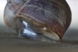
If you’ve ever pressed a picture-hanging strip onto the wall only to realize it’s slightly off-center, you know the disappointment behind adhesion as we typically experience it: it may be strong, but it’s mostly irreversible. While you can un-stick the used strip from the wall, you can’t turn its stickiness back on to adjust its placement; you have to start over with a new strip or tolerate your mistake. Beyond its relevance to interior decorating, durable, reversible adhesion could allow for reusable envelopes, gravity-defying boots, and more heavy-duty industrial applications like car assembly.

Such adhesion has eluded scientists for years but is naturally found in snail slime. A snail’s epiphragm — a slimy layer of moisture that can harden to protect its body from dryness — allows the snail to cement itself in place for long periods of time, making it the ultimate model in adhesion that can be switched on and off as needed. In a new study, Penn Engineers demonstrate a strong, reversible adhesive that uses the same mechanisms that snails do.

This study is a collaboration between Penn Engineering, Lehigh University’s Department of Bioengineering, and the Korea Institute of Science and Technology.

Read the full story on Penn Engineering’s Medium blog. 

Low-Dose Radiation CT Scans Could Be Improved by Machine Learning

Machine learning is a type of artificial intelligence growing more and more popular for applications in bioengineering and therapeutics. Based on learning from patterns in a way similar to the way we do as humans, machine learning is the study of statistical models that can perform specific tasks without explicit instructions. Now, researchers at Rensselaer Polytechnic Institute (RPI) want to use these kinds of models in computerized tomography (CT) scanning by lowering radiation dosage and improving imaging techniques.

A recent paper published in Nature Machine Intelligence details the use of modularized neural networks in low-dose CT scans by RPI bioengineering faculty member Ge Wang, Ph.D., and his lab. Since decreasing the amount of radiation used in a scan will also decrease the quality of the final image, Wang and his team focused on a more optimized approach of image reconstruction with machine learning, so that as little data as possible would be altered or lost in the reconstruction. When tested on CT scans from Massachusetts General Hospital and compared to current image reconstruction methods for the scans, Wang and his team’s method performed just as well if not better than scans performed without the use of machine learning, giving promise to future improvements in low-dose CT scans.

A Mind-Controlled Robotic Arm That Requires No Implants

A new mind-controlled robotic arm designed by researchers at Carnegie Mellon University is the first successful noninvasive brain-computer interface (BCI) of its kind. While BCIs have been around for a while now, this new design from the lab of Bin He, Ph.D.,  a Trustee Professor and the Department Head of Biomedical Engineering at CMU, hopes to eliminate the brain implant that most interfaces currently use. The key to doing this isn’t in trying to replace the implants with noninvasive sensors, but in improving noisy EEG signals through machine learning, neural decoding, and neural imaging. Paired with increased user engagement and training for the new device, He and his team demonstrated that their design enhanced continuous tracking of a target on a computer screen by 500% when compared to typical noninvasive BCIs. He and his team hope that their innovation will help make BCIs more accessible to the patients that need them by reducing the cost and risk of a surgical implant while also improving interface performance.

People and Places

Daeyeon Lee, professor in the Department of Chemical and Biomolecular Engineering and member of the Bioengineering Graduate Group Faculty here at Penn, has been selected by the U.S. Chapter of the Korean Institute of Chemical Engineers (KIChE) as the recipient of the 2019 James M. Lee Memorial Award.

KIChE is an organization that aims “to promote constructive and mutually beneficial interactions among Korean Chemical Engineers in the U.S. and facilitate international collaboration between engineers in U.S. and Korea.”

Read the full story on Penn Engineering’s Medium blog.

We would also like to congratulate Natalia Trayanova, Ph.D., of the Department of Biomedical Engineering at Johns Hopkins University on being inducted into the Women in Tech International (WITI) Hall of Fame. Beginning in 1996, the Hall of Fame recognizes significant contributions to science and technology from women. Trayanova’s research specializes in computational cardiology with a focus on virtual heart models for the study of individualized heart irregularities in patients. Her research helps to improve treatment plans for patients with cardiac problems by creating virtual simulations that help reduce uncertainty in either diagnosis or courses of therapy.

Finally, we would like to congratulate Andre Churchwell, M.D., on being named Vanderbilt University’s Chief Diversity Officer and Interim Vice Chancellor for Equity, Diversity, and Inclusion. Churchwell is also a professor of medicine, biomedical engineering, and radiology and radiological sciences at Vanderbilt, with a long career focused in cardiology.

César de la Fuente on 35 Innovators Under 35 List

Cesar de la Fuente-Nunez, PhD

César de la Fuente, assistant professor in the Perelman School of Medicine and in the School of Engineering and Applied Science, was named one of MIT Technology Review’s “35 Innovators Under 35” for 2019.

“It’s part of our ethos that technology can and should be a force for good. Our annual list of 35 innovators under 35 is a way of putting faces on that idea,” reads the 2019 award announcement. “This year’s list shows that even in our hard, cynical world, there are still lots of smart people willing to dedicate their lives to the idea that technology can make a safer, fairer world.”

De la Fuente was named in the list’s “Pioneers” category for his work researching antibiotics with a computational approach. Using algorithms, he creates artificial antibiotics to better understand how bacteria will evolve and how scientists can optimize treatments. De la Fuente, who was also recently featured in GEN’s Top 10 Under 40 list, further expands his search for medical solutions by extensively studying a variety of proteins, searching for molecules to develop into antimicrobials.

Included in the honor of being named on the 2019 Innovators List is an invitation for de la Fuente to speak at the EmTech MIT conference in September, an event that reflects on the potential impacts of the year’s biggest developments.

Read MIT Technology Review’s coverage of de la Fuente’s pioneering work and learn more about de la Fuente’s research on his lab website.

Originally posted on the Penn Engineering Medium Blog.

Lee Bassett and Andrew Tsourkas Awarded Grainger Foundation Grant for Interdisciplinary Research

Lee Bassett and Andrew Tsourkas

By Lauren Salig

The National Academy of Engineering (NAE) has awarded two Penn Engineers with The Grainger Foundation Frontiers of Engineering Grant for Advancement of Interdisciplinary Research. Lee Bassett, assistant professor in the Department of Electrical and Systems Engineering, and Andrew Tsourkas, professor and undergraduate chair in the Department of Bioengineering, will be using the $30,000 award to kick-start their research collaboration.

The NAE describes the Frontiers of Engineering program as one that “brings together outstanding early-career engineers from industry, academia, and government to discuss pioneering technical work and leading-edge research in various engineering fields and industry sectors. The goal is to facilitate interactions and exchange of techniques and approaches across fields and facilitate networking among the next generation of engineering leaders.”

Bassett and Tsourkas fit the grant’s description, as their proposed research requires them to combine their different areas of expertise to push the state of the art in engineering. The pair plans to engineer a new class of nanoparticles that can sense and differentially react to particular chemicals in their biochemical environment. This new class of nanoparticles could allow scientists to better study cellular processes and could eventually have important applications in medicine, potentially allowing for more personalized diagnoses and targeted treatment of disease.

To design and create this type of nanoparticle is no small task. The research demands Bassett’s background in engineering quantum-mechanical systems for use as environmental sensors, and Tsourkas’ ability to apply these properties to nanoscale “theranostic” agents, which are designed to target treatments based on a patient’s specific diagnostic test results.

By combining forces, Bassett and Tsourkas hope to introduce a new nanoparticle tool into their fields and to connect even more people in their different areas to promote future interdisciplinary work.

Originally posted on the Penn Engineering Medium Blog.

Week in BioE (June 28, 2019)

by Sophie Burkholder

Innovations in Vascularization Could Lead to a New Future in Bioprinting

We may be one step closer to 3D-printing organs for transplants thanks to innovations in vascularization from researchers at Rice University and Washington University. Jordan Miller, Ph.D., a Penn Bioengineering alumnus, now an assistant professor of bioengineering at Rice, worked with his colleague Kelly Stevens, Ph.D., an assistant professor of the bioengineering department at Washington, to develop 3D-printed networks that mimicked the vascularized pathways for the transport of blood, lymph, and other fluids in the body. Their work appeared on a recent cover of Science, featuring a visual representation of the 3D-printed vessels in vasculature meant to mirror that of the human lung.

Relying heavily on open source 3D-printing, Miller and Stevens, along with collaborators from a handful of other institutions and start-ups, found ways to model dynamic vasculature systems similar to heart valves, airways systems, and bile ducts to keep 3D-printed tissue viable. The video below demonstrates the way the team successfully modeled vasculature in a small portion of the lung by designing a net-like structure around a sack of air. But Miller, a long-time supporter of open source printing and bioprinting, hopes that this is merely one step closer to what he sees as the ultimate goal of allowing for all organs to be bioprinted. Having that sort of power would reduce the complex issues that come with organ transplants, from organ availability to compatibility, and bring an end to a health issue that affects the over 100,000 people on the organ transplant waiting list.

A Combination of Protein Synthesis and Spectrometry Improve Cell Engineering

One goal of modern medicine is to create individualized therapeutics by figuring out a way to control cell function to perform specific tasks for the body without disrupting normal cell function. Balancing these two goals often proves to be one of the greatest difficulties of this endeavor in the lab, but researchers at Northwestern University found a way to combine the two functions at once in methods they’re calling cell-free protein synthesis and self-assembled monolayer desorption ionization (SAMDI) mass spectrometry. This innovation in the combination of the two methods accelerates the trial and error process that comes with engineering cells for a specific need, allowing researchers to cover a lot more ground in determining what works best in a smaller amount of time.

Leading the study are Milan Mrksich, Ph.D., a Henry Wade Rogers Professor of Biomedical Engineering at Northwestern, and Michael Jewett, Ph.D., a Charles Deering McCormick Professor of Teaching Excellence and co-director of the Center for Synthetic Biology at Northwestern. Together, they hope to continue to take advantage of the factory-like qualities of cell operations in order to use cells from any organisms to our advantage as needed. By helping to reduce the amount of time spent on trial and error, this study brings us one step closer to a world of efficient and individualized medicine.

Non-Invasive Sensory Stimulation as New Way of Treating Alzheimer’s

What if we could reduce the effects of Alzheimer’s disease with a non-invasive therapy comprised of only sensory inputs of light and sound? A recent study between Georgia Tech and MIT tries to make that possible. Alzheimer’s patients often have a larger than normal number of amyloid plaques in their brains, which is a naturally occurring protein that in excess can disrupt neurological function. The treatment —  designed in part by Abigail Paulson, a graduate student in the lab of Annabelle Singer, Ph.D., assistant professor of Biomedical Engineering at Georgia Tech and Emory University — uses a combination of light and sound to induce gamma oscillations in brain waves of mice with high amounts of these amyloid plaques. Another lead author of the study is Anthony Martorell, a graduate student in the Tsai Lab at MIT, where Singer was a postdoctoral researcher.

This new approach is different from other non-invasive brain therapies for memory improvement, as tests demonstrated that it had the power to not only reach the visual cortex, but that it also had an effect on the memory centers in the hippocampus. An innovation like this could bring about a more widespread form of treatment for Alzheimer’s patients, as the lack of a need for surgery makes it far more accessible. Singer hopes to continue the project in the future by looking at how these sensory stimulations affect the brain throughout a variety of processes, and more importantly, if the therapy can be successfully applied to human patients.

NIH Grant Awarded to Marquette Biomedical Engineering Professor for Metal Artifact Reduction Techniques in CT Scans

Taly Gilat-Shmidt, Ph.D., an associate professor of biomedical engineering at Marquette University, recently received a $1.4 million grant from the National Institute of Health to improve methods for radiation treatment through metal artifact reduction techniques. When patients have some sort of metal that can’t be removed, such as an orthopaedic implant like a hip or knee replacement, it can interfere with the imaging process for CT scans and lead to inaccuracies by obscuring some tissue in the final images. These inaccuracies can lead to difficulty in devising treatment plans for patients who require radiation, as CT scans are often used to assess patients and determine which line of treatment is most appropriate. Gilat-Schmidt hopes to use the grant to implement tested algorithms to help reduce this variability in imaging that comes from metal implants.

People and Places

Activities for Community Education in Science (ACES), founded by Penn chemistry graduate students in 2014, aims to inspire interest and provide a positive outlook in STEM for kids and their families. The biannual event provides students grades 3–8 with an afternoon of demonstrations, experiments, and hands-on activities focused on physics and chemistry.

After an explosive opening demonstration, more than 70 students made their way between experiments in small groups, each participating in different experiments based on their age.

Read the rest of this story on Penn Today.

The Society of Women Engineers (SWE) is a non-profit organization serving as one of the world’s largest advocates for women in engineering and technology over the past six decades. With a mission to empower women to become the next leading engineers of the world, SWE is just one of many agents hoping to bring more diversity to the field. Our chapter of SWE at Penn focuses particularly on professional development, local educational outreach, and social activities across all general body members. In a new article from SWE Magazine, the organization collected social media responses from the public on the women engineers we should all know. With a diverse list of engineers from both the past and present, the article helps bring to light just how much even a handful of women contributed to the field of engineering already.


Chip Diagnostics receives the JPOD @ Philadelphia QuickFire Challenge Award

By Erica K. Brockmeier

Chip Diagnostics is the awardee of the JPOD @ Philadelphia QuickFire Challenge sponsored by Johnson & Johnson Innovation — JLABS. The Challenge was designed to accelerate healthcare innovation and commercialization within the greater Philadelphia area.

David Issadore (center) was announced as the awardee of the JPOD @ Philadelphia QuickFire Challenge by Katherine Merton (right), head of JLABS New York City, Boston, and Philadelphia, at last week’s BIO 2019 International convention. (Photo: Johnson & Johnson Innovation)

Chip Diagnostics is a Philadelphia-based device company founded in 2016 based on research from the lab of David Issadore, Assistant Professor of Bioengineering and Electrical and Systems Engineering in the School of Engineering and Applied Science. The startup combines microelectronics, microfluidics, and nanomaterials with the aim to better diagnose cancer. The company is developing technologies and digital assays for minimally-invasive early cancer detection and screening that can be done using mobile devices.

There has been a long interest in diagnosing cancer using blood tests by looking for proteins, cells, or DNA molecules shed by tumors, but these tests have not worked well for many cancers since the molecules shed tend to be either nonspecific or very rare.

Issadore’s group aims to target different particles called exosomes: Tiny particles shed by cells that contain similar proteins and RNA as the parent cancer cell. The problem, explains Issadore, is that because of the small size of the exosomes, conventional methods such as microscopy and flow cytometry wouldn’t work. “As an engineering lab, we saw an opportunity to build devices on a nanoscale that could specifically sort the cancer exosomes versus the background exosomes of other cells,” he explains.

After Issadore was approached by the IP group at PCI Ventures in the early stages of their research, Chip Diagnostics has since made huge strides as a company. Now, as the awardee of the JPOD @ Philadelphia QuickFire Challenge, Chip Diagnostics will receive $30,000 in grant funding to further develop the first-in-class, ultra-high-definition exosomal-based cancer diagnostic. The award also includes one year of residency at Pennovation Works as well as access to educational programs and mentoring provided by Johnson & Johnson Family of Companies global network of experts.

Originally posted on the Penn Engineering Medium Blog. Continue reading at Penn Today.

Week in BioE (June 14, 2019)

by Sophie Burkholder

Bio-inspiration Informs New Football Helmet Design from IUPUI Students

Art, design, biology, and engineering all interact with each other in a recent design for a football helmet from two students one of media arts and the other of engineering at the Indiana University – Purdue University Indianapolis. Directed by Lecturer in Media Arts and Science Zebulun Wood, M.S., and Associate Professor of Mechanical and Energy Engineering and Assistant Professor of Biomedical Engineering Andres Tovar, Ph.D., the students found inspiration in biological structures like a pomelo peel, nautilus shell, and woodpecker skull to create energy-absorbing helmet liners. The resulting design took these natural concussion-reducing structures and created compliant mechanism lattice-based liners the replace the foam traditionally placed in between two harder shells of a typical helmet. Their work not only exemplifies the benefits of bio-inspiration, but demonstrates the way that several different domains of study can overlap in the innovation of a new product.

Study of Mechanical Properties of Hyaluronic Acid Could Help Inform Current Debates Over Treatment Regulation for Osteoarthritis

Arthritis is an extremely common condition, especially in older patients, in which inflammation of the joints can cause high amounts of stiffness and pain. Osteoarthritis in particular is the result of the degradation of flexible tissue between the bones of a joint, which increases friction in joint motion. A common treatment of this form of arthritis is the injection of hyaluronic acid, which is meant to provide joint lubrication, and decreases this friction between bones. Recently, however, there has been a debate over hyaluronic acid’s classification by the FDA and whether it should remain based on the knowledge of the mechanical actions of the acid in treatment for osteoarthritis or if potential chemical action of the acid should be considered as well.

Because of limited ways of testing the mechanical properties of the acid, many researchers felt that there could be more to hyaluronic acid’s role in pain relief for arthritic patients. But Lawrence Bonassar, Ph.D., the Daljit S. and Elaine Sarkaria Professor in Biomedical Engineering at the Meinig School of Bioengineering of Cornell University, had another idea. With his lab, he created a custom-made tribometer to measure the coefficient of friction of a given lubricant by rubbing a piece of cartilage back and forth across a smooth glass plate. The research demonstrated that hyaluronic acid’s ability to reduce the coefficient of friction aligned with patients’ pain relief. Bonassar and his team hope that these results will demonstrate the heavy contribution of mechanical action that hyaluronic acid has in osteoarthritis treatment, and help bring an end to the debate over its FDA classification.

A New Way of Mapping the Heart Could Lead to Better Understanding of Contractile Activity

Though reduced contractions in certain regions of the heart can be an indicator of a certain condition, there is currently no way to directly measure contractile activity. This is why Cristian Linte, Ph.D., an Associate Professor of Biomedical Engineering in the Kate Gleason College of Engineering at the Rochester Institute of Technology (RIT), hopes to create a map of the heart that can quantify contraction power. In collaboration with Niels Otani, Ph.D., an Associate Professor in the School of Mathematics at RIT, Linte plans to use an $850,000 grant from the National Science Foundation to achieve a more comprehensive understanding of the heart through both medical imaging and mechanical modeling. The group hopes that their approach will lead to not only a better way to diagnose certain heart conditions and diseases, but also open up understanding of active contraction, passive motion, and the stresses within the heart walls that underlie each.

Celebrity Cat Lil Bub Helps Penn and German Researchers Draw Public Attention to Genetics

Lil Bub’s unique appearance has garnered millions of online fans, and now, an avenue for researchers to talk about genetics. (Photo Courtesy of Mike Bridavsky)

In 2015, a group of curious researchers set out to sequence the genome of a celebrity cat named Lil Bub. They were hoping to understand the genetics behind Lil Bub’s extra toes and unique skeletal structure, which contribute to her heart-warming, kitten-like appearance. However, an equally important goal of their “LilBUBome” project was to invite the general public into the world of genetics.

Orsolya “Uschi” Symmons, a postdoctoral researcher at Penn in Associate Professor of Bioengineering Arjun Raj’s lab, led the research team along with Darío Lupiáñez at the Max-Delbrück Center for Molecular Medicine in Berlin, and Daniel Ibrahim at the Max Planck Institute for Molecular Geneticsin Berlin. Lil Bub’s owner, Mike Bridavsky, also contributed to the project.

Because of Lil Bub’s online fame, the project garnered attention from her fans and the media, all hoping to discover the secret to Lil Bub’s charm. As early as 2015, Gizmodo’s Kiona Smith-Strickland reported on the team’s intentions to sequence Lil Bub’s genome, and, since then, many have been awaiting the results of the LilBUBome.

To read more of this story, visit Penn Engineering’s Medium Blog.

People and Places

The Alfred P. Sloan Foundation awarded a six-year grant to Barnard College and Columbia University’s School of Engineering and Applied Science to support graduate education for women in engineering. The funding will go towards a new five-year program that enables Barnard students to attain both a B.A. and M.S. in one year after their traditional four years of undergraduate education. The program will offer M.S. degrees in chemical engineering, biomedical engineering, and industrial engineering and operations research, and is one of the first of its kind for women’s colleges.

We would like to congratulate Jean Paul Allain, Ph.D., on being named the first head of the new Ken and Mary Alice Lindquist Department of Nuclear Engineering at Penn State. Allain, who is currently a Professor and head of graduate programs in the University of Illinois at Urbana-Champaign’s Department of Nuclear, Plasma, and Radiological Engineering, conducts research in models of particle-surface interactions. In addition to being head of the new department at Penn State, Allain will also hold a position as a Professor of Biomedical Engineering at the university.

We would also like to congratulate Andrew Douglas, Ph.D., on his appointment as the Vice Provost for Faculty Affairs at Johns Hopkins University. Douglas currently holds the position of Vice Dean for Faculty at the Whiting School of Engineering, and has joint appointments in Mechanical and Biomedical Engineering. Douglas’s research at Hopkins focuses on mechanical properties and responses of compliant biological tissue and on the nonlinear mechanics of solids, with a focus on soft tissues and organs like the heart and tongue.

César de la Fuente Named One of GEN’s ‘Top 10 Under 40’

Cesar de la Fuente-Nunez, PhD

César de la Fuente, assistant professor in the Departments of Psychiatry and Microbiology in the Perelman School of Medicine and the Department of Bioengineering in the School of Engineering and Applied Science, has been listed as one of the top 10 emerging professionals in his field under the age of 40 by GEN, a publication that covers genetic engineering and biotechnology news. The list recognizes up-and-coming leaders in the field of life sciences, both in industry and academia.

De la Fuente, who started at Penn earlier this year, was recognized because he “is pioneering the computerization of biological systems for the development of transformative biotechnologies designed to solve societal grand challenges such as antibiotic resistance.”

Read the full story at the Penn Engineering Medium blog.

Week in BioE (May 31, 2019)

by Sophie Burkholder

Vector Flow Imaging Helps Visualize Blood Flow in Pediatric Hearts

A group of biomedical engineers at the University of Arkansas used a new ultrasound-based imaging technique called vector flow imaging to help improve the diagnosis of congenital heart disease in pediatric patients. The study, led by associate professor of biomedical engineering Morten Jensen, Ph.D., collaborated with cardiologists at the local Children’s Hospital in Little Rock to produce images of the heart in infants to help potentially diagnose congenital heart defects. Though the use of vector flow imaging has yet to be developed for adult patients, this type of imaging could possibly provide more detail about the direction of blood flow through the heart than traditional techniques like echocardiography do. In the future, the use of both techniques could provide information about both the causes and larger effects of heart defects in patients.

Using Stem Cells to Improve Fertility in Leukemia Survivors

One of the more common side effects of leukemia treatment in female patients is infertility, but researchers at the University of Michigan want to change that. Led by associate professor of biomedical engineering Ariella Shikanov, Ph.D., researchers in her lab found ways of increasing ovarian follicle productivity in mice, which directly relates to the development of mature eggs. The project involves the use of adipose-derived stem cells, that can be found in human fat tissue, to surround the follicles in an ovary-like, three-dimensional scaffold.  Because the radiation treatments for leukemia and some other cancers are harmful to follicles, increasing their survival rate with this stem cell method could reduce the rate of infertility in patients undergoing these treatments. Furthermore, this new approach is innovative in its use of a three-dimensional scaffold as opposed to a two-dimensional one, as it stimulates follicle growth in all directions and thus helps to increase the follicle survival rate.

Penn Engineers Look at How Stretching & Alignment of Collagen Fibers Help Cancer Cells Spread

Cancer has such a massive impact on people’s lives that it might be easy to forget that the disease originates at the cellular level. To spread and cause significant damage, individual cancer cells must navigate the fibrous extracellular environment that cells live in, an environment that Penn Engineer Vivek Shenoy has been investigating for years.

Shenoy is the Eduardo D. Glandt President’s Distinguished Professor with appointments in Materials Science and Engineering, Mechanical Engineering and Applied Mechanics, and Bioengineering. He is also the Director of the Center for Engineering MechanoBiology (CEMB), one of the NSF’s twelve Science and Technology Centers.

Shenoy’s most recent study on cancer’s mechanical environment was led by a postdoctoral researcher in his lab, Ehsan Ban. Paul Janmey, professor in Physiology and Bioengineering, and colleagues at Stanford University also contributed to the study. Shenoy also received the Heilmeier Award this March and delivered the Heilmeier Award Lecture in April.

Read the rest of this story on Penn Engineering’s Medium Blog.

Controlled Electrical Stimulation Can Prevent Joint Replacement Infections

Joint replacements are one of the most common kinds of surgery today, but they still require intense post-operative therapy and have a risk of infection from the replacement implant. These infections are usually due to the inflammatory response that the body has to any foreign object, and can become serious and life-threatening if left untreated. Researchers at the University of Buffalo Jacobs School of Medicine and Biomedical Sciences hope to offer a solution to preventing infections through the use of controlled electrical stimulation. Led by Mark Ehrensberger, Ph.D., Kenneth A. Krackow, M.D., and Anthony A. Campagnari, Ph.D., the treatment system uses the electrical signal to create an antibacterial environment at the interface of the body and the implant. While the signal does not prevent infections completely, these antibacterial properties will prevent infections from worsening to a more serious level. Patented as the Biofilm Disruption Device TM, the final product uses two electrode skin patches and a minimally invasive probe that delivers the electrical signal directly to the joint-body interface. The researchers behind the design hope that it can help create a more standard way of effectively treating joint replacement infections.

People and Places

TBx: Gabriel Koo, Ethan Zhao, Daphne Cheung, and Shelly Teng

For their senior design project, four bioengineering seniors Gabriel Koo, Ethan Zhao, Daphne Cheung, and Shelly Teng created a low-cost tuberculosis diagnostic that they called TBx. Using their knowledge of the photoacoustic effect of certain dyes, the platform the group created can detect the presence of lipoarabinomannan in patient urine. The four seniors presented TBx at the Rice360 Design Competition in Houston, Texas this spring, which annually features student-designed low-cost global health technologies.

Dan Huh’s Space-based Organ-on-a-Chip Experiments Featured in WIRED

By Lauren Salig

SpaceX launched its 17th resupply mission to the International Space Station on May 4, with bioengineering professor Dan Huh’s organ-on-a-chip experiments in tow.

Dan Huh, the Wilf Family Term Assistant Professor in the Department of Bioengineering, researches human organs and the diseases that infect them by engineering devices made of living cells that act as stand-ins for organs. Huh’s lab has developed imitations of many organs, including the placenta and the eye, but it’s his lung-on-a-chip and his bone-marrow-on-a-chip that are reaching unprecedented heights as part of a new experiment taking place at the International Space Station (ISS).

On May 4, SpaceX launched a ISS-bound cargo capsule carrying Huh’s organ-on-a-chip experiments, which will remain in space for a month. Once back on Earth, the chips that spent time in space will be compared to control chips from Huh’s lab that are being monitored in parallel. Huh’s team is looking to see how being in space affects bacterial infections in lungs and white blood cell behavior in bone marrow. The researchers’ hope is that their studies will reveal important information about how human organs function both in space and on Earth.

Daniel Oberhaus of WIRED wrote an article describing the multiple organs-on-a-chip experiments being conducted at the ISS, including the two experiments headed by Huh:

Dan Huh is a bioengineer at the University of Pennsylvania and the lead researcher on the lung tissue chip headed to the ISS. This lung chip models a human airway and will be infected with Pseudomonas aeruginosa, a species of bacteria that had previously been found on the ISS. On Earth this bacteria is usually associated with respiratory infections, which are one of the leading types of illness on long-duration missions to the ISS.

Huh says scientists still know very little about why astronauts’ immune response seems to become suppressed in orbit, and the tissue chips are aimed at building a better understanding of the phenomenon.


Originally posted at the Penn Engineering Medium Blog.

Read the entire article at WIRED.

Penn Engineers: Cells Require Gene Expression Feedback to Keep Moving

By Lauren Salig

When cells move throughout the body, they do so by dragging themselves, using molecular “arms” to pull themselves closer to where they need to be while unlatching themselves from the area they’re moving away from. In a recent study, Penn Engineers looked at a few mechanobiological factors that help regulate cells’ migration towards their destination, providing new insight into the gene expression feedback loops that keep them from getting stuck.

Joel Boerckel and Devon Mason

The research was led by Joel Boerckel, Assistant Professor of Orthopaedic Surgery in the Perelman School of Medicine and in Bioengineering in Penn Engineering, and bioengineering graduate student Devon Mason. Co-authors include bioengineering graduate student Joseph Collins and researchers from the University of Notre Dame, Indiana University and Purdue University.

The study was published in the Journal of Cell Biology.

Read the full story at the Penn Engineering Medium Blog.