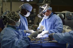
When the immune system detects a foreign pathogen, a cascade of chemical signals call white blood cells to the scene. Neutrophils are the most common and abundant type of these cells and while they start accumulating at the site of an infection within minutes, they are essentially at the mercy of the circulatory system’s one-way flow of traffic to get them where they need to go.
Now, research from the University of Pennsylvania’s School of Engineering and Applied Science shows how these cells can be coaxed to fight the direction of blood flow, crawling upstream along the walls of veins and arteries.
The in vitro study suggested that this technique could get neutrophils to the sites of infections faster when they are restricted to the direction of blood flow.
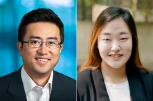
Alexander Buffone and Daniel Hammer
Daniel A. Hammer, Alfred G. and Meta A. Ennis Professor in the Department of Bioengineering, and Alexander Buffone, Jr., a research associate in his lab, led the research. Nicholas R. Anderson, a graduate student in the Hammer lab, also contributed to the study.
As technology and hands-on activities continue to become a larger part of education at all levels, a new movement of do-it-yourself projects is on the rise. Known as the “MakerSpace Movement,” the idea is that with the use of devices like 3-D printers, laser cutters, and simple circuitry materials, students, classes and communities can apply topics discussed in the classroom to real-life projects. Especially popular among STEM educators, the MakerSpace Movement is one that’s taken over labs in engineering schools around the country. Here at Penn, our own Stephenson Foundation Bioengineering Educational Lab and Bio-MakerSpace is equipped with all of the tools needed to bring student designs to fruition. In particular, the Stephenson Lab is the only lab on Penn’s campus that is open to all students and has both mechanical and electrical rapid prototyping equipment, as well as tools for biological and chemistry work.
Though Penn helps to fund the lab’s operation, many of the technologies and materials used in the Stephenson Lab and Bio-MakerSpace to help students throughout different class and independent projects are actually relatively affordable. Sevile Mannickarottu, Director of the Educational Laboratories, recently presented a paper describing the innovations and opportunities available to students through the MakerSpace attributes of the lab.
The Stephenson Lab mostly looks to support bioengineering majors, particularly in their lab courses and seniors design projects, but also encourages students of all disciplines to use the space for whatever MakerSpace-inspired ideas they might have, whether it be fixing a bike or measuring EMG signals for use in a mechanical engineering design.
Believe it or not, however, some of the best parts of the Bio-MakerSpace can actually be purchased for a total of under $1500. Though that number is probably far beyond the individual budget of most students, it might be more affordable for a student club or dorm floor that receives additional funding from Penn. While the idea of building a MakerSpace from nothing might sound intimidating, the popularity of the movement actually helps to provide a wide range of technology and affordable options.
One of the hallmarks of the MakerSpace at the Stephenson Lab, and of any MakerSpace, is the 3-D printer. Certainly, the highest quality 3-D printers on the market are incredibly expensive, but the ones used in the Stephenson Lab are actually only $750 per printer. Even better, most spools of the PLA filaments used in printers like this one can be found online for under a price of $30 each. With access to free CAD-modeling services like OpenScad and SketchUp, all you need is a computer to start 3-D printing on your own.
But if you can’t afford a 3-D printer, or want to add more electric components to the plastic designs the printer can make, the Stephenson Lab also has NI myDAQ devices, external power sources, wires, resistors, voltage meters, Arduino kits, and other equipment that can all be purchased by students for less than $500.
The most expensive device is the NI myDAQ, which costs $200 for students, but $400 for everyone else. With access to software that includes a digital multimeter, oscilloscope, function generator, Bode analyzer, and several other applications, the myDAQ is essential to any project that involves data with electronic signals. But even without the myDAQ, components like breadboards, wire cutters, resistors, voltage regulators, and all of the other basic elements of circuitry can typically be found online for a total price of under $100.
The Stephenson Lab also provides students with Arduino Kits, which are a combination of hardware and software in circuitry and programming that can be purchased for under $100 from the Arduino website. With sensors, breadboards, and other essential circuit elements, the Arduino Kits also allow users to control their designs through a software code that corresponds to hands-on setup. Particularly for those new to understanding the relationship between codes and circuitry, an Arduino Kit can be a great place to start.
Using all of these items, you can easily start your own MakerSpace for under $1500, especially if you can take advantage of student pricing. At the heart of the MakerSpace movement is the notion that anyone, anywhere can bring their own ideas and innovations to reality with the right equipment. So if you have a project in mind, get started on building your own MakerSpace, with these tools or your own — it’s cheaper than you’d think!
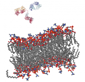
Getting a complex protein like an antibody through the membrane of a cell without damaging either is a long-standing challenge in the life sciences. Penn Engineers have found a plug-and-play solution that makes antibodies compatible with the delivery vehicles commonly used to ferry nucleic acids across that barrier.
For almost any conceivable protein, corresponding antibodies can be developed to block it from binding or changing shape, which ultimately prevents it from carrying out its normal function. As such, scientists have looked to antibodies as a way of shutting down proteins inside cells for decades, but there is still no consistent way to get them past the cell membrane in meaningful numbers.
Now, Penn Engineering researchers have figured out a way for antibodies to hitch a ride with transfection agents, positively charged bubbles of fat that biologists routinely use to transport DNA and RNA into cells. These delivery vehicles only accept cargo with a highly negative charge, a quality that nucleic acids have but antibodies lack. By designing a negatively charged amino acid chain that can be attached to any antibody without disrupting its function, they have made antibodies broadly compatible with common transfection agents.
Beyond the technique’s usefulness towards studying intracellular dynamics, the researchers conducted functional experiments with antibodies that highlight the technique’s potential for therapeutic applications. One antibody blocked a protein that decreases the efficacy of certain drugs by prematurely ejecting them from cells. Another blocked a protein involved in the transcription process, which could be an even more fundamental way of knocking out proteins with unwanted effects.
The researchers’ experiments involved making synthetic tissues with artificial “cells.” The fibrin network that surrounds these beads pull on them when compressed; by changing the number of beads in their experimental tissues, the researchers could suss out how cell-fiber interplay contributes to the tissue’s overall properties.
Tissue gets stiffer when it’s compressed. That property can become even more pronounced with injury or disease, which is why doctors palpate tissue as part of a diagnosis, such as when they check for lumps in a cancer screening. That stiffening response is a long-standing biomedical paradox, however: tissue consists of cells within a complex network of fibers, and common sense dictates that when you push the ends of a string together, it loosens tension, rather than increasing it.
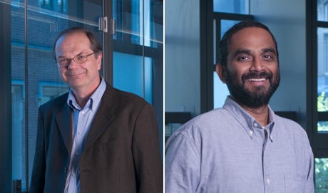
Now, in a study published in Nature, University of Pennsylvania’s School of Engineering and Applied Science researchers have solved this mystery by better understanding the mechanical interplay between that fiber network and the cells it contains.
The researchers found that when tissue is compressed, the cells inside expand laterally, pulling on attached fibers and putting more overall tension on the network. Targeting the proteins that connect cells to the surrounding fiber network might therefore be the optimal way of reducing overall tissue stiffness, a goal in medical treatments for everything from cancer to obesity.
Paul Janmey and Vivek Shenoy
The study was led by Paul Janmey, Professor in the Perelman School of Medicine’s Department of Physiology and in Penn Engineering’s Department of Bioengineering, and Vivek Shenoy, Eduardo D. Glandt President’s Distinguished Professor in Penn Engineering’s Department of Materials Science and Engineering, Mechanical Engineering and Applied Mechanics, and Bioengineering, along with Anne van Oosten and Xingyu Chen, graduate students in Janmey’s and Shenoy’s labs. Van Oosten is now a postdoctoral fellow at Leiden University in The Netherlands.
Shenoy is Director of Penn’s Center for Engineering Mechanobiology, which studies how physical forces influence the behavior of biological systems; Janmey is the co-director of one of the Center’s working groups, organized around the question, “How do cells adapt to and change their mechanical environment?”
Together, they have been interested in solving the paradox surrounding tissue stiffness.
The blue circle is the global symbol for diabetes. Wikimedia Commons.
Diabetes is one of the more common diseases among Americans today, with the American Diabetes Association estimating that approximately 9.5 percent of the population battles the condition today. Though symptoms and causes may vary across types and patients, diabetes generally results from the body’s inability to produce enough insulin to keep blood sugar levels in check. A new experimental treatment from the lab of Sha Jin, Ph.D., a biomedical engineering professor at Binghamton University, aims to use about $1.2 million in recent federal grants to develop a method for pancreatic islet cell transplantation, as those are the cells responsible for producing insulin.
But the catch to this new approach is that relying on healthy donors of these islet cells won’t easily meet the vast need for them in diabetic patients. Sha Jin wants to use her grants to consider the molecular mechanisms that can lead pluripotent stem cells to become islet-like organoids. Because pluripotent stem cells have the capability to evolve into nearly any kind of cell in the human body, the key to Jin’s research is learning how to control their mechanisms and signaling pathways so that they only become islet cells. Jin also wants to improve the eventual culture of these islet cells into three-dimensional scaffolds by finding ways of circulating appropriate levels of oxygen to all parts of the scaffold, particularly those at the center, which are notoriously difficult to accommodate. If successful in her tissue engineering efforts, Jin will not only be able to help diabetic patients, but also open the door to new methods of evolving pluripotent stem cells into mini-organ models for clinical testing of other diseases as well.
A Treatment to Help Others See Better
Permanently crossed eyes, a medical condition called strabismus, affects almost 18 million people in the United States, and is particularly common among children. For a person with strabismus, the eyes don’t line up to look at the same place at the same time, which can cause blurriness, double vision, and eye strain, among other symptoms. Associate professor of bioengineering at George Mason University, Qi Wei, Ph.D., hopes to use almost $2 million in recent funding from the National Institute of Health to treat and diagnose strabismus with a data-driven computer model of the condition. Currently, the most common method of treating strabismus is through surgery on one of the extraocular muscles that contribute to it, but Wei wants her model to eventually offer a noninvasive approach. Using data from patient MRIs, current surgical procedures, and the outcomes of those procedures, Wei hopes to advance and innovate knowledge on treating strabismus.
A Newly Analyzed Brain Mechanism Could be the Key to Stopping Seizures
Among neurological disorders, epilepsy is one of the most common. An umbrella term for a lot of different seizure-inducing conditions, many versions of epilepsy can be treated pharmaceutically. Some, however, are resistant to the drugs used for treatment, and require surgical intervention. Bin He, Ph. D., the Head of the Department of Biomedical Engineering at Carnegie Mellon University, recently published a paper in collaboration with researchers at Mayo Clinic that describes the way that seizures originating at a single point in the brain can be regulated by what he calls “push-pull” dynamics within the brain. This means that the propagation of a seizure through the brain relies on the impact of surrounding tissue. The “pull” he refers to is of the surrounding tissue towards the seizure onset zone, while the “push” is what propagates from the seizure onset zone. Thus, the strength of the “pull” largely dictates whether or not a seizure will spread. He and his lab looked at different speeds of brain rhythms to perform analysis of functional networks for each rhythm band. They found that this “push-pull” mechanism dictated the propagation of seizures in the brain, and suggest future pathways of treatment options for epilepsy focused on this mechanism.
Hyperspectral Imaging Might Provide New Ways of Finding Cancer
A new method of imaging called hyperspectral imaging could help improve the prediction of cancerous cells in tissue specimens. A recent study from a University of Texas Dallas team of researchers led by professor of bioengineering Baowei Fei, Ph.D., found that a combination of hyperspectral imaging and artificial intelligence led to an 80% to 90% level of accuracy in identifying the presence of cancer cells in a sample of 293 tissue specimens from 102 patients. With a $1.6 million grant from the Cancer Prevention and Research Institute of Texas, Fei wants to develop a smart surgical microscope that will help surgeons better detect cancer during surgery.
Fei’s use of hyperspectral imaging allows him to see the unique cellular reflections and absorptions of light across the electromagnetic spectrum, giving each cell its own specific marker and mode of identification. When paired with artificial intelligence algorithms, the microscope Fei has in mind can be trained to specifically recognize cancerous cells based on their hyperspectral imaging patterns. If successful, Fei’s innovations will speed the process of diagnosis, and potentially improve cancer treatments.
People and Places
A group of Penn engineering seniors won the Pioneer Award at the Rothberg Catalyzer Makerthon led be Penn Health-Tech that took place from October 19-20, 2019. SchistoSpot is a senior design project created by students Vishal Tien (BE ‘20), Justin Swirbul (CIS ‘20), Alec Bayliff (BE ‘20), and Bram Bruno (CIS ‘20) in which the group will design a low-cost microscopy dianostic tool that uses computer vision capabilities to automate the diagnosis of schistosomiasis, which is a common parasitic disease. Read about all the winners here.
Virginia Tech University will launch a new Cancer Research Initiative with the hope of creating an intellectual community across engineers, veterinarians, biomedical researchers, and other relevant scientists. The initiative will focus not only on building better connections throughout departments at the university, but also in working with local hospitals like the Carilion Clinic and the Children’s National Hospital in Washington, D.C. Through these new connections, people from all different areas of science and engineering and come together to share ideas.
Associate Professor of Penn Bioengineering Dani Bassett, Ph.D., recently sat down with the Penn Integrates Knowledge University Professor Duncan Watts, Ph.D., for an interview published in Penn Engineering. Bassett discusses the origins of network science, her research in small-world brain networks, academic teamwork, and the pedagogy of science and engineering. You can read the full interview here.
An all-female group of researchers from Northern Illinois University developed a device for use by occupational therapists that can capture three-dimensional images of a patient’s hand, helping to more accurately measure the hand or wrist’s range of motion. The group presented the abstract for their design at this year’s meeting of the Biomedical Engineering Society here in Philadelphia, where Penn students and researchers presented as well.
NB: Penn Bioengineering would like to congratulate one of its current Senior Design teams (Alec Bayliff, Bram Bruno, Justin Swirbul, and Vishal Then) which took home the $500 Pioneer Award at this year’s Rothberg Catalyzer competition this past weekend! Keep reading for more information on the competition, awards, and winners.
Penn Health-Tech’s Rothberg Catalyzer is a two-day makerthon that challenges interdisciplinary student teams to prototype and pitch medical devices that aim to address an unmet clinical need.
The Catalyzer’s third competition was held last weekend and was won by MAR Designs, a team of Mechanical Engineering and Applied Mechanics graduate students: Rebecca Li, Ariella Mansfield and Michael Sobrepera.
MAR Designs took home the top prize of $10,000 for their project, an orthotic device that children with cerebral palsy can more comfortably wear as they sleep.
According to the team’s presentation, existing wrist orthoses “improve function and treat/prevent spasticity. However, patients report that these devices are uncomfortable which leads to lack of compliance and may also prevent patient’s eligibility for surgeries.” MAR Designs’ device initially allows full range of motion, but gradually straightens the wrist as the child is falling asleep.
In second place was Splash Throne. Team members Greg Chen, Nik Evitt, Jake Crawford and Meghan Lockwood proposed a toilet safety frame intended for elderly users. Embedded sensors track basic health information, like weight and heart-rate, as part of a preventative health routine.
Integrated Product Design students Jonah Arheim, Laura Ceccacci, Julia Lin and Alex Wan took third place with ONESCOPE, an untethered, hands-free laproscope designed to make minimally-invasive surgeries faster and safer.
Finally, SchistoSpot took home the Catalyzer’s Pioneer Award. Bioengineering and Computer and Information Science seniors Alec Bayliff, Bram Bruno, Justin Swirbul and Vishal Then designed a low-cost microscopy system that can aid in the diagnosis of the parasitic disease schistosomiasis by detecting eggs in urine samples, eliminating the need for a hospital visit.
The event was made possible by a three-year donation by scientist and entrepreneur Jonathan Rothberg, with the intent of inspiring the next generation of healthcare innovators.
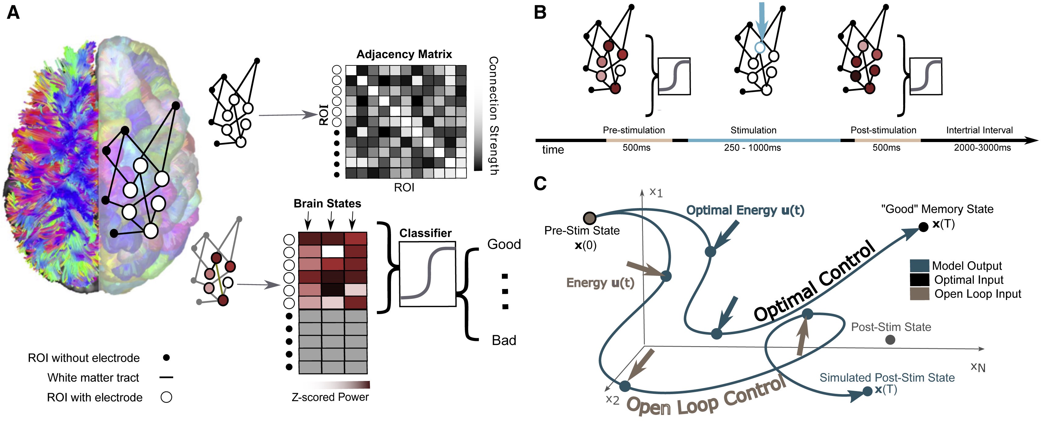
The researchers’ model involves mapping the connections between different regions of an individual’s brain while they performed a basic memory task, then using that data to predict how electrical stimulation in one region would affect activity throughout the network. Individuals’ improved performance on the same memory task after stimulation suggests the model could eventually be generalized toward a variety of stimulation therapies.
Brain stimulation, where targeted electrical impulses are directly applied to a patient’s brain, is already an effective therapy for depression, epilepsy, Parkinson’s and other neurological disorders, but many more applications are on the horizon. Clinicians and researchers believe the technique could be used to restore or improve memory and motor function after an injury, for example, but progress is hampered by how difficult it is to predict how the entire brain will respond to stimulation at a given region.
In an effort to better personalize and optimize this type of therapy, researchers from the University of Pennsylvania’s School of Engineering and Applied Science and Perelman School of Medicine, as well as Thomas Jefferson University Hospital and the University of California, Riverside, have developed a way to model how a given patient’s brain activity will change in response to targeted stimulation.
To test the accuracy of their model, they recruited a group of study participants who were undergoing an unrelated treatment for severe epilepsy, and thus had a series of electrodes already implanted in their brains. Using each individual’s brain activity data as inputs for their model, the researchers made predictions about how to best stimulate that participant’s brain to improve their performance on a basic memory test.
The participants’ brain activity before and after stimulation suggest the researchers’ models have meaningful predictive power and offer a first step towards a more generalizable approach to specific stimulation therapies.
Danielle Bassett and Jennifer Stiso.
The study, published in the journal Cell Reports, was led by Danielle Bassett, J. Peter Skirkanich Professor in Penn Engineering’s Department of Bioengineering, and Jennifer Stiso, a neuroscience graduate student in Penn Medicine and a member of Bassett’s Complex Systems Lab.
A New Sprayable Gel Can Help Prevent Surgical Adhesions
Adhesions are a common kind of scar tissue that can occur after surgery, and though usually not painful, they have the potential to result in complications like chronic pain or decreased heart efficiency, depending on where the scar tissue forms. Now, a sprayable gel developed by researchers at Stanford University will help to prevent adhesions from forming during surgical procedures. The gel, called PNP 1:10 in reference to its polymer-nanoparticle structure, has a similar stiffness to mayonnaise and achieves an ideal balance of slipperiness and stickiness that allows it to adhere easily to tissue of irregular shapes and surfaces. The flexible gel will actually dissolve in the body after two weeks, which is about how long most adhesions take to heal. Though lead author Lyndsay Stapleton, M.S., and senior authors Joseph Woo, M.D., and Eric Appel, Ph.D., have only tested the gel in rats and sheep so far, they hope that human applications are not too far in the future.
Learning to Understand Blood Clots in a New Model
Blood clots are the source of some of the deadliest human conditions and diseases. When a clot forms, blood flow can be interrupted, cutting off supply to the brain, heart, or other vital organs, resulting in potentially serious damage to the mind and body. For patients with certain bleeding disorders, clotting or the lack thereof can hold tremendous importance in surgery, and affect some of the typical procedures of a given operation. To help plan for such situations, researchers at the University of Buffalo created an in vitro model to help better illustrate the complex fluid mechanics of blood flow and clotting. Most importantly, this new model better demonstrates the role of shear stress in blood flow, and the way that it can affect the formation or destruction of blood clots – an aspect that current clinical devices often overlook. Led by Ruogang Zhao, Ph.D., the model can allow surgeons and hematologists to consider the way that certain chemical or physical treatments can affect clot formation on a patient-to-patient basis. The two key factors of the model are its incorporation of blood flow, and its relationship to shear stress, with clot stiffness through microfabrication technology using micropillars as force sensors for the stiffness. Going forward, Zhao and his research team hope to test the model on more patients, to help diversify the different bleeding disorders it can exhibit.
Training the Next Generation of Researchers
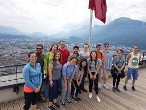
REACT 2019 students and Grenoble summer program interns, including undergraduate Rebecca Zappala (third from left, front), pose in front of the Chartreuse Mountains after completing a challenging ropes course. (Photo: Hermine Vincent)
Rebecca Zappala, a junior from Miami, Florida who is majoring in bioengineering, worked in Grenoble this summer on new ways to harvest water from fog. She describes her research project as a “futuristic” way to collect water and says that she’s thankful for the opportunity to work on her first independent research project through the Research and Education in Active Coatings Technology (REACT) program.
After learning the technical skills she needed for her project, Zappala spent her summer independently working on new ways to modify her material’s properties while working closely with her French PI and a post-doc in the lab. She was surprised to see how diverse the lab was, with researchers working on everything from biomolecular research to energy in the same space.
“I learned a lot,” she says about being in such an interdisciplinary setting. “I hadn’t been part of a research team before, and I got a lot of exposure to things that I wouldn’t have been exposed to otherwise.”
Virginia Tech Course Addresses the Needs of Wounded Veterans
A new course at Virginia Tech encourages students to apply engineering skills to real-life problems in the biomedical world by designing medical devices or other applications to assist veterans suffering from serious injuries or illnesses. Funded by the National Institute of Health, faculty from the Department of Biomedical Engineering and Mechanics hope that the course will allow students to see how theoretical knowledge from the classroom actually works in a clinical setting, and to understand how different stakeholder interests factor into designing a real device. What makes this new class unique from other traditional design-focused courses at other universities is its level of patient interaction. Students at Virginia Tech who choose to take this class will have the chance to gain input from field professionals like clinicians and engineers from the Salem Veterans Affairs Medical Center, while also being able to get direct feedback from the patients that the devices will actually help. Beginning in the spring of 2020, students can take the new course, and volunteer in the veterans clinics to gain even more experience with patients.
People and Places
Sevile Mannickarottu, the Director of the Educational Laboratories in Penn’s Department of Bioengineering and recent recipient of the Staff Recognition Award from the School of Engineering and Applied Sciences, presented a paper to highlight the Stephenson Foundation Bioengineering Educational Lab and Bio-Makerspace at the 126th annual conference of the American Society for Engineering Education. Thanks to the dedication of Mannickarottu and the lab staff to creating a space for simultaneous education and innovation, the Bioengineering Lab continues to be a hub for student community and projects of all kinds.
A week-long program for high school girls interested in STEM allows students to explore ideas and opportunities in the field through lab tours, guest speakers, and hands-on challenges. A collaboration across the University of Virginia Department of Biomedical Engineering, Charlottesville Women in Tech, and St. Anne’s Belfield School, the program gave this year’s students a chance to design therapies for children with disorders like hemiplegia and cerebral palsy, in the hopes that these interactive design challenges will inspire the girls to pursue future endeavors in engineering.
We would like to congratulate Nancy Albritton, Ph.D., on her appointment as the next Frank & Julie Jungers Dean of the College of Engineering at the University of Washington. Albritton brings both a deep knowledge of the research-to-marketplace pipeline and experience in the development of biomedical microdevices and pharmacoengineering to the new position.
We would also like to congratulate Jeffrey Brock, Ph.D., on his appointment as the dean of the Yale School of Engineering and Applied Science. Already both a professor of mathematics and a dean of science in the Faculty of Arts and Sciences at Yale, Brock’s new position will help him to foster collaborations across different departments of academia and research in science and engineering.
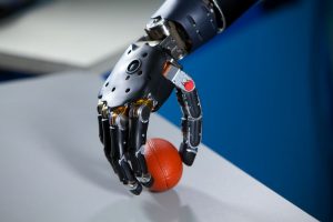
After nearly fifteen years of work, a new high-tech prosthetic arm from researchers at the University of Utah allows hand amputees to pluck grapes, pick up eggs without breaking them, and even put on their wedding rings. Named after Luke Skywalker’s robotic hand in the Star Wars saga, the LUKE Arm includes sensors that better mimic the way the human body sends information to the brain, allowing users to distinguish between soft and hard surfaces and to perform more complicated tasks. The arm relies heavily on an electrode array invented by University of Utah biomedical engineering professor Richard A. Normann, Ph.D., which is a bundle of microelectrodes that enable a computer to read signals from connected nerves in the user’s forearm.
But the biggest innovation in the use of these electrode arrays for the LUKE Arm is in the way they allow the prosthetic to mimic the sense of feeling on the surface of an object that indicates how much pressure should be applied when handling it. Gregory Clark, Ph.D., an associate professor of biomedical engineering at the University of Utah and the leader of the LUKE Arm project, says the key to improving these functions in the prosthetic was by more closely mimicking the path that this information takes to the brain, as opposed to merely what comprises that sensory information. In the future, Clark hopes to improve upon the LUKE Arm by including more inputs, like one for temperature data, and on making them more portable by eliminating the device’s need for computer connection.
Philly Voice Recognizes the Cremins Lab’s Innovations in Light-Activated Gene-Folding
While technological advancements over the past few decades have opened doors to understanding the topological structures of DNA, we still have far more to learn about how these structures impact and contribute to genome function. But here at Penn, the Cremins Laboratory in 3D Epigenomes and Systems Neurobiology hopes to fix that. Led by Jennifer E. Phillips-Cremins, Ph.D., members of the lab use light-activated dynamic looping (LADL) to better understand the way that genome topological properties and folding can affect protein translation. Cremins and her lab use this technique to force specific genome folds to interact with each other, and create temporary DNA loops that can then be bound together in the presence of blue light for certain proteins in the Arabidopsis plant. Using the data from these tests, researchers can better understand the genome structure-function relationships, and hopefully open the door to new treatments for diseases in which expression or mis-expression of certain genes is the cause.
Artificial Cells Can Deliver Molecules Better than the Real Thing
From pills to vaccines, ways to deliver drugs into the body have been constantly evolving since the early days of medicine.
Now, a new study from an interdisciplinary team led by researchers at the University of Pennsylvania provides a new platform for how drugs could be delivered to their targets in the future. Their work was published in the Proceedings of the National Academy of Sciences.
The research focuses on a dendrimersome, a compartment with a lamellar structure and size that mimic a living cell. It can be thought of as the shipping box of the cellular world that carries an assortment of molecules as cargo.
The scientists found that these dendrimersomes, which have a multilayered, onion-like structure, were able to “carry” high concentrations of molecules that don’t like water, which is common in pharmaceutical drugs. They were also able to carry these molecules more efficiently than other commercially available vessels. Additionally, the building block of the cell-like compartment, a janus dendrimer, is classified as an amphiphile, meaning it contains molecules that don’t like water and also molecules that are soluble in water, like lipids, that make up natural membranes.
“This is a different amphiphile that makes really cool self-assembled onions into which we were able to load a bunch of molecular cargos,” says co-author Matthew Good.
In a recent review of over 5,000 sleep studies, biomedical engineering researchers at the University of Texas at Austin found a connection between water-based passive body heating and sleep onset latency, efficiency, and quality. Using meta-analytical tools to compare all of the studies and patient data, lead author and Ph.D. candidate Shahab Haghayegh and his team found that a warm bath in the temperature range of 104-109 degrees Fahrenheit taken 1-2 hours before bed has the ability to improve all three considered sleep categories. This makes sense considering that our body’s Circadian rhythms govern both our sleep cycles and temperature, bringing us to a higher temperature during the day and a lower one at night during sleep. In fact, this lowering of body temperature before sleep is what helps to trigger the onset of sleep, so taking a warm bath and allowing your body to cool down from it before going to sleep enhances the body’s own efforts of naturally cooling down before we go to bed. With this new and comprehensive review, those who suffer from poor sleep quality may soon find solace in temperature regulation therapy systems.
People & Places
With the recent 50th anniversary of the first moon landing by Americans Neil Armstrong, Buzz Aldrin, and Michael Collins in 1969, ABC News looked back at one of the women involved in the project. Judy Sullivan was a biomedical engineer at the time of the project, and served as the lead engineer of the biomedical system for Apollo 11. In this role, she led studies on the astronauts’ breathing rates and sensor capabilities for the devices being sent into space to help the astronauts monitor their health. For the Apollo 11 mission and a lot of Sullivan’s early work at NASA, she worked on teams of all men, as women were often encouraged to become teachers, secretaries, or homemakers over other professions. Today, Sullivan says she’s thrilled that women have more career options to choose from, and wants to continue seeing more women getting involved in math and science.
We would like to congratulate Sanjay Kumar, M.D., Ph.D., on his appointment as the new Department Chair of Bioengineering at the University of California, Berkeley. Since joining the faculty in 2005, Kumar has received several prestigious awards including the NSF Career Award, the NIH Director’s New Innovator Award, the Presidential Early Career Award for Scientists and Engineers, and the Berkeley student-voted Outstanding Teacher Award.
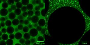
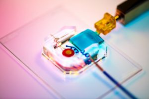
Rachel Young, a graduate student in Huh’s lab, holds up the new eye-on-a-chip device. The latest iteration of the lab’s eye-on-a-chip has a mechanical eyelid to simulate blinking, and was used to test an experimental drug for dry eye disease. By incorporating human cells into an engineered scaffolding, the eye-on-a-chip has many of the benefits of testing on living subjects, while minimizing risks and ethical concerns.
People who spend eight or more hours a day staring at a computer screen may notice their eyes becoming tired or dry, and, if those conditions are severe enough, they may eventually develop dry eye disease (DED). DED is a common disease with shockingly few FDA-approved drug options, partially because of the difficulties of modeling the complex pathophysiology in human eyes. Enter the blinking eye-on-a-chip: an artificial human eye replica constructed in the laboratory of Penn Engineering researchers.
This eye-on-a-chip, complete with a blinking eyelid, is helping scientists and drug developers to improve their understanding and treatment of DED, among other potential uses. The research, published in Nature Medicine, outlines the accuracy of the eye-on-a-chip as an organ stand-in and demonstrates its utility as a drug testing platform.
They collaborated with Vivian Lee, Vatinee Bunya and Mina Massaro-Giordano from the Department of Ophthalmology in Penn’s Perelman School of Medicine, as well as with Vivek Shenoy, Eduardo D. Glandt President’s Distinguished Professor in Penn Engineering’s Department of Materials Science and Engineering. Other collaborators included Woo Byun, Andrei Georgescu and Yoon-suk Yi, members of Huh’s lab, and Farid Alisafaei, a member of Shenoy’s lab.
Huh’s lab specializes in creating organs-on-a-chip that provide microengineered in vitro platforms to mimic their in vivo counterparts, including lung and bone marrow proxies launched into space this May to study astronaut illness. The lab has spent years fine-tuning its eye-on-a-chip, which earned them the 2018 Lush Prize for its promise in animal-free testing of drugs, chemicals, and cosmetics.
In this study, Huh and Seo focused on engineering an eye model that could imitate a healthy eye and an eye with DED, allowing them to test an experimental drug without risk of human harm.
The Huh lab’s eye-on-a-chip attached to a motorized, gelatin-based eyelid. Blinking spreads tears over the corneal surface, and so was a critical aspect to replicate in the researchers’ model of dry eye disease. cells. The cells of the cornea grow on the inner circle of scaffolding, dyed yellow, and the cells of the conjunctiva grow on the surrounding red circle. Artificial tears are supplied by a tear duct, dyed blue.
To construct their eye-on-a-chip, Huh’s team starts with a porous scaffold engineered with 3D printing, about the size of a dime and the shape of a contact lens, on which they grow human eye cells. The cells of the cornea grow on the inner circle of scaffolding, dyed yellow, and the cells of the conjunctiva, the specialized tissue covering the white part of human eyes, grow on the surrounding red circle. A slab of gelatin acts as the eyelid, mechanically sliding over the eye at the same rate as human blinking. Fed by a tear duct, dyed blue, the eyelid spreads artificial tear secretions over the eye to form what is called a tear film.
“From an engineering standpoint, we found it interesting to think about the possibility of mimicking the dynamic environment of a blinking human eye. Blinking serves to spread tears and generate a thin film that keeps the ocular surface hydrated. It also helps form a smooth refractive surface for light transmission. This was a key feature of the ocular surface that we wanted to recapitulate in our device,” says Huh.
For people with DED, that tear film evaporates faster than it’s replenished, resulting in inflammation and irritation. A common cause of DED is the reduced blinking that occurs during excessive computer usage, but people can develop the disease for other reasons as well. DED affects about 14 percent of the world’s population but has been notably difficult to develop new treatments for, with 200 failed clinical drug trials since 2010 and only two currently available FDA-approved drugs for treatment.
Huh’s lab has been considering the drug-testing potential of organs-on-a-chip since their initial conceptualization, and, because of its surface-level area of impact, DED seemed the perfect place to start putting their eye model to the test. But before they started a drug trial, the team had to ensure their model could really imitate the conditions of DED.
“Initially, we thought modeling DED would be as simple as just keeping the culture environment dry. But as it turns out, it’s an incredibly complex multifactorial disease with a variety of sub-types,” Huh says. “Regardless of type, however, there are two core mechanisms that underlie the development and progression of DED. First, as water evaporates from the tear film, salt concentration increases dramatically, resulting in hyperosmolarity of tears. And second, with increased tear evaporation, the tear film becomes thinner more rapidly and often ruptures prematurely, which is referred to as tear film instability. The question was: Is our model capable of modeling these core mechanisms of dry eye?”
The answer, after much experimentation, was yes. The team evoked DED conditions in their eye-on-a-chip by cutting their device’s artificial blinking in half and carefully creating an enclosed environment that simulated the humidity of real-life conditions. When put to the test against real human eyes, both healthy and with DED, the corresponding eye-on-a-chip models proved their similarity to the actual organ on multiple clinical measures. The eyes-on-a-chip mimicked actual eyes’ performance in a Schirmer strip, which tests liquid production; in an osmolarity test, which looks at tear film salt content; and in a keratography test, which evaluates the time it takes for a tear film to break up.
Having confirmed their eye-on-a-chip’s ability to mirror the performance of a human eye in normal and DED-inducing settings, Huh’s team turned to the pharmaceutical industry to find a promising DED drug candidate to test-drive their model. They landed on an upcoming drug based on lubricin, a protein primarily found in the lubricating fluid that protects joints.
“When people think of DED, they normally treat it as a chronic disease driven by inflammation,” says Huh, “but there’s now increasing evidence suggesting that mechanical forces are important for understanding the pathophysiology of DED. As the tear film becomes thinner and more unstable, friction between the eyelids and the ocular surface increases, and this can damage the epithelial surface and also trigger adverse biological responses such as inflammation. Based on these observations, there is emerging interest in developing ophthalmic lubricants as a topical treatment for dry eye. In our study, we used an lubricin-based drug that is currently undergoing clinical trials. When we tested this drug in our device, we were able to demonstrate its friction-lowering effects, but, more importantly, using this model we discovered its previously unknown capacity to suppress inflammation of the ocular surface.”
By comparing the testing results of their models of a healthy eye, an eye with DED, and an eye with DED plus lubricin, Huh and Seo were able to further scientists’ understanding of how lubricin works and show the drug’s promise as a DED treatment.
Similarly, the process of building a blinking eye-on-a-chip pushed forward scientists’ understanding of the eye itself, providing insights into the role of mechanics in biology. Collaborating with Shenoy, director of the Center for Engineering MechanoBiology, the team’s attention was drawn to how the physical blinking action was affecting the cells they cultivated to engineer an artificial eye on top of their scaffolding.
“Initially, the corneal cells start off as a single layer, but they become stratified and form multiple layers as a result of differentiation, which happens when these cells are cultured at the air-liquid interface. They also form tight cell-cell junctions and express a set of markers during differentiation,” Huh says. “Interestingly, we found out that mechanical forces due to blinking actually help the cells differentiate more rapidly and more efficiently. When the corneal cells were cultured under air in the presence of blinking, the rate and extent of differentiation increased significantly in comparison to static models without blinking. Based on this result, we speculate that blink-induced physiological forces may contribute to differentiation and maintenance of the cornea.”
In other words, human cornea cells growing on the scientists’ scaffold more quickly became specialized and efficient at their particular jobs when the artificial eyelid was blinking on top of them, suggesting that mechanical forces like blinking contribute significantly to how cells function. These types of conceptual advances, coupled with drug discovery applications, highlight the multifaceted value that engineered organs-on-a-chip can contribute to science.
Huh and Seo’s eye-on-a-chip is still just dipping its toes into the field of drug testing, but this first step is a victory that represents years of work refining their artificial eye to reach this level of accuracy and utility.
“Although we have just demonstrated proof-of-concept,” says Seo, “I hope our eye-on-a-chip platform is further advanced and used for a variety of applications besides drug screening, such as testing of contact lenses and eye surgeries in the future.”
“We are particularly proud of the fact that our work offers a great and rare example of interdisciplinary efforts encompassing a broad spectrum of research activities from design and fabrication of novel bioengineering systems to in vitro modeling of complex human disease to drug testing,” says Huh. “I think this is what makes our study unique and representative of innovation that can be brought about by organ-on-a-chip technology.”
This work was supported by the National Institutes of Health through grants 1DP2HL127720–0, R01EY026972 and K08EY025742–01, the National Science Foundation through grants CMMI:15–48571, and Research to Prevent Blindness.


 As technology and hands-on activities continue to become a larger part of education at all levels, a new movement of do-it-yourself projects is on the rise. Known as the “MakerSpace Movement,” the idea is that with the use of devices like 3-D printers, laser cutters, and simple circuitry materials, students, classes and communities can apply topics discussed in the classroom to real-life projects. Especially popular among STEM educators, the MakerSpace Movement is one that’s taken over labs in engineering schools around the country. Here at Penn, our own
  Believe it or not, however, some of the best parts of the Bio-MakerSpace can actually be purchased for a total of under $1500. Though that number is probably far beyond the individual budget of most students, it might be more affordable for a student club or dorm floor that receives additional funding from Penn. While the idea of building a MakerSpace from nothing might sound intimidating, the popularity of the movement actually helps to provide a wide range of technology and affordable options.