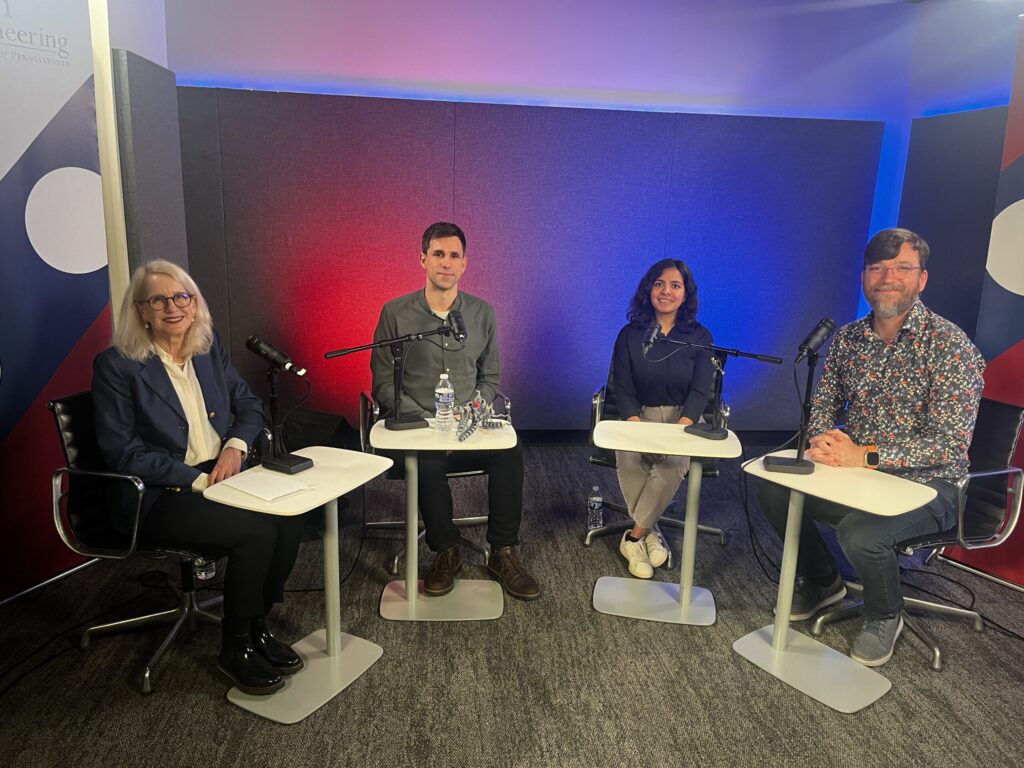
Susan Davidson, Cesar de la Fuente, Surbhi Goel and Chris Callison-Burch speak on AI in Engineering in episode 4 of the Innovation & Impact podcast.
With AI technologies finding their way into every industry, important questions must be considered by the research community: How can deep learning help identify new drugs? How can large language models disseminate information? Where and how are researchers using AI in their own work? And, how are humans anticipating and defending against potential harmful consequences of this powerful technology?
In this episode of Innovation & Impact, host Susan Davidson, Weiss Professor in Computer and Information Science (CIS), speaks with three Penn Engineering experts about leveraging AI to advance scientific discovery and methods to protect its users. Panelists include:
Chris Callison-Burch, Associate Professor in CIS, who researches the applications of large language models and AI tools in current and future real-world problems with a keen eye towards safety and ethical use of AI;
Surbhi Goel, Magerman Term Assistant Professor in CIS, who works at the intersection of theoretical computer science and machine learning. Her focus on developing theoretical foundations for modern machine learning paradigms expands the possibilities of deep learning; and
Cesar de la Fuente, Presidential Assistant Professor in Bioengineering, Psychiatry and Microbiology with a secondary appointment in Chemical and Biomolecular Engineering, who leads research on technology in the medical field, using computers to find antibiotics in extinct organisms and identify pre-clinical candidates to advance drug discovery.
Each episode of Penn Engineering’s Innovation & Impact podcast shares insight from leading experts at Penn and Penn Engineering on science, technology and medicine.
Patients being treated for B-cell non-Hodgkin’s Lymphoma (NHL) who are part of minority populations may not have equal access to cutting-edge CAR T cell therapies, according to a new analysis led by researchers from the Perelman School of Medicine and published in NEJM Evidence.
CAR T cell therapy is a personalized form of cancer therapy that was pioneered at Penn Medicine and has brought hope to thousands of patients who had otherwise run out of treatment options. Six different CAR T cell therapies have been approved since 2017 for a variety of blood cancers, including B-cell NHL that has relapsed or stopped responding to treatment. Image: iStock/PeopleImages
“CAR T cell therapy represents a major leap forward for blood cancer treatment, with many patients living longer than ever before, but its true promise can only be realized if every patient in need has access to these therapies,” says lead author Guido Ghilardi, a postdoctoral fellow in the laboratory of senior author Marco Ruella, an assistant professor of hematology-oncology and scientific director of the Lymphoma Program. “From the scientific perspective, we’re constantly working in the laboratory to make CAR T cell therapy work better, but we also want to make sure that when a groundbreaking treatment like this becomes available, it reaches all patients who might be able to benefit.”
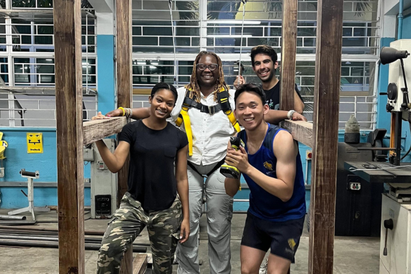
Students test the GaitMate harness and structure as a tool to help recovering patients walk.
Penn students have been building their knowledge and hands-on experience in places all over the world through Penn Global Seminars. Last May, “Robotics and Rehabilitation” brought Penn students back to the tropical island of Jamaica to collaborate with local university students and make an impact on recovery and quality of life for patients in Kingston and beyond.
Course leaders Camillo Jose (CJ) Taylor, Raymond S. Markowitz President’s Distinguished Professor in Computer and Information Science (CIS), and Michelle J. Johnson, Associate Professor of Physical Medicine and Rehabilitation at the Perelman School of Medicine and Associate Professor in Bioengineering (BE) and Mechanical Engineering and Applied Mechanics (MEAM) at Penn Engineering, brought the first cohort of students to the island in 2019.
“CJ and I are both Jamaicans by birth,” says Johnson. “We were both excited to introduce the next generation of engineers to robotics, rehabilitation and the process of culturally sensitive design in a location that we are personally connected to.”
As they built relationships with colleagues at the University of West Indies, Mona (UWI, Mona) and the University of Technology, Jamaica (UTECH), both Johnson and Taylor worked to tie the goals of the course to the location.
“In the initial iteration of the course, our goal was to focus on the applications of robotics to rehabilitation in a developing country where it is necessary to create solutions that are cost effective and will work in under-resourced settings,” says Taylor.
Taylor and Johnson wanted to make the course a regular offering, however, due to COVID-related travel restrictions, it wasn’t until last spring that they were able to bring it back. But when they did, they made up for lost time and expanded the scope of the course to include solving health problems for both people and the environment.
“While we started with a focus on people, we realized that the health and quality of life of a community is also impacted by the health of the environment,” says Taylor. “Jamaica has rich terrestrial and marine ecosystems, but those resources need to be monitored and regulated. We ventured into developing robotics tools to make environmental monitoring more effective and cost-friendly.”
One of those student-invented tools was a climate survey drone called “BioScout.”
“Our aim was to create a drone to monitor the ecosystem and wildlife in Jamaica,” says Rohan Mehta, junior in Systems Science and Engineering. “We wanted to help researchers and rangers who need to monitor wildlife and inspect forest sectors without entering and disturbing territories, but there were no available drones that met all of the following criteria necessary for the specific environment: affordable, modular, water-resistant and easy to repair. So we made our own.”
Another team of students created a smart buoy to reduce overfishing. The buoy was equipped with an alarm that goes off when fishermen get too close to a no-fishing zone.
Five other student teams dove into projects aligned to the original goals of the course. Their devices addressed patients’ decreased mobility due to diabetes, strokes and car accidents. These projects were sponsored by the Sir John Golding Rehabilitation Center.
One of which, the GaitMate, was engineered to help stroke patients who had lost partial muscle control regain their ability to walk.
“We developed a device that supports a patient’s weight and provides sensory feedback to help correct their form and gait as they walk on a treadmill, ultimately enhancing the recovery process and providing some autonomy to the patient,” says Taehwan Kim, senior in BE. “The device is also relatively cheap and simple, making it an option for a wide variety of physical therapy needs in Jamaica and other countries.”
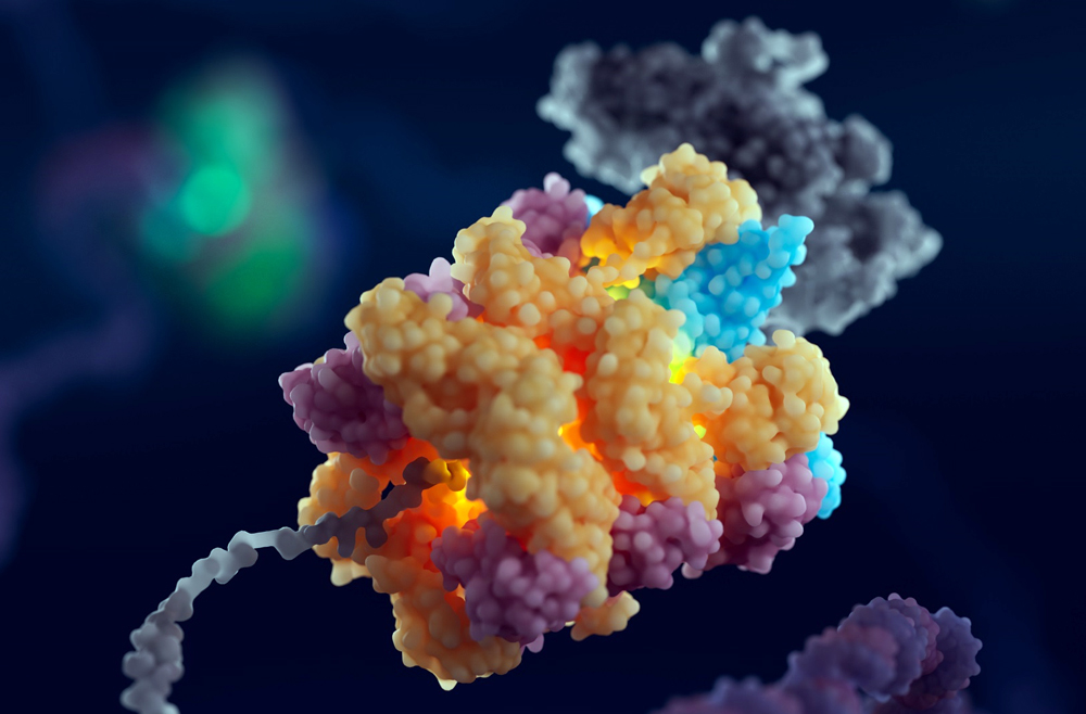
Illustration of the 55LCC complex. (Image: Courtesy of Cameron Baines/Phospho Biomedical Animation)
When cells in the human body divide, they must first make accurate copies of their DNA. The DNA replication exercise is one of the most important processes in all living organisms and is fraught with risks of mutation, which can lead to cell death or cancer. Now, findings from biologists from the Perelman School of Medicine and from the University of Leeds have identified a multiprotein “machine” in cells that helps govern the pausing or stopping of DNA replication to ensure its smooth progress. Illustration of the 55LCC complex. (Image: Courtesy of Cameron Baines/Phospho Biomedical Animation)
The discovery, published in Cell, advances the understanding of DNA replication, helps explain a puzzling set of genetic diseases, and could inform the development of future treatments for neurologic and developmental disorders.
“We’ve found what appears to be a critical quality-control mechanism in cells,” says senior co-corresponding author Roger Greenberg, the J. Samuel Staub, M.D. Professor in the department of Cancer Biology, director of the Penn Center for Genome Integrity, and director of basic science at the Basser Center for BRCA at Penn Medicine. “Trillions of cells in our body divide every single day, and this requires accurate replication of our genomes. Our work describes a new mechanism that regulates protein stability in replicating DNA. We now know a bit more about an important step in this complex biological process.”
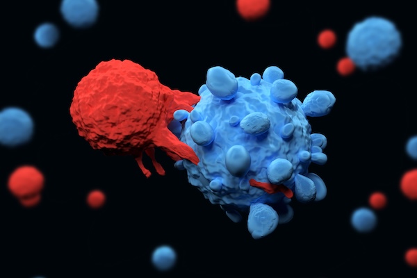
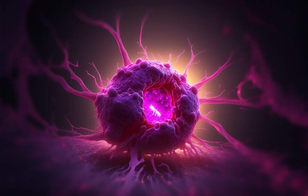
Visualization of a CAR T cell (in red) attacking a cancer cell (in blue) (Meletios Varras via Getty Images)
For patients with certain types of cancer, CAR T cell therapy has been nothing short of life changing. Developed in part by Carl June, Richard W. Vague Professor at Penn Medicine, and approved by the Food and Drug Administration (FDA) in 2017, CAR T cell therapy mobilizes patients’ own immune systems to fight lymphoma and leukemia, among other cancers.
However, the process for manufacturing CAR T cells themselves is time-consuming and costly, requiring multiple steps across days. The state of the art involves extracting patients’ T cells, then activating them with tiny magnetic beads, before giving the T cells genetic instructions to make chimeric antigen receptors (CARs), the specialized receptors that help T cells eliminate cancer cells.
Now, Penn Engineers have developed a novel method for manufacturing CAR T cells, one that takes just 24 hours and requires only one step, thanks to the use of lipid nanoparticles (LNPs), the potent delivery vehicles that played a critical role in the Moderna and Pfizer-BioNTech COVID-19 vaccines.
In a new paper in Advanced Materials, Michael J. Mitchell, Associate Professor in Bioengineering, describes the creation of “activating lipid nanoparticles” (aLNPs), which can activate T cells and deliver the genetic instructions for CARs in a single step, greatly simplifying the CAR T cell manufacturing process. “We wanted to combine these two extremely promising areas of research,” says Ann Metzloff, a doctoral student in Bioengineering and NSF Graduate Research Fellow in the Mitchell lab and the paper’s lead author. “How could we apply lipid nanoparticles to CAR T cell therapy?”
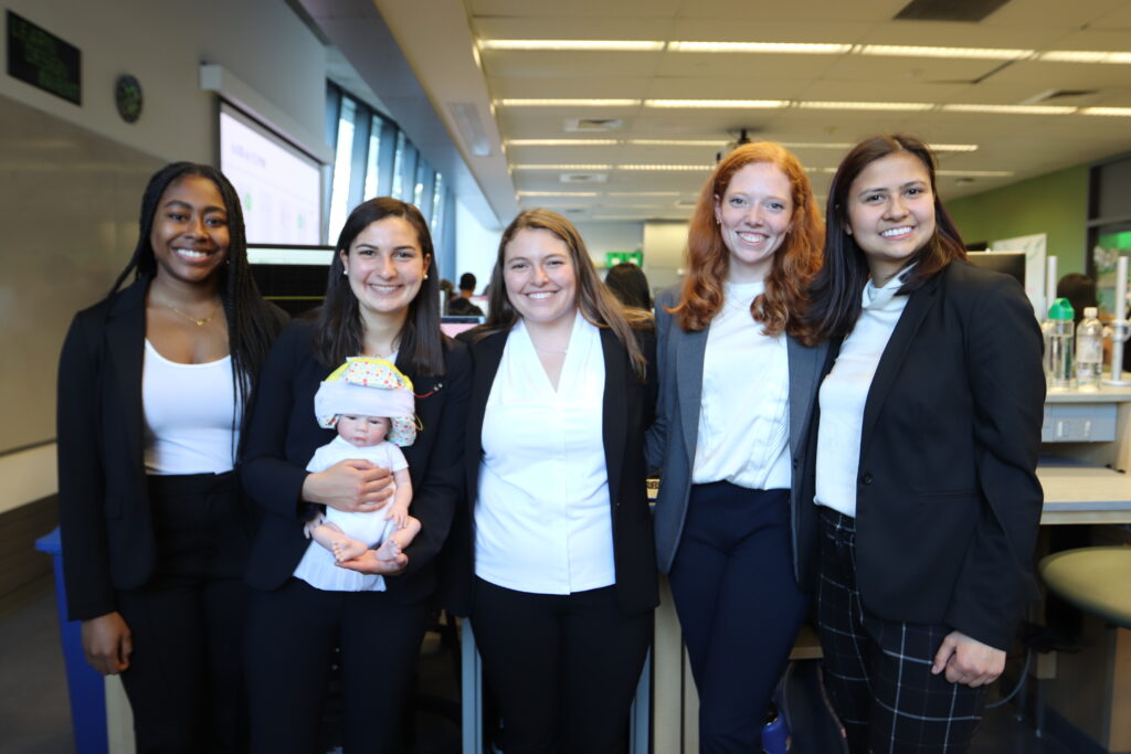
Members of Team Sonura: Tifara Boyce, Gabriela Cano, Gabriella Daltoso, Sophie Ishiwari, & Caroline Magro (credit: Penn BE Labs)
In April 2023, three President’s Prize-winning teams were selected from an application pool of 76 to develop post-graduation projects that make a positive, lasting difference in the world. Each project received $100,000 and a $50,000 living stipend per team member.
The winning projects include Sonura, the winner of the President’s Innovation Prize (PIP), who are working to improve infant development by reducing harsh noise exposure in neonatal intensive care units. To accomplish this, they’ve developed a noise-shielding beanie that can also relay audio messages from parents.
Sonura, a bioengineering quintet, developed a beanie that shields newborns from the harsh noise environments present in neonatal intensive care units (NICUs)—a known threat to infant wellbeing—and also supports cognitive development by relaying audio messages from their parents.
Since graduating from the School of Engineering and Applied Science, the team of Tifara Boyce, Gabriela Cano, Gabriella Daltoso, Sophie Ishiwari, and Caroline Magro, has collaborated with more than 50 NICU teams nationwide. They have been helped by the Intensive Care Nursery (ICN) at the Hospital of the University of Pennsylvania (HUP), which shares Sonura’s goal of reducing NICU noise. “Infant development is at the center of all activities within the HUP ICN,” note Daltoso and Ishiwari. “Even at the most granular level, like how each trash can has a sign urging you to shut it quietly, commitment to care is evident, a core tenet we strive to embody as we continue to grow.”
An initial challenge for the team was the inability to access the NICU, crucial for understanding how the beanie integrates with existing workflows. Collaboration with the HUP clinical team was key, as feedback from a range of NICU professionals has helped them refine their prototype.
In the past year, the team has participated in the University of Toronto’s Creative Destruction Lab and the Venture Initiation Program at Penn’s Venture Lab, and received funding from the Pennsylvania Pediatric Device Consortium. “These experiences have greatly expanded our perspective,” Cano says.
With regular communication with mentors from Penn Engineering and physicians from HUP, Children’s Hospital of Philadelphia, and other institutes, Sonura is looking ahead as they approach the milestone of completing the FDA’s regulatory clearance process within the year. They will begin piloting their beanie with the backing of NICU teams, further contributing to neonatal care.
Read the full story and watch a video about Sonura’s progress in Penn Today.
Read more stories featuring Sonura in the BE Blog.
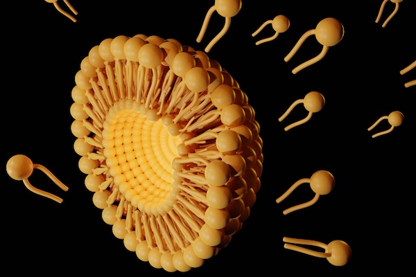
Like space shuttles using booster rockets to breach the atmosphere, lipid nanoparticles (LNPs) equipped with the new molecule more successfully deliver medicinal payloads. (Love Employee via Getty Images)
Inspired by the design of space shuttles, Penn Engineering researchers have invented a new way to synthesize a key component of lipid nanoparticles (LNPs), the revolutionary delivery vehicle for mRNA treatments including the Pfizer-BioNTech and Moderna COVID-19 vaccines, simplifying the manufacture of LNPs while boosting their efficacy at delivering mRNA to cells for medicinal purposes.
In a paper in Nature Communications, Michael J. Mitchell, Associate Professor in the Department of Bioengineering, describes a new way to synthesize ionizable lipidoids, key chemical components of LNPs that help protect and deliver medicinal payloads. For this paper, Mitchell and his co-authors tested delivery of an mRNA drug for treating obesity and gene-editing tools for treating genetic disease.
Previous experiments have shown that lipidoids with branched tails perform better at delivering mRNA to cells, but the methods for creating these molecules are time- and cost-intensive. “We offer a novel construction strategy for rapid and cost-efficient synthesis of these lipidoids,” says Xuexiang Han, a postdoctoral student in the Mitchell Lab and the paper’s co-first author.
Bispecific T cell engagers are emerging as a powerful class of immunotherapy to treat cancer but are sometimes hindered by unwanted outcomes, such as on-target, off-tumor toxicity; cytokine release syndrome; and neurotoxicity. Now, researchers Penn researchers have developed a novel “switchable” bispecific T cell engager that mitigates these negative effects by co-opting a drug already approved by the FDA. (Image: iStock / CIPhotos)
In the ever-evolving battle against cancer, immunotherapy presents a turning point. It began with harnessing the body’s immune system to fight cancer, a concept rooted more than a century ago but only gaining significant momentum in recent years. Pioneering this shift were therapies like CAR T cell therapy, which reprograms a patient’s T cells to attack cancer cells. Within this domain, bispecific T cell engagers, or bispecific antibodies, have emerged as effective treatments for many blood-borne cancers in the clinic and are being evaluated for solid tumor therapy.
These antibodies simultaneously latch onto both a cancer cell and a T cell, effectively bridging the gap between the two. This proximity triggers the T cells to unleash their lethal arsenal, thereby killing the cancer cells. However, bispecific T cell engagers, like many cancer therapies, face hurdles such as cell-specific targeting limitations, known as on-target off-tumor toxicity, which means the tumor is correctly targeted but so are other healthy cells in the body, leading to healthy tissue damage. Moreover, bispecific antibodies may also lead to immune system overactivation, a precursor for cytokine release syndrome (CRS), and neurotoxicity.
Now, researchers led by Michael Mitchell of the University of Pennsylvania have found a way to circumvent many of these deleterious effects by developing a bispecific T cell nanoengager that is equipped with an “off switch.” Their findings are published in Nature Biomedical Engineering.
“We’re excited to show that bispecific antibodies can be tweaked in a way that allows us to tap into their powerful cancer-killing potential without inducing toxicity to healthy tissues,” says Mitchell, associate professor of bioengineering at Penn’s School of Engineering and Applied Science. “This new controllable drug-delivery mechanism, which we call switchable bispecific T cell nanoengagers, or SiTEs, adds this switchable component to the antibody via administering an FDA-approved small-molecule drug, amantadine.”
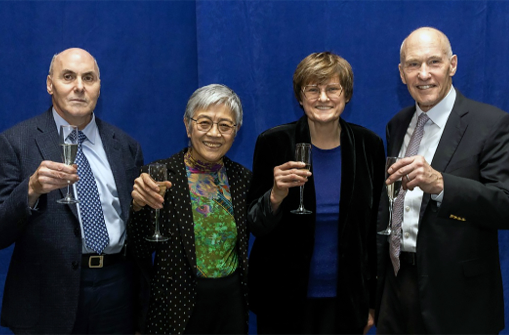
(From left to right) Breakthrough Prize recipients Drew Weissman, Virginia M-Y Lee, Katalin Karikó, and Carl June at a reception on Feb. 13. (Image: Courtesy of Penn Medicine News)
In popular culture, scientific discovery is often portrayed in “Eureka!” moments of sudden realization: a lightbulb moment, coming sometimes by accident. But in real life—and in Penn Medicine’s rich history as a scientific innovator for more than 250 years—scientific breakthroughs can never truly be distilled down to a single, “ah-ha” moment. They’re the result of years of hard work, perseverance, and determination to keep going, despite repeated, often discouraging, barriers and setbacks.
“Research is [like taking], four, or six, or eight steps back, and then a little stumble forward,” said Drew Weissman, MD, PhD, the Roberts Family Professor of Vaccine Research. “You keep doing that over and over and somehow, rarely, you can get to the top of the step.”
For Weissman and his research partner, Katalin Karikó, PhD, an adjunct professor of Neurosurgery, that persistence—documented in thousands of news stories across the globe—led to the mRNA technology that enabled two lifesaving COVID-19 vaccines, earning the duo numerous accolades, including the highest scientific honor, the 2023 Nobel Prize in Medicine.
Weissman and Karikó were also the 2022 recipients of the Breakthrough Prize in Life Sciences, the world’s largest science awards, popularly known as the “Oscars of Science.” Founded in 2012 by a group of web and tech luminaries including Google co-founder Sergey Brin and Meta CEO Mark Zuckerberg, the Breakthrough Prizes recognize “the world’s top scientists working in the fundamental sciences—the disciplines that ask the biggest questions and find the deepest explanations.” With six total winners, including four from the Perelman School of Medicine (PSOM), Penn stands alongside Harvard and MIT as the institutions whose researchers have been honored with the most Breakthrough Prizes.
Virginia M.–Y. Lee, PhD, the John H. Ware 3rd Professor in Alzheimer’s Research, was awarded the Prize in 2020 for discovering how different forms of misfolded proteins can move from cell to cell and lead to neurodegenerative disease progression. Carl June, MD, the Richard W. Vague Professor in Immunotherapy, is the most recent recipient and will be recognized at a star-studded red-carpet event in April for pioneering the development of CAR T cell therapy, which programs patients’ own immune cells to fight their cancer.
The four PSOM Breakthrough Prize recipients were honored on Tuesday, Feb. 13, 2024, when a new large-scale installation was unveiled in the lobby of the Biomedical Research Building to celebrate each laurate and their life-changing discoveries. During a light-hearted panel discussion, the honorees shared how a clear purpose, dogged determination, and a good sense of humor enabled their momentum forward.
Weissman presented the Department of Bioengineering’s 2022 Herman P. Schwan Distinguished Lecture: “Nucleoside-modified mRNA-LNP therapeutics.” Read more stories featuring Weissman in the BE Blog here.
In the human body, the lungs and their vasculature can be likened to a building with an intricate plumbing system. The lungs’ blood vessels are the pipes essential for transporting blood and nutrients for oxygen delivery and carbon dioxide removal. Much like how pipes can get rusty or clogged, disrupting normal water flow, damage from respiratory viruses, like SARS-CoV-2 or influenza, can interfere with this “plumbing system.”
In a recent study, researchers looked at the critical role of vascular endothelial cells in lung repair. Their work, published in Science Translational Medicine, was led by Andrew Vaughan of the University of Pennsylvania’s School of Veterinary Medicine and shows that, by using techniques that deliver vascular endothelial growth factor alpha (VEGFA) via lipid nanoparticles (LNPs), that they were able to greatly enhance modes of repair for these damaged blood vessels, much like how plumbers patch sections of broken pipes and add new ones.
“While our lab and others have previously shown that endothelial cells are among the unsung heroes in repairing the lungs after viral infections like the flu, this tells us more about the story and sheds light on the molecular mechanisms at play,” says Vaughan, assistant professor of biomedical sciences at Penn Vet. “Here we’ve identified and isolated pathways involved in repairing this tissue, delivered mRNA to endothelial cells, and consequently observed enhanced recovery of the damaged tissue. These findings hint at a more efficient way to promote lung recovery after diseases like COVID-19.”
They found VEGFA’s involvement in this recovery, while building on work in which they used single cell RNA sequencing to identify transforming growth factor beta receptor 2 (TGFBR2) as a major signaling pathway. The researchers saw that when TGFBR2 was missing it stopped the activation of VEGFA. This lack of signal made the blood vessel cells less able to multiply and renew themselves, which is vital for the exchange of oxygen and carbon dioxide in the tiny air sacs of the lungs.
“We’d known there was a link between these two pathways, but this motivated us to see if delivering VEGFA mRNA into endothelial cells could improve lung recovery after disease-related injury,” says first author Gan Zhao, a postdoctoral researcher in the Vaughan Lab.
“LNPs have been great for vaccine delivery and have proven incredibly effective delivery vehicles for genetic information. But the challenge here was to get the LNPs into the bloodstream without them heading to the liver, which is where they tend to congregate as its porous structure lends favor to substances passing from the blood into hepatic cells for filtration,” says Mitchell, an associate professor of bioengineering at Penn Engineering and a coauthor of the paper. “So, we had to devise a way to specifically target the endothelial cells in the lungs.”
Lulu Xue, a postdoctoral researcher in the Mitchell Lab and a co-first author of the paper, explains that they engineered the LNP to have an affinity for lung endothelial cells, this is known as extra hepatic delivery, going beyond the liver.