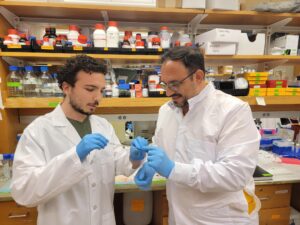
Matthew Aronson (left), Ph.D. student in Bioengineering, and Riccardo Gottardi, Assistant Proessor in Bioengineering and Pediatrics.
Riccardo Gottardi, Assistant Professor in Pediatrics in the Perelman School of Medicine and in Bioengineering in the School of Engineering and Applied Science, has been named a “Young Innovator of Cellular and Molecular Bioengineering” by Cellular and Molecular Bioengineering, the official journal of the Biomedical Engineering Society (BMES). Gottardi is Chief Scientist in the Pediatric Airway Frontier Program at the Children’s Hospital of Philadelphia (CHOP). He leads the Bioengineering and Biomaterials (Bio2) Lab, and was recognized here for his research to prevent subglottic stenosis in children.
Gottardi’s work in subglottic stensosis, a severe narrowing of the airway in response to intubation, was recently profiled in CHOP’s Cornerstone Blog. CHOP’s award press release describes Gottardi’s innovative treatment:
“Prior studies by Dr. Gottardi’s lab used in vitro models to demonstrate that incorporating AMPs into polymer-coated tubes can inhibit bacterial growth and modulate the upper-airway microbiome. In a recent study in Cellular and Molecular Engineering, led by [Bioengineering] PhD student Matthew Aronson of the Gottardi Lab, the researchers went a step further and used both ex vivo and in vivo models to show how their patent-pending antimicrobial peptide-eluting endotracheal tube (AMP-ET) effectively targeted the local airway microbiota, reducing inflammation and resolving stenosis.
‘I am honored to be recognized by Cellular and Molecular Engineering for this exciting and notable award,” Dr. Gottardi said. “We are hopeful that our airway innovation will show similar success in human trials, so that we can improve outcomes for intubated pediatric patients.’”
A research team led by engineers at the University of Pennsylvania and Northwestern University scientists has created a new synthetic biology approach, or a “QR code for cancer cells,” to follow tumor cells over time, finding there are meaningful differences in why a cancer cell dies or survives in response to anti-cancer therapies.
Remarkably, what fate cancer cells choose after months of therapy is “entirely predictable” based on seemingly small, yet important, differences that appear even before treatment begins. The researchers also discovered the reason is not genetics, contrary to beliefs held in the field.
The study outlined the team’s new technology platform that developed a QR code for each of the millions of cells for scientists to find and use later — much like tagging swans in a pond. The QR code directs researchers to a genome-wide molecular makeup of these cells and provides information about how they’ve reacted to cancer treatment.
“We think this work stands to really change how we think about therapy resistance,” said Arjun Raj, co-senior author and Professor in Bioengineering in the School of Engineering and Applied Science at the University of Pennsylvania. “Rather than drug-resistant cells coming in just one flavor, we show that even in highly controlled conditions, different ‘flavors’ can emerge, raising the possibility that each of these flavors may need to be treated individually.”
In the study, the lab and collaborators sought to apply synthetic biology tools to answer a key question in cancer research: What makes certain tumors come back a few months or years after therapy? In other words, could the lab understand what causes some rare cells to develop therapeutic resistance to a drug?
“There are many ways cells become different from each other,” said Yogesh Goyal, the co-senior author at Northwestern University. “Our lab asks, how do individual cells make decisions? Understanding this in the context of cancer is all the more exciting because there’s a clinically relevant dichotomy: A cell dies or becomes resistant when faced with therapies.”
Using the interdisciplinary team, the scientists put the before-and-after cloned cells through a whole genome sequencing pipeline to compare the populations and found no systematic underlying genetic mutations to investigate the hypothesis. Raj and Goyal helped develop the QR code framework, FateMap, that could identify each unique cell that seemed to develop resistance to drug therapy. “Fate” refers to whether a cell dies or survives (and if so, how), and the scientists “map” the cells across their lifespan, prior to and following anti-cancer therapy. FateMap is the result of work from several research institutions, and it applies an amalgamation of concepts spanning several disciplines, including synthetic biology, genome engineering, bioinformatics, machine learning and thermodynamics.
“Some are different by chance — just as not all leaves on a tree look the same — but we wanted to determine if that matters,” Goyal said. “The cell biology field has a hard time defining if differences have meaning.”
Traumatic brain injury (TBI) has disabled 1 to 2% of the population, and one of their most common disabilities is problems with short-term memory. Electrical stimulation has emerged as a viable tool to improve brain function in people with other neurological disorders.
Now, a new study in the journal Brain Stimulation shows that targeted electrical stimulation in patients with traumatic brain injury led to an average 19% boost in recalling words.
Led by University of Pennsylvania psychology professor Michael Jacob Kahana, a team of neuroscientists studied TBI patients with implanted electrodes, analyzed neural data as patients studied words, and used a machine learning algorithm to predict momentary memory lapses. Other lead authors included Wesleyan University psychology professor Youssef Ezzyat and Penn research scientist Paul Wanda.
“The last decade has seen tremendous advances in the use of brain stimulation as a therapy for several neurological and psychiatric disorders including epilepsy, Parkinson’s disease, and depression,” Kahana says. “Memory loss, however, represents a huge burden on society. We lack effective therapies for the 27 million Americans suffering.”
Michael Kahana is the Edmund J. and Louise W. Kahn Term Professor of Psychology at the University of Pennsylvania. He is a member of the Penn Bioengineering Graduate Group.
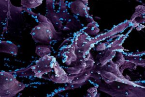
Artificial intelligence is a new addition to the infectious disease researcher’s toolbox. Yet in merely half a decade, AI has accelerated progress on some of the most urgent issues in medical science and public health. Researchers in this field blend knowledge of life sciences with skill in computation, chemistry and design, satisfying decades-long appeals for interdisciplinary tactics to treat these disorders and stop their spread.
Diseases are “infectious” when they are caused by organisms, including parasites, viruses, bacteria and fungi. People and animals can contract infectious diseases from their environments or food, or through interactions with one another. Some, but not all, are contagious.
Infectious diseases are an intractable global challenge, posing problems that continue to grow in severity even as science has offered a steady pace of solutions. The world continues to become more interconnected, bringing people into new kinds and levels of relation, and the climate crisis is throwing environmental and ecological networks out of balance. Diseases that were once treatable by drugs have become resistant, and new drug discovery is more costly than ever. Uneven resource distribution means that certain parts of the world are perennial hotspots for diseases that others never fear.
In the paper, de la Fuente and co-authors assess the progress, limitations and promise of research in AI and infectious diseases in three major areas of inquiry: anti-infective drug discovery, infection biology, and diagnostics for infectious diseases.
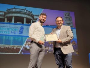
Paul Gehret (left) and Riccardo Gottardi, PhD, at Biofabrication 2022, the International Conference on Biofabrication.
Bioengineering researchers at Children’s Hospital of Philadelphia are developing a less invasive and quicker method to create cartilage implants as an alternative to the current treatment for severe subglottic stenosis, which occurs in 10 percent of premature infants in the U.S.
Subglottic stenosis is a narrowing of the airway, in response to intubation. Severe cases require laryngotracheal reconstruction that involves grafting cartilage from the rib cage with an invasive surgery. With grant support from the National Institutes of Health, Riccardo Gottardi, PhD, who leads the Bioengineering and Biomaterials (Bio2) Lab at CHOP, is refining a technology called Meniscal Decellularized scaffold (MEND). Working with a porcine model meniscus, the researchers remove blood vessels and elastin fibers to create pathways that allow for recellularization. Dr. Gottardi and his team then harvest ear cartilage progenitor cells (CPCs) with a minimally invasive biopsy, combine them with MEND, and create cartilage implants that could be a substitute for the standard laryngotracheal reconstruction.
While laryngotracheal reconstruction in the adult population has a success rate of up to 96%, success rates in children range from 75% to 85%, and children often require revision surgery due to a high incidence of restenosis. The procedure also involves major surgery to remove cartilage from the rib cage, which is more difficult for childrens’ smaller bodies.
“Luckily not many children suffer from severe subglottic stenosis, but for those who do, it is really serious,” said Dr. Gottardi, who also is assistant professor in the Department of Pediatrics and Department of Bioengineering at CHOP and the University of Pennsylvania. “With our procedure, we have an easily accessible source for the cartilage and the cells, providing a straightforward and noninvasive treatment option with much potential.”
Riccardo Gottardi is an Assistant Professor in the Department of Pediatrics, Division of Pulmonary Medicine in the Perelman School of Medicine and in the Department of Bioengineering in the School of Engineering and Applied Science. He also holds an appointment in the Children’s Hospital of Philadelphia (CHOP).
Paul Gehret is a Ph.D. student in Bioengineering, an Ashton Fellow and a NSF Fellow. His research focuses on leveraging decellularized cartilage scaffolds and novel cell sources to reconstruct the pediatric airway.
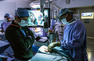
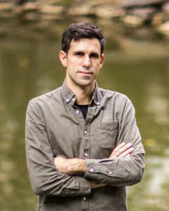
Kyle Vining, who is jointly appointed in the School of Dental Medicine and the School of Engineering and Applied Science, hopes that his research will help to push forward the state of clinical dentistry.
“During my training, I saw that there was overlap where I could do clinical work and science at the same time, and so that’s what I’ve been doing ever since,” Vining says. “As far back as middle school, I always wanted to be a biomedical engineer, and then the clinical side became interesting to me because I didn’t want to only do the theoretical or research side of things. I also wanted the hands-on, practical interaction of a skilled profession.”
The benefits of a dual career: Variety and opportunities to give back
Vining finds that wearing two hats offers the best of both worlds: opportunities to help both individual patients and to contribute to scientific and clinical progress.
“On the dentistry side, what I enjoy is getting to see patients, solving clinical problems, and trying to perform the best treatment I can; it has this rapid pace, which is kind of exciting and keeps you motivated,” Vining says. “And then research allows me to explore my interests and think about making an impact more broadly, not just in dentistry, but in medicine or in the world in general.”
Vining says dental school was demanding, yet a good time to explore his varied interests. He says he’d encourage others to pursue dentistry with an interdisciplinary approach. “Having exposure to different fields or different knowledge while you’re a student is really good for students and the profession in general,” he says.
The path towards a dual career
Vining first delved into research as a biomedical engineering undergraduate at Northwestern University. “I had the opportunity to work in a materials science lab studying the chemistry of surfaces. We would use molecules to modify the properties and surfaces that environments or cells could interact with,” he says.
Then, as a student at the University of Minnesota School of Dentistry, Vining realized that this same materials science research had many applications in dentistry. While in dental school, Vining conducted independent research in a materials science lab and also took the opportunity to do a yearlong fellowship in a cell and developmental biology lab at the National Institutes of Health.
Vining credits this fellowship with launching him towards a Ph.D., which he completed in bioengineering at Harvard in 2020. After earning his Ph.D., Vining conducted research at the Dana-Farber Cancer Institute prior to joining Penn.
Using biomaterials to understand how cells and tissues interact
Vining’s research at Penn aims to understand how the biophysical properties of materials impact cellular processes such as inflammation and fibrosis.
“Fibrosis is a physical change in tissues that produces a scar-like matrix that can inhibit healing, impair cancer treatment, and in general is not compatible with tissues regeneration,” Vining says. “There’s been a lot of effort on antifibrotic drugs, but we’re trying to look at fibrosis a little bit differently. Instead of directly inhibiting fibrosis, we’re trying to understand its consequences for the immune system because the immune system can be hijacked and become detrimental for your tissues.”
Through a better understanding the feedback loop between fibrotic tissue and the immune system, Vining hopes to design interventions to facilitate wound healing and tissue remodeling during restorative dental procedures and for treating diseases including head and neck cancer.
He’s also investigating how biomaterials like the resin used in tooth fillings interact with dental tissues. “Dental fillings rely on decades-old technologies that have been grandfathered in and contain toxic monomers that are not safe for biological systems,” Vining says. “We found a biocompatible resin chemistry that supports cells in vitro, and we’re trying to apply this to new types of dental fillings that promote repair or generation of dental tissues.”
Fostering interdisciplinary collaborations at Penn
“Dr. Vining is an ideal fit for the vision and mission of the CiPD,” says Penn Dental’s Hyun (Michel) Koo, co-founder and co-director of the CiPD. “With a secondary appointment in the School of Engineering, he will be instrumental in continuing to strengthen our engineering collaborations and teaching our students to work across disciplines to advance research, training, and entrepreneurship in this realm.”
Ultimately, Vining says it was Penn’s scientific community and the opportunities for interdisciplinary collaborations that drew him here.
“It was very apparent that there were a lot of potential research paths to pursue here and a lot of opportunities for collaborations,” Vining says. “One of the most exciting things for me so far has been meeting with faculty, whether it’s at Penn Medicine, the School of Engineering, Wharton, Penn Dental, or the Veterinary School. These meetings have already opened up new projects and collaborations.”
The collaboration sparked when Vining saw Mitchell present on a new technology that uses lipid nanoparticles to bind and target bone marrow cells at the 2022 CiPD first annual symposium. “It got me thinking because the dentin inside of teeth is a mineralized tissue very similar to bone, and the pulp inside the dentin is analogous to bone marrow tissue,” Vining says.
Rather than being the single disease class many people refer to, “cancer” is a blanket term that covers more than 100 distinct diseases, many of which have little in common aside from originating with rapidly dividing cells. Since different cancers demand different treatments, it follows that any given new therapy emerging from any institution would be likely to be a new cancer treatment.
But why so many in just this five-year period?
The volume of new cancer treatments makes sense, says Abramson Cancer Center (ACC) director Robert Vonderheide, attributing the flurry of new cancer drug approvals to a recent “explosion” in knowledge about cancer biology.
“Much of that knowledge is about the immune system’s ability to attack cancer, which people seriously doubted until about 20 years ago. As soon as we had a clinical validation for this Achilles heel in cancer, the dam burst for ideas about other ways to exploit that vulnerability to come forward,” he says. “The first drug that came out to activate the immune system inspired the rest of the field to find the next drug, and the one after that. We as a field have moved from serendipity and empiricism to science-driven drug design.”
The first CAR T cell therapy approval invigorated Penn faculty interested in finding new ways to harness the immune system to fight cancer.
“An approval like that makes what you’re working on more of a reality,” says Avery Posey, an assistant professor of systems pharmacology and translational therapeutics in the Perelman School of Medicine, whose lab team spends much of its time trying to identify more specific antigens for solid tumors and also studies ways to optimize engineered donor T cells. “It brings a new perspective, showing that your work is more than basic research and can actually become drugs that impact patients’ lives. That’s a real motivator to keep pushing forward.”
Honing new immunotherapies is a priority among Penn researchers, but not every recently approved new cancer treatment or detection tool developed at the institution engages the immune system. Faculty have explored and introduced widely varying approaches to improving the standard of care for cancer patients.
Presented at the biennial American Peptide Symposium, the Makineni Lectureship Award recognizes an individual who has made a recent contribution of unusual merit to research in the field of peptide science, and is intended to acknowledge original and singular discoveries.
Established in 2003 by an endowment by PolyPeptide Laboratories and Murray and Zelda Goodman, this lectureship honors Rao Makineni, a long-time supporter of peptide science, peptide scientists, and the American Peptide Society.
For decades, LGBTQ+ patients have faced stringent requirements to donate blood—most gay and bisexual men were not allowed to donate at all. Now, however, many more of them will be able to give this selfless gift. The U.S. Food and Drug Administration, which regulates blood donation in this country, has reworked the donor-screening criteria, and in the process opened the door to donation for more Americans.
The previous restriction on accepting blood from men who have sex with men (MSM) dates back to the early days of the AIDS epidemic, when blood donations weren’t able to be screened for HIV, leading to cases of transfusion-transmitted HIV. In 1985, the FDA instituted a lifetime ban on blood donation for MSM, effectively preventing gay and bisexual men from donating. (Also included were women who have sex with MSM.)
Twenty years later, the agency rescinded the ban—but added a restriction that only MSM who had been abstinent from sex for at least one year could donate. In 2020, the FDA shortened the “deferral” period to 90 days of abstinence. While the changes were welcome news for those who had been unable to donate, they still prevented many MSM from giving blood. As he wrote in an op-ed for the Philadelphia Inquirer last year, Kevin B. Johnson,the David L. Cohen University Professor with appointments in the School of Engineering and Applied Science, the Perelman School of Medicine, and Annenberg School for Communication, was one of them. He and his husband were shocked to learn when they went to donate blood during a shortage early in the COVID-19 pandemic, that despite being married and monogamous for close to 17 years, they could not donate unless they were celibate for three months.
“It is time to move quickly to a policy under which all donors are evaluated equally and fairly, and to encourage local blood collection facilities to comply with that policy,” Johnson wrote last year.
Now, such changes are underway. As the pandemic wound down, the FDA moved forward with plans to re-evaluate its donation criteria. The first big change was removal of an indefinite ban on people who lived in or spent significant amounts of time in the United Kingdom, Ireland, and France, a measure that aimed to protect the U.S. blood supply against Creutzfeldt-Jakob disease (CJD; also known as “mad cow disease”), a terminal brain condition caused by hard-to-detect prions that occurred in those countries in the 1980s and 1990s.
Extensive and careful evaluation of epidemiological studies and statistical analysis has shown that the risk of CJD transmission is no longer a concern. The changes to eligibility for LGBTQ+ patients are related to advances in medical and social science, and have also been very thoroughly studied to ensure that the changes will maintain the safety of the blood supply without being discriminatory.
“In the decades since HIV was first recognized, there have been advances in testing methods for detection of the virus, changes in how we process blood products, public health advances, and extensive study of the evolving risk of disease transmission given these advances,” says Sarah Nassau,vice chair of pathology and laboratory medicine at Lancaster General Hospital.
They also draw on rethinking the reliability of the guidelines. For example, while the rules partially or fully prevented gay and bisexual men from donating blood, they did not erect similar barriers to other people engaging in anal sex, or people who have multiple partners.
“Specifying the sexual orientation of the person rather than a behavior in which they engaged was discriminatory and not evidence based,” points out Judd David Flesch, vice chief of inpatient operations in the Department of Medicine at Penn Presbyterian Medical Center and co-director of the Penn Medicine Program for LGBT Health.
Kevin Johnson is the David L. Cohen University of Pennsylvania Professor in the Departments of Biostatistics, Epidemiology and Informatics and Computer and Information Science. As a Penn Integrates Knowlegde (PIK) University Professor, Johnson also holds appointments in the Departments of Bioengineering and Pediatrics, as well as in the Annenberg School of Communication.
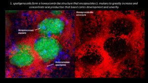
Collaborating researchers from the University of Pennsylvania School of Dental Medicine and the Adams School of Dentistry and Gillings School of Global Public Health at the University of North Carolina have discovered that a bacterial species called Selenomonas sputigena can have a major role in causing tooth decay.
Scientists have long considered another bacterial species, the plaque-forming, acid-making Streptococcus mutans, as the principal cause of tooth decay—also known as dental caries. However, in the study, published in Nature Communications, the Penn Dental Medicine and UNC researchers showed that S. sputigena, previously associated only with gum disease, can work as a key partner of S. mutans, greatly enhancing its cavity-making power.
“This was an unexpected finding that gives us new insights into the development of caries, highlights potential future targets for cavity prevention, and reveals novel mechanisms of bacterial biofilm formation that may be relevant in other clinical contexts,” says study co-senior author Hyun (Michel) Koo, a professor in the Department of Orthodontics and Divisions of Pediatrics and Community Oral Health and co-director of the Center for Innovation & Precision Dentistry at Penn Dental Medicine.
The other two co-senior authors of the study were Kimon Divaris, professor at UNC’s Adams School of Dentistry, and Di Wu, associate professor at the Adams School and at the UNC Gillings School of Global Public Health.
“This was a perfect example of collaborative science that couldn’t have been done without the complementary expertise of many groups and individual investigators and trainees,” Divaris says.
Michel Koo is a professor in the Department of Orthodontics and divisions of Community Oral Health and Pediatric Dentistry in Penn Dental Medicine and co-director of the Center for Innovation & Precision Dentistry. He is a member of the Penn Bioengineering Graduate Group.

 A research team led by engineers at the