Primary Projects From Ghana: Group 1

Throughout the Spring 2017 semester, our professor, Dr. David Issadore, taught us (a class of eight undergraduates students and one graduate student) about microfluidics and point-of-care diagnostics. The next phase of the course was to come up with a new diagnostic for pediatric tuberculosis. At the end of the semester, our final assignments included submitting an NIH Research Project Grant (R01) proposal and giving a 20-minute presentation for our devices. These assignments greatly prepared us for our trip to Ghana, as we were able to ask questions and get feedback on our proposed devices by speaking to healthcare professionals at Ghanaian hospitals, clinics, and research facilities. The semester course was mainly focused on the technical design of our devices, which enabled us to hone in on the practical and real-world implementation of the devices while in Ghana. This week, the BE Blog will publish our summaries.

Fecal Diagnostics for Pediatric Tuberculosis

Katharine Cocherl ’20, Kathleen Givan ’20, Kaila Helm ’20, Hope McMahon ’18, David Pontoriero ’18

In order to address the numerous diagnostic problems specific to pediatric tuberculosis in low-resource settings, we have designed a device that uses a fecal sample rather than the current method of sputum samples. Because many children cannot produce sputum samples with the required quality and quantity of sputum, we decided to use stool samples. This noninvasive substitute will ideally allow us to collect all the bacteria swallowed by the patient. The bacterium that causes the disease, Mycobacterium tuberculosis (MTB), is very hardy and has been found to appear in fecal matter. However, this method may be difficult because there are many other substances in fecal matter that need to be removed. By filtering out these impurities, the presence of the bacteria can be detected.

The device we designed is essentially a disposable cartridge that separates  virulent TB bacteria from all other fecal material. This collection can be performed with no power and minimal technician input and can be obtained in any desired volume. The total operation time is predicted to be 90 minutes.

ghana group 1
The figure shows an overview of the steps (a-h) for use of the fecal diagnostic for pediatric TB (click to enlarge).

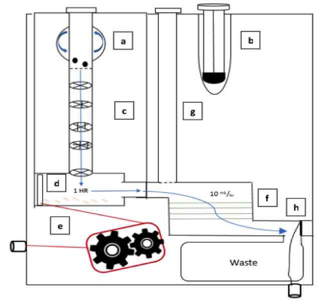
The first aim of our project is to identify a target protein on the surface of the bacteria so that the bacteria can be isolated from the solution. Next, the MTB will be enriched from fecal samples with a single-use filtration device so that a final sample can be provided in a similar form as a sputum sample. This final sample can then be used for smear microscopy, in which technicians look for the presence of the bacteria under a microscope, or for use with the GeneXpert. The GeneXpert is an automated diagnostic test that can identify MTB DNA and resistance to the most potent TB drug, rifampicin. These devices have been distributed to labs and hospitals across Ghana, but they are not yet widely used for general diagnostics.

Because the number of GeneXperts available and the infrastructure supporting them are increasing, we are hopeful that, in the near future, our diagnostic will be able to be used in conjunction with this technology. Upon integration with the GeneXpert system, our device would be able to increase sample specificity for the underserved demographic of pediatric TB patients. In addition, as technology becomes available in smaller, more local clinics, we foresee lower travel burdens for families and lower operational costs for healthcare facilities.

We are beyond grateful for the opportunity to engage with Ghana’s medical system. Before traveling to Ghana, we created a proposal for our fecal diagnostic for pediatric TB. After learning more about the current medical system and infrastructure in place, we were able to revise our ideas in a meaningful way. It is our hope that one day a project of this magnitude can come to fruition.

Macrophages Engineered Against Cancer Cells

macrophages Discher
Dennis Discher, Ph.D.

Dennis E. Discher, Ph.D., Robert D. Bent Professor in the Department of Chemical and Biomolecular Engineering and a secondary faculty member in the Department of Bioengineering, was the lead author on a recent study that showed that engineered macrophages (a type of immune cell) could be injected into mice, circulate through their bodies, and invade solid tumors in the mice, engulfing human cancers cells in the tumors.

According to Cory Alvey, a graduate student in pharmacology who works in Professor Discher’s lab and the first author on the paper, said, “Combined with cancer-specific targeting antibodies, these engineered macrophages swarm into solid tumors and rapidly drive regression of human tumors without any measurable toxicity.”

Read more here.

This Week in BioE (July 13, 2017)

Devices and Drug Delivery

Abdominal surgery can lead to complications when the intestines are accidentally damaged. One key point in the surgery is during wound closure, when the surgeon must place the final sutures without knowing where the underlying intestine is located. A new material designed by bioengineers can be inserted into the abdomen and protect the intestines from perforations by the surgical needle. Key features of this material include its flexibility to fit into the small incision made during laprascopy and its ability to dissolve naturally within hours upon insertion into the abdominal cavity.  Before it dissolves, the material is tough enough to protect the intestines from puncture, allowing the surgeon to close the incision with much less risk of perforation. So the material is ready when it is needed and disappears soon thereafter.

New materials appear frequently to perform the functions of naturally occurring biological tissues. For decades, several researchers attempted to re-create cartilage outside of the body. Although these artificial cartilage tissues may contain all of the right ‘ingredients’ — i.e., molecules and cells — the tissue is commonly not strong enough to withstand the forces normally experienced by the target tissue.  Recently, researchers invented a process to load the cartilage tissue surrogate while it was fabricated, a departure from the traditional process in which the tissue substitute is mechanically loaded after it is built. Both techniques are designed to make the artificial tissue stronger.  However, this subtle new design step to mechanically load during fabrication makes the cartilage substitute six times stronger than any existing manufacturing technique, raising the possibility that we can build tissue outside the body for use inside the body.

emu egg
An emu egg

Finally, the field is constantly discovering new ways to use historical observations in science. One example was an observation from the analysis of 19,000-year-old DNA from an emu egg, made possible because the DNA was protected from degradation by the calcified material present in the eggshell. This scientific observation to understand the origin of the species inspired bioengineer Bill Murphy at the University of Wisconsin-Madison to create a new method to protect proteins from degradation by incorporating these proteins into mineralized materials. Reminiscent of the mineralized matrix found in the emu eggshell that protected DNA for 19,000 years, the charged mineralized matrix stabilizes the protein structure and significantly improves the stability of the protein. By designing the mineralized material to degrade slowly, this work shows that one can stabilize and release therapeutic proteins over much longer periods than previously possible.

Technological Advances in Cancer Diagnosis and Treatment

Despite tremendous advances in diagnosis and treatment, cancer remains a major public health threat. Surgery is often a key part of cancer treatment, but tumor removal is complicated by the difficulty in producing surgical margins that are free of cancer cells. If cancer cells remain in the margins, it is common for the cancer to return. However, a team of researchers at the University of Washington developed a light-sheet microscope capable of imaging these surgical margins quickly – about 30 minutes. The technology could go a long way toward reducing or eliminating the 20% to 40% of cases of breast cancer in which relapse occurs.

While breast cancer remains the most common cancer among women, proliferation of HPV has resulted in a steadily increasing rate of cervical cancer over past decades. Early screening here is a key to successful treatment, but gynecological examinations are uncomfortable for many women. Failure to schedule a follow-up colposcopy is common following an abnormal Pap smear, resulting in persistently higher rates. A pocket colposcope developed by Duke Biomedical Engineering Professor Nimmi Ramanujam could close this gap in treatment. Although the colposcope must still be rigorously tested, a small group of 15 volunteers who tested the device reported that it was 80% accurate.

New BioE dept at Lehigh

Lehigh University in Bethlehem, Pa., has announced the creation of a Department of Bioengineering. Anand Jagota, a professor formerly in the Department of Chemical Engineering, founded the department and will act as its first chair. In addition to Professor Jagota, 16 professors form the core department faculty.

Welcome to the club, Lehigh!

Mind Control and an Ethical Appeal

mind control brain
A “wiring diagram of the human brain,” produced using diffusion MRI scans of the brain.

A group of four scholars from the University of Pennsylvania, including Bioengineering professor Danielle Bassett, have issued a call in the journal Nature Human Behaviour for greater safeguards for patients as treatments in the field of neuroscience evolve and come ever closer to resembling “mind control.”

“While we don’t believe,” Bassett said, “that the science-fiction idea of mind control, totally overriding a person’s autonomy, will ever be possible, new brain-focused therapies are becoming more specific, targeted and effective at manipulating individuals’ mental states. As these techniques and technologies mature, we need systems in place to make sure they are applied such that they maximize beneficial effects and minimize unwanted side effects.”

Read more at the Penn News Web Site.

New Faculty: Interview With Alex Hughes

Alex new faculty
Alex Hughes, Ph.D.

As noted earlier this week, Penn BE will be bringing in three new faculty members over the coming academic year, starting with Alex Hughes, who will start in the fall semester. Here’s the first of our series of podcasts with the new faculty, to come each Friday this month. Enjoy!

(P.S. Apologies for the rough version of the audio. We are still learning!)

This Week in BioE (July 6, 2017)

Bioengineering of Genes and DNA

Since Watson and Crick published their initial studies detailing the double helix structure of DNA in the early 1960s, what we know about genetics and the nucleic acids underlying them has grown enormously. Consequently, what bioengineering can do with DNA and genes continually expands.

One fascinating bioengineering field that emerged in the past decade was DNA origami, which uses the well-established binding across DNA elements to create three-dimensional structures out of linear DNA sequences. Recent work has utilized this feature of DNA construction to make machines, rather than just parts, out of DNA.

Yonggang Ke, Ph.D., of Georgia Tech/Emory’s Department of Biomedical Engineering, constructed machines made of DNA that consist of arrays of units that can “switch” between “settings” by changing shape. A change in shape of one unit of an array can cause the other units in the array to shift; these changes are stimulated by inserting a previously deleted strand of DNA into the array. Although it has been known for some time that DNA could be used to store and transmit information, Dr. Ke’s research team proved for the first time that these arrays could be shaped physically into machines in the shapes of rectangles and tubes.

genes dna
DNA under a microscope

While we learn more about how to make DNA-based devices, we are also creating new technologies to manipulate DNA more rapidly.  Scientists at Rutgers and Harvard developed a process whereby thousands of genes could be cloned at one time to create enormous libraries of proteins. To achieve this goal, the authors used a technology called LASSO (long-adapter single-strand oligonucleotide) probes, which they have already used to clone a library using a human microbiome sample.

Instead of the traditional process of cloning one gene at a time, the team led by Professor Biju Parekkadan, Ph.D. at Rutgers, invented a technology to clone hundred of genes simultaneously. These cloned DNA segments are much longer than the length of DNA cloned with standard techniques, allowing us to test the functional significance of these much longer DNA segments.  The technology could impact a number of scientific fields because we will finally learn how long stretches of protein function — some parts may degrade other proteins, while other parts will interact and modify other proteins (e.g., phosphorylation, a key process in epigenetics). These new discoveries can be key for discovering new ways to engineer proteins and to manufacture new drugs that mimic the function of nature’s DNA products.

Using Sweat as a Biosensor

While the field learns more about the molecular-level control of DNA, we are also taking advantage of new micro- and nanoscale manufacturing processes to capture diagnostic information from easily accessible body fluids. Many clinical diagnostics use chemical measurements from blood to diagnose a disease or to take corrective action. This is not an ideal procedure because it requires either the collection of blood at a laboratory or the repeated collection of small blood volumes through a pinprick.  Either one hurts.

Bioengineers at the University of Texas at Dallas developed a wearable diagnostic device to detect cortisol, glucose, and IL-6 in body sweat, eliminating any painful needle sticks.  Its transmissions vary, but if optimized, the device could replace the painful and inconvenient practice of sticking one’s finger to obtain a drop of blood for glucose testing, which many patients with diabetes must do several times per day. Although insulin pumps have been available for some time, these are invasive devices that must be worn at all times.

New Faculty Joining Penn Bioengineering

We are thrilled to announce the successful recruitment of three (!) new faculty members to the department. We conducted a national faculty search and could not decide on one — we wanted all three of our finalists!  We are very happy that they chose Penn and think we can provide an amazing environment for their education and research programs.

new faculty hughes
Alex Hughes, Ph.D.

Alex Hughes, Ph.D., will join us in the Spring 2018 semester. Dr. Hughes comes to us from the University of California, San Francisco (UCSF), where he is a postdoctoral fellow. Alex’s research regards determining what he calls the “design rules” underlying how cells assemble into tissues during development, both to better understand these tissues and to engineer methods to build them from scratch

new faculty bugaj
Lukasz Bugaj, Ph.D.

Lukasz Bugaj, Ph.D., will arrive in the Spring 2018 semester. Dr. Bugaj is also coming here from UCSF following a postdoc, and his work is in the field of optogenetics — a scientific process whereby light is used to alter protein conformation, thereby giving one a tool to manipulate cells. In particular, Lukasz’s research has established the ability to induce proteins to cluster ‘on demand’ using light, and he wants to use these and other new technologies he invented to study cell signaling in stem cells and in cancer.

new faculty mitchell
Mike Mitchell, Ph.D.

Mike Mitchell, Ph.D., will also join us in the Spring 2018 semester after finishing his postdoctoral fellowship at MIT in the Langer Lab. In his research, Dr. Mitchell seeks to engineer cells in the bone marrow and blood vessels as a way of gaining control over how and why cancer metastasizes. Mike’s work has already had impressive results in animal models of cancer. His lab will employ tools and concepts from cellular engineering, biomaterials science, and drug delivery to fundamentally understand and therapeutically target complex biological barriers in the body.

In the coming month, we’ll feature podcasts of interview with each of the new faculty members, as well as with Konrad Kording, so be sure to keep an eye out for those.

And to our new faculty, welcome to Penn!

Setton Named WashU BME Chair

Setton
Lori A. Setton, Ph.D.

Lori A. Setton, Ph.D., a major innovator in the field of tissue regeneration and repair and a member of the Penn Bioengineering Departmental Advisory Board, has been named chair of the Department of Biomedical Engineering at Washington University in St. Louis. Her appointment begins on August 1.

An alumna of Princeton (BSE) and Columbia (MS, Ph.D.) with degrees in mechanical engineering, Dr. Setton was on the faculty at Duke until 2015, when she moved to WashU. Over the last decade or so, she has coauthored nearly 150 peer-reviewed research papers in the field of biomedical engineering, establishing a sterling reputation as a scientist and researcher.

In addition to her distinguished career in research and academia, she is also the current president of BMES, where she has been a pioneer in fostering greater diversity within the field, both in instituting a partnership with the National Society for Black Engineers (NSBE) and as a mentor at Duke, where the introduction of a mentoring program to increase diversity among under-represented minorities has been particularly successful.

“We are thrilled that Lori was recognized with this significant leadership opportunity,” said David Meaney, Ph.D., chair of the Bioengineering Department at Penn. “As a key academic on our department advisory board, Lori’s incisive input on Penn Bioengineering has been invaluable as we grow and change as a department. I know she will be an outstanding leader for WashU.”