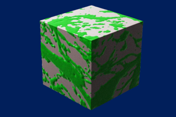
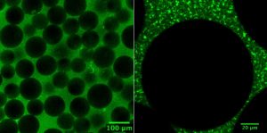
Bicontinuous materials, like this representation of a cube of gelatin and hyaluronic acid, have greater internal surface area, allowing cells to travel faster between two points. (Credit: Karen Xu)
One of the most important but least understood aspects of healing is cell migration, or the process of cells moving from one part of the body to another. “If you are an ambulance out in the woods,” says Karen Xu, an M.D/Ph.D. student in Medicine and Bioengineering, “and there are no paths for you to move forward, it will be a lot harder for you to get to a site that needs you.”
Earlier this year, Xu co-authored a paper in Nature Communications describing a new cue to help cells get to where they need to go: a material made chiefly of hyaluronic acid and gelatin, two gooey substances commonly found outside cells in joints and connective tissue.
“Hundreds of thousands of people tear their meniscus every year,” says Robert Mauck, Mary Black Ralston Professor in Orthopaedic Surgery in Penn Medicine and Professor in Bioengineering at Penn Engineering and one of Xu’s advisors, as well as a senior author on the paper. “This material could potentially speed up their recovery.”
What makes the material — known as a hydrogel due to its blend of gelatinous matter and water — unique is that the combination of hyaluronic acid and gelatin forms a complex network of paths, providing cells many different ways to travel between two points.
This property is known as bicontinuity, and is exemplified by two discrete continuous phases that are each connected throughout the entire volume of the material (for example with a sponge, with phases of cellulose and air; in the hydrogel, this is comprised of gelatin and hyaluronic acid) resulting in a dizzying array of patterns that dramatically increase the surface area inside the material.
To test the hydrogel’s efficacy, Xu and her collaborators — including co-advisor Jason Burdick, formerly the Robert D. Bent Professor in Bioengineering at Penn Engineering and now the Bowman Endowed Professor at the University of Colorado Boulder, and the paper’s other senior author — first created several different versions of the hydrogel to find the sweet spot at which the constituents formed the bicontinuous structure and had the highest internal surface area. “We found that a precise combination of the various hydrogel components and control over their mixing was needed to form the bicontinuous structure,” says Burdick.
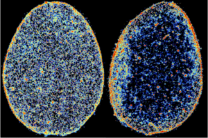
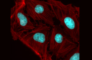
In these super-resolution images of tendon cell nuclei, the color coding represents chromatin density map, from low density in blue to high density in red. Comparing a healthy human tendon cell nucleus (left) to one diagnosed with tendinosis (right) shows that disease alters the spatial localization and compaction of chromatin.
In a recent study published in Nature Biomedical Engineering, the team detailed what they found when they closely observed the nucleus of cells inside connective tissues deteriorating as a result of tendinosis, which is the chronic condition that results from a tendon repeatedly suffering small injuries that don’t heal correctly. Using the latest super-resolution imaging techniques, they found that the tendon cells involved in maintaining the tissue’s structure in a diseased microenvironment improperly reorder their chromatin — the DNA-containing material that chromosomes are composed of — when attempting to repair.
This and other findings highlighted in the report point to the possibility of new treatments, such as small-molecule therapies, that could restore order to the affected cells.
“Interestingly, we were able to explain the role of mechanical forces on the 3-D organization of chromatin by developing a theory that integrates fundamental thermodynamic principles (physics) with the kinetics of epigenetic regulation (biology),” said study co-author and CEMB Director Vivek Shenoy in a news release from Penn Medicine News.
The CEMB, one of 18 active interdisciplinary research centers funded by the National Science Foundation’s Science and Technology Center (STC) program, brings together dozens of researchers from Penn Engineering and the Perelman School of Medicine, as well as others spread across campus and at partner institutions around the world.
With its funding recently renewed for another five years, the CEMB has entered into a new phase of its mission, centered on the nascent concept of “mechanointelligence,” which is exemplified by studies like this one. While mechanobiology is the study of the physical forces that govern the behavior of cells and their communication with their neighbors, mechanointelligence adds another layer of complexity: attempting to understand the forces that allow cells to sense, remember and adapt to their environments.
Ultimately, harnessing these forces would allow researchers to help multicellular organisms — plants, animals and humans — better adapt to their environments as well.
This story originally appeared in Penn Engineering Today.
Vivek Shenoy is Eduardo D. Glandt President’s Distinguished Professor in Materials Science and Engineering, Bioengineering, and in Mechanical Engineering and Applied Mechanics.
The dynamics governing mechanointelligence vary greatly along time- and length-scales, so detailed models of individual cells and their components are necessary to connect the effects of their physical environments to the downstream effects those forces have on biological processes.
The National Science Foundation’s Science and Technology Center (STC) program is its flagship funding mechanism for organizing interdisciplinary research on cutting-edge topics. Penn’s Center for Engineering MechanoBiology (CEMB) is one of the 18 active STCs, bringing together dozens of researchers from Penn Engineering and the Perelman School of Medicine, as well as others spread across campus and at partner institutions around the world.
With its NSF funding now renewed for another five years, the Center is entering into a new phase of its mission, centered on the nascent concept of “mechanointelligence.”
Mechanobiology is the study of the physical forces that govern the behavior of cells and their communication with their neighbors. Mechanointelligence adds another layer of complexity, attempting to understand the forces that allow cells to sense, remember and adapt to their environments.
Ultimately, harnessing these forces would allow researchers to help multicellular organisms — plants, animals and humans — better adapt to their environments as well.
“Mechanointelligence is a key element of a cell’s ability to survive and reproduce,” says CEMB Director and Eduardo D. Glandt President’s Distinguished Professor Vivek Shenoy. “Just like with complex organisms, a cell’s ‘fitness’ depends on its environment, and adapting means rewiring how its genes are expressed.”
Vivek Shenoy is Eduardo D. Glandt President’s Distinguished Professor in Materials Science and Engineering, Bioengineering and Mechanical Engineering and Applied Mechanics.
Dani S. Bassett, J. Peter Skirkanich Professor in Bioengineering and in Electrical and Systems Engineering
Bassett runs the Complex Systems lab which tackles problems at the intersection of science, engineering, and medicine using systems-level approaches, exploring fields such as curiosity, dynamic networks in neuroscience, and psychiatric disease. They are a pioneer in the emerging field of network science which combines mathematics, physics, biology and systems engineering to better understand how the overall shape of connections between individual neurons influences cognitive traits.
Jason Burdick, Ph.D.
Jason A. Burdick, Robert D. Bent Professor in Bioengineering
Burdick runs the Polymeric Biomaterials Laboratory which develops polymer networks for fundamental and applied studies with biomedical applications with a specific emphasis on tissue regeneration and drug delivery. The specific targets of his research include: scaffolding for cartilage regeneration, controlling stem cell differentiation through material signals, electrospinning and 3D printing for scaffold fabrication, and injectable hydrogels for therapies after a heart attack.
César de la Fuente, Ph.D.
César de la Fuente, Presidential Assistant Professor in Bioengineering and Chemical & Biomedical Engineering in Penn Engineering and in Microbiology and Psychiatry in the Perelman School of Medicine
De la Fuente runs the Machine Biology Group which combines the power of machines and biology to prevent, detect, and treat infectious diseases. He pioneered the development of the first antibiotic designed by a computer with efficacy in animals, designed algorithms for antibiotic discovery, and invented rapid low-cost diagnostics for COVID-19 and other infections.
Carl June, M.D.
Carl H. June, Richard W. Vague Professor in Immunotherapy in the Perelman School of Medicine and member of the Bioengineering Graduate Group
June is the Director for the Center for Cellular Immunotherapies and the Parker Institute for Cancer Therapy and runs the June Lab which develops new forms of T cell based therapies. June’s pioneering research in gene therapy led to the FDA approval for CAR T therapy for treating acute lymphoblastic leukemia (ALL), one of the most common childhood cancers.
Vivek Shenoy, Ph.D.
Vivek Shenoy, Eduardo D. Glandt President’s Distinguished Professor in Bioengineering, Mechanical Engineering and Applied Mechanics (MEAM), and in Materials Science and Engineering (MSE)
Shenoy runs the Theoretical Mechanobiology and Materials Lab which develops theoretical concepts and numerical principles for understanding engineering and biological systems. His analytical methods and multiscale modeling techniques gain insight into a myriad of problems in materials science and biomechanics.
The highly anticipated annual list identifies researchers who demonstrated significant influence in their chosen field or fields through the publication of multiple highly cited papers during the last decade. Their names are drawn from the publications that rank in the top 1% by citations for field and publication year in the Web of Science™ citation index.
Bassett and Burdick were both on the Highly Cited Researchers list in 2019 and 2020.
The methodology that determines the “who’s who” of influential researchers draws on the data and analysis performed by bibliometric experts and data scientists at the Institute for Scientific Information™ at Clarivate. It also uses the tallies to identify the countries and research institutions where these scientific elite are based.
David Pendlebury, Senior Citation Analyst at the Institute for Scientific Information at Clarivate, said: “In the race for knowledge, it is human capital that is fundamental and this list identifies and celebrates exceptional individual researchers who are having a great impact on the research community as measured by the rate at which their work is being cited by others.”
The full 2021 Highly Cited Researchers list and executive summary can be found online here.
A colorized microscope image of an osteosarcoma shows how cellular fibers can transfer physical force between neighboring nuclei, influencing genes. The Penn Anti-Cancer Engineering Center will study such forces, looking for mechanisms that could lead to new treatments or preventative therapies.
Advances in cell and molecular technologies are revolutionizing the treatment of cancer, with faster detection, targeted therapies and, in some cases, the ability to permanently retrain a patient’s own immune system to destroy malignant cells.
However, there are fundamental forces and associated challenges that determine how cancer grows and spreads. The pathological genes that give rise to tumors are regulated in part by a cell’s microenvironment, meaning that the physical push and pull of neighboring cells play a role alongside the chemical signals passed within and between them.
The Penn Anti-Cancer Engineering Center (PACE) will bring diverse research groups from the School of Engineering and Applied Science together with labs in the School of Arts & Sciences and the Perelman School of Medicine to understand these physical forces, leveraging their insights to develop new types of treatments and preventative therapies.
The Center’s founding members are Dennis Discher, Robert D. Bent Professor with appointments in the Departments of Chemical and Biomolecular Engineering (CBE), Bioengineering (BE) and Mechanical Engineering and Applied Mechanics (MEAM), and Ravi Radhakrishnan, Professor and chair of BE with an appointment in CBE.
Discher, an expert in mechanobiology and in delivery of cells and nanoparticles to solid tumors, and Radhakrishnan, an expert on modeling physical forces that influence binding events, have long collaborated within the Physical Sciences in Oncology Network. This large network of physical scientists and engineers focuses on cancer mechanisms and develops new tools and trainee opportunities shared across the U.S. and around the world.
Lukasz Bugaj, Alex Hughes, Jenny Jiang, Bomyi Lim, Jennifer Lukes and Vivek Shenoy (Clockwise from upper left).
Among the PACE Center’s initial research efforts are studies of the genetic and immune mechanisms associated with whether a tumor is solid or liquid and investigations into how physical stresses influence cell signaling.
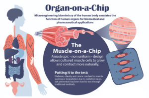
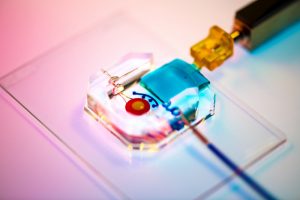
Bioengineering’s Dan Huh and colleagues have developed a number of organ-on-a-chip devices to simulate how human cells grow and perform in their natural environments. Their latest is a muscle-on-a-chip, which carefully captures the directionality of muscle cells as they anchor themselves within the body. See the full infographic at the bottom of this story. (Illustration by Melissa Pappas).
Studying drug effects on human muscles just got easier thanks to a new “muscle-on-a-chip,” developed by a team of researchers from Penn’s School of Engineering and Applied Science and Inha University in Incheon, Korea.
Muscle tissue is essential to almost all of the body’s organs, however, diseases such as cancer and diabetes can cause muscle tissue degradation or “wasting,” severely decreasing organ function and quality of life. Traditional drug testing for treatment and prevention of muscle wasting is limited through animal studies, which do not capture the complexity of the human physiology, and human clinical trials, which are too time consuming to help current patients.
An “organ-on-a-chip” approach can solve these problems. By growing real human cells within microfabricated devices, an organ-on-a-chip provides a way for scientists to study replicas of human organs outside of the body.
Using their new muscle-on-a-chip, the researchers can safely run muscle injury experiments on human tissue, test targeted cancer drugs and supplements, and determine the best preventative treatment for muscle wasting.
Dan Huh, Ph.D.
This research was published in Science Advances and was led by Dan Huh, Associate Professor in the Department of Bioengineering, and Mark Mondrinos, then a postdoctoral researcher in Huh’s lab and currently an Assistant Professor of Biomedical Engineering at Tulane University. Their co-authors included Cassidy Blundell and Jeongyun Seo, former Ph.D. students in the Huh lab, Alex Yi and Matthew Osborn, then research technicians in the Huh lab, and Vivek Shenoy, Eduardo D. Glandt President’s Distinguished Professor in the Department of Materials Science and Engineering. Lab members Farid Alisafaei and Hossein Ahmadzadeh also contributed to the research. The team collaborated with Insu Lee and professors Sun Min Kim and Tae-Joon Jeon of Inha University.
In order to conduct meaningful drug testing with their devices, the research team needed to ensure that cultured structures within the muscle-on-a-chip were as close to the real human tissue as possible. Critically, they needed to capture muscle’s “anisotropic,” or directionally aligned, shape.
“In the human body, muscle cells adhere to specific anchor points due to their location next to ligament tissue, bones or other muscle tissue,” Huh says. “What’s interesting is that this physical constraint at the boundary of the tissue is what sculpts the shape of muscle. During embryonic development, muscle cells pull at these anchors and stretch in the spaces in between, similar to a tent being held up by its poles and anchored down by the stakes. As a result, the muscle tissue extends linearly and aligns between the anchoring points, acquiring its characteristic shape.”
The team mimicked this design using a microfabricated chip that enabled similar anchoring of human muscle cells, sculpting three-dimensional tissue constructs that resembled real human skeletal muscle.
The researchers’ experiments involved making synthetic tissues with artificial “cells.” The fibrin network that surrounds these beads pull on them when compressed; by changing the number of beads in their experimental tissues, the researchers could suss out how cell-fiber interplay contributes to the tissue’s overall properties.
Tissue gets stiffer when it’s compressed. That property can become even more pronounced with injury or disease, which is why doctors palpate tissue as part of a diagnosis, such as when they check for lumps in a cancer screening. That stiffening response is a long-standing biomedical paradox, however: tissue consists of cells within a complex network of fibers, and common sense dictates that when you push the ends of a string together, it loosens tension, rather than increasing it.
Now, in a study published in Nature, University of Pennsylvania’s School of Engineering and Applied Science researchers have solved this mystery by better understanding the mechanical interplay between that fiber network and the cells it contains.
The researchers found that when tissue is compressed, the cells inside expand laterally, pulling on attached fibers and putting more overall tension on the network. Targeting the proteins that connect cells to the surrounding fiber network might therefore be the optimal way of reducing overall tissue stiffness, a goal in medical treatments for everything from cancer to obesity.
Paul Janmey and Vivek Shenoy
The study was led by Paul Janmey, Professor in the Perelman School of Medicine’s Department of Physiology and in Penn Engineering’s Department of Bioengineering, and Vivek Shenoy, Eduardo D. Glandt President’s Distinguished Professor in Penn Engineering’s Department of Materials Science and Engineering, Mechanical Engineering and Applied Mechanics, and Bioengineering, along with Anne van Oosten and Xingyu Chen, graduate students in Janmey’s and Shenoy’s labs. Van Oosten is now a postdoctoral fellow at Leiden University in The Netherlands.
Shenoy is Director of Penn’s Center for Engineering Mechanobiology, which studies how physical forces influence the behavior of biological systems; Janmey is the co-director of one of the Center’s working groups, organized around the question, “How do cells adapt to and change their mechanical environment?”
Together, they have been interested in solving the paradox surrounding tissue stiffness.
Rachel Young, a graduate student in Huh’s lab, holds up the new eye-on-a-chip device. The latest iteration of the lab’s eye-on-a-chip has a mechanical eyelid to simulate blinking, and was used to test an experimental drug for dry eye disease. By incorporating human cells into an engineered scaffolding, the eye-on-a-chip has many of the benefits of testing on living subjects, while minimizing risks and ethical concerns.
People who spend eight or more hours a day staring at a computer screen may notice their eyes becoming tired or dry, and, if those conditions are severe enough, they may eventually develop dry eye disease (DED). DED is a common disease with shockingly few FDA-approved drug options, partially because of the difficulties of modeling the complex pathophysiology in human eyes. Enter the blinking eye-on-a-chip: an artificial human eye replica constructed in the laboratory of Penn Engineering researchers.
This eye-on-a-chip, complete with a blinking eyelid, is helping scientists and drug developers to improve their understanding and treatment of DED, among other potential uses. The research, published in Nature Medicine, outlines the accuracy of the eye-on-a-chip as an organ stand-in and demonstrates its utility as a drug testing platform.
They collaborated with Vivian Lee, Vatinee Bunya and Mina Massaro-Giordano from the Department of Ophthalmology in Penn’s Perelman School of Medicine, as well as with Vivek Shenoy, Eduardo D. Glandt President’s Distinguished Professor in Penn Engineering’s Department of Materials Science and Engineering. Other collaborators included Woo Byun, Andrei Georgescu and Yoon-suk Yi, members of Huh’s lab, and Farid Alisafaei, a member of Shenoy’s lab.
Huh’s lab specializes in creating organs-on-a-chip that provide microengineered in vitro platforms to mimic their in vivo counterparts, including lung and bone marrow proxies launched into space this May to study astronaut illness. The lab has spent years fine-tuning its eye-on-a-chip, which earned them the 2018 Lush Prize for its promise in animal-free testing of drugs, chemicals, and cosmetics.
In this study, Huh and Seo focused on engineering an eye model that could imitate a healthy eye and an eye with DED, allowing them to test an experimental drug without risk of human harm.
The Huh lab’s eye-on-a-chip attached to a motorized, gelatin-based eyelid. Blinking spreads tears over the corneal surface, and so was a critical aspect to replicate in the researchers’ model of dry eye disease. cells. The cells of the cornea grow on the inner circle of scaffolding, dyed yellow, and the cells of the conjunctiva grow on the surrounding red circle. Artificial tears are supplied by a tear duct, dyed blue.
To construct their eye-on-a-chip, Huh’s team starts with a porous scaffold engineered with 3D printing, about the size of a dime and the shape of a contact lens, on which they grow human eye cells. The cells of the cornea grow on the inner circle of scaffolding, dyed yellow, and the cells of the conjunctiva, the specialized tissue covering the white part of human eyes, grow on the surrounding red circle. A slab of gelatin acts as the eyelid, mechanically sliding over the eye at the same rate as human blinking. Fed by a tear duct, dyed blue, the eyelid spreads artificial tear secretions over the eye to form what is called a tear film.
“From an engineering standpoint, we found it interesting to think about the possibility of mimicking the dynamic environment of a blinking human eye. Blinking serves to spread tears and generate a thin film that keeps the ocular surface hydrated. It also helps form a smooth refractive surface for light transmission. This was a key feature of the ocular surface that we wanted to recapitulate in our device,” says Huh.
For people with DED, that tear film evaporates faster than it’s replenished, resulting in inflammation and irritation. A common cause of DED is the reduced blinking that occurs during excessive computer usage, but people can develop the disease for other reasons as well. DED affects about 14 percent of the world’s population but has been notably difficult to develop new treatments for, with 200 failed clinical drug trials since 2010 and only two currently available FDA-approved drugs for treatment.
Huh’s lab has been considering the drug-testing potential of organs-on-a-chip since their initial conceptualization, and, because of its surface-level area of impact, DED seemed the perfect place to start putting their eye model to the test. But before they started a drug trial, the team had to ensure their model could really imitate the conditions of DED.
“Initially, we thought modeling DED would be as simple as just keeping the culture environment dry. But as it turns out, it’s an incredibly complex multifactorial disease with a variety of sub-types,” Huh says. “Regardless of type, however, there are two core mechanisms that underlie the development and progression of DED. First, as water evaporates from the tear film, salt concentration increases dramatically, resulting in hyperosmolarity of tears. And second, with increased tear evaporation, the tear film becomes thinner more rapidly and often ruptures prematurely, which is referred to as tear film instability. The question was: Is our model capable of modeling these core mechanisms of dry eye?”
The answer, after much experimentation, was yes. The team evoked DED conditions in their eye-on-a-chip by cutting their device’s artificial blinking in half and carefully creating an enclosed environment that simulated the humidity of real-life conditions. When put to the test against real human eyes, both healthy and with DED, the corresponding eye-on-a-chip models proved their similarity to the actual organ on multiple clinical measures. The eyes-on-a-chip mimicked actual eyes’ performance in a Schirmer strip, which tests liquid production; in an osmolarity test, which looks at tear film salt content; and in a keratography test, which evaluates the time it takes for a tear film to break up.
Having confirmed their eye-on-a-chip’s ability to mirror the performance of a human eye in normal and DED-inducing settings, Huh’s team turned to the pharmaceutical industry to find a promising DED drug candidate to test-drive their model. They landed on an upcoming drug based on lubricin, a protein primarily found in the lubricating fluid that protects joints.
“When people think of DED, they normally treat it as a chronic disease driven by inflammation,” says Huh, “but there’s now increasing evidence suggesting that mechanical forces are important for understanding the pathophysiology of DED. As the tear film becomes thinner and more unstable, friction between the eyelids and the ocular surface increases, and this can damage the epithelial surface and also trigger adverse biological responses such as inflammation. Based on these observations, there is emerging interest in developing ophthalmic lubricants as a topical treatment for dry eye. In our study, we used an lubricin-based drug that is currently undergoing clinical trials. When we tested this drug in our device, we were able to demonstrate its friction-lowering effects, but, more importantly, using this model we discovered its previously unknown capacity to suppress inflammation of the ocular surface.”
By comparing the testing results of their models of a healthy eye, an eye with DED, and an eye with DED plus lubricin, Huh and Seo were able to further scientists’ understanding of how lubricin works and show the drug’s promise as a DED treatment.
Similarly, the process of building a blinking eye-on-a-chip pushed forward scientists’ understanding of the eye itself, providing insights into the role of mechanics in biology. Collaborating with Shenoy, director of the Center for Engineering MechanoBiology, the team’s attention was drawn to how the physical blinking action was affecting the cells they cultivated to engineer an artificial eye on top of their scaffolding.
“Initially, the corneal cells start off as a single layer, but they become stratified and form multiple layers as a result of differentiation, which happens when these cells are cultured at the air-liquid interface. They also form tight cell-cell junctions and express a set of markers during differentiation,” Huh says. “Interestingly, we found out that mechanical forces due to blinking actually help the cells differentiate more rapidly and more efficiently. When the corneal cells were cultured under air in the presence of blinking, the rate and extent of differentiation increased significantly in comparison to static models without blinking. Based on this result, we speculate that blink-induced physiological forces may contribute to differentiation and maintenance of the cornea.”
In other words, human cornea cells growing on the scientists’ scaffold more quickly became specialized and efficient at their particular jobs when the artificial eyelid was blinking on top of them, suggesting that mechanical forces like blinking contribute significantly to how cells function. These types of conceptual advances, coupled with drug discovery applications, highlight the multifaceted value that engineered organs-on-a-chip can contribute to science.
Huh and Seo’s eye-on-a-chip is still just dipping its toes into the field of drug testing, but this first step is a victory that represents years of work refining their artificial eye to reach this level of accuracy and utility.
“Although we have just demonstrated proof-of-concept,” says Seo, “I hope our eye-on-a-chip platform is further advanced and used for a variety of applications besides drug screening, such as testing of contact lenses and eye surgeries in the future.”
“We are particularly proud of the fact that our work offers a great and rare example of interdisciplinary efforts encompassing a broad spectrum of research activities from design and fabrication of novel bioengineering systems to in vitro modeling of complex human disease to drug testing,” says Huh. “I think this is what makes our study unique and representative of innovation that can be brought about by organ-on-a-chip technology.”
This work was supported by the National Institutes of Health through grants 1DP2HL127720–0, R01EY026972 and K08EY025742–01, the National Science Foundation through grants CMMI:15–48571, and Research to Prevent Blindness.
Vector Flow Imaging Helps Visualize Blood Flow in Pediatric Hearts
A group of biomedical engineers at the University of Arkansas used a new ultrasound-based imaging technique called vector flow imaging to help improve the diagnosis of congenital heart disease in pediatric patients. The study, led by associate professor of biomedical engineering Morten Jensen, Ph.D., collaborated with cardiologists at the local Children’s Hospital in Little Rock to produce images of the heart in infants to help potentially diagnose congenital heart defects. Though the use of vector flow imaging has yet to be developed for adult patients, this type of imaging could possibly provide more detail about the direction of blood flow through the heart than traditional techniques like echocardiography do. In the future, the use of both techniques could provide information about both the causes and larger effects of heart defects in patients.
Using Stem Cells to Improve Fertility in Leukemia Survivors
One of the more common side effects of leukemia treatment in female patients is infertility, but researchers at the University of Michigan want to change that. Led by associate professor of biomedical engineering Ariella Shikanov, Ph.D., researchers in her lab found ways of increasing ovarian follicle productivity in mice, which directly relates to the development of mature eggs. The project involves the use of adipose-derived stem cells, that can be found in human fat tissue, to surround the follicles in an ovary-like, three-dimensional scaffold. Because the radiation treatments for leukemia and some other cancers are harmful to follicles, increasing their survival rate with this stem cell method could reduce the rate of infertility in patients undergoing these treatments. Furthermore, this new approach is innovative in its use of a three-dimensional scaffold as opposed to a two-dimensional one, as it stimulates follicle growth in all directions and thus helps to increase the follicle survival rate.
Penn Engineers Look at How Stretching & Alignment of Collagen Fibers Help Cancer Cells Spread
Cancer has such a massive impact on people’s lives that it might be easy to forget that the disease originates at the cellular level. To spread and cause significant damage, individual cancer cells must navigate the fibrous extracellular environment that cells live in, an environment that Penn Engineer Vivek Shenoy has been investigating for years.
Shenoy’s most recent study on cancer’s mechanical environment was led by a postdoctoral researcher in his lab, Ehsan Ban. Paul Janmey, professor in Physiology and Bioengineering, and colleagues at Stanford University also contributed to the study. Shenoy also received the Heilmeier Award this March and delivered the Heilmeier Award Lecture in April.
Controlled Electrical Stimulation Can Prevent Joint Replacement Infections
Joint replacements are one of the most common kinds of surgery today, but they still require intense post-operative therapy and have a risk of infection from the replacement implant. These infections are usually due to the inflammatory response that the body has to any foreign object, and can become serious and life-threatening if left untreated. Researchers at the University of Buffalo Jacobs School of Medicine and Biomedical Sciences hope to offer a solution to preventing infections through the use of controlled electrical stimulation. Led by Mark Ehrensberger, Ph.D., Kenneth A. Krackow, M.D., and Anthony A. Campagnari, Ph.D., the treatment system uses the electrical signal to create an antibacterial environment at the interface of the body and the implant. While the signal does not prevent infections completely, these antibacterial properties will prevent infections from worsening to a more serious level. Patented as the Biofilm Disruption Device TM, the final product uses two electrode skin patches and a minimally invasive probe that delivers the electrical signal directly to the joint-body interface. The researchers behind the design hope that it can help create a more standard way of effectively treating joint replacement infections.
People and Places
TBx: Gabriel Koo, Ethan Zhao, Daphne Cheung, and Shelly Teng
For their senior design project, four bioengineering seniors — Gabriel Koo, Ethan Zhao, Daphne Cheung, and Shelly Teng — created a low-cost tuberculosis diagnostic that they called TBx. Using their knowledge of the photoacoustic effect of certain dyes, the platform the group created can detect the presence of lipoarabinomannan in patient urine. The four seniors presented TBx at the Rice360 Design Competition in Houston, Texas this spring, which annually features student-designed low-cost global health technologies.
Tulane Researchers Use Cancer Imaging Technique to Help Detect Preeclampsia
Preeclampsia is potentially life-threatening pregnancy disorder that typically occurs in about 200,000 expectant mothers every year. With symptoms of high blood pressure, swelling of the hands and feet, and protein presence in urine, preeclampsia is usually treatable if diagnosed early enough. However, current methods for diagnosis involve invasive procedures like cordocentesis, a procedure which takes a sample of fetal blood.
Researchers at Tulane School of Medicine led by assistant professor of bioengineering Carolyn Bayer, Ph.D., hope to improve diagnostics for preeclampsia with the use of spectral photoacoustic imaging. Using this technique, Bayer’s team noticed a nearly 12 percent decrease in placental oxygenation in rats with induced preeclampsia when compared to normal pregnant rats after only two days. If success in using this imaging technology continues at the clinical level, Bayer plans to find more applications of it in the detection and diagnosis of breast and ovarian cancers as well.
New CRISPR-powered device detects genetic mutations in minutes
This new chip eliminates the long and expensive amplification process involved in the typical polymerase chain reaction (PCR) used to read DNA sequences. In doing so, the CRISPR-Chip is much more of a point-of-care diagnostic, having the ability to quickly detect a given mutation or sequence when given a pure DNA sample. Led by Kiana Aran, Ph.D., the research team behind the CRISPR-Chip hopes that this new combination of nanoelectronics and modern biology will allow for a new world of possibilities in personalized medicine.
New Method of Brain Stimulation Might Alleviate Symptoms of Depression
Depression is one of the most common mental health disorders in the United States, with nearly 3 million cases every year. For most patients suffering from depression, treatment involves prolonged psychotherapy, antidepressant medication, or even electroconvulsive therapy in extreme cases. Now, scientists at the University of North Carolina School of Medicine study the use of transcranial alternating current stimulation (tACS) to alleviate symptoms of depression.
Led by Flavio Frohlich, Ph.D., who has an adjunct appointment in biomedical engineering, this team of researchers based this new solution on information from each patient’s specific alpha oscillations, which are a kind of wave that can be detected by an electroencephalogram (EEG). Those who suffer from depression tend to have imbalanced alpha oscillations, so Frohlich and his team sought to use tACS to restore this balance in those patients. After seeing positive results from data collected two weeks after patients in a clinical trial receives the tACS treatment, Frohlich hopes that future applications will include treatment for even more mental health disorders and psychiatric illnesses.
University of Utah Researchers Receive Grant to Improve Hearing Devices for Deaf Patients
Engineers at the University of Utah are part of team that recently received a $9.7 million grant from the National Institute of Health (NIH) to design new implantable hearing devices for deaf patients, with the hope to improve beyond the sound quality of existing devices. The work will build upon a previous project at the University of Utah called the Utah Electrode Array, a brain-computer interface originally developed by Richard Normann, Ph.D., that can send and receive neural impulses to and from the brain. This new device will differ from a typical cochlear implant because the Utah Electrode Array assembly will be attached directly to the auditory nerve instead of the cochlea, providing the patient with a much higher resolution of sound.
People & Places
Vivek Shenoy, Eduardo D. Glandt President’s Distinguished Scholar in the Department of Materials Science and Engineering and Secondary Faculty in Bioengineering, has been named the recipient of the 2018–19 George H. Heilmeier Faculty Award for Excellence in Research for “for pioneering multi-scale models of nanomaterials and biological systems.”
The Heilmeier Award honors a Penn Engineering faculty member whose work is scientifically meritorious and has high technological impact and visibility. It is named for George H. Heilmeier, a Penn Engineering alumnus and advisor whose technological contributions include the development of liquid crystal displays and whose honors include the National Medal of Science and Kyoto Prize.
We would also like to congratulate Jay Goldberg, Ph.D., from Marquette University on his election as a fellow to the National Academy of Inventors. Nominated largely for his six patents involving medical devices, Goldberg also brings this innovation to his courses. One in particular called Clinical Issues in Biomedical Engineering Design allows junior and senior undergraduates to observe the use of technology in clinical settings like the operating room, in an effort to get students thinking about how to improve the use of medical devices in these areas.


 A group of biomedical engineers at the University of Arkansas used a
 Preeclampsia is potentially life-threatening pregnancy disorder that typically occurs in about 200,000 expectant mothers every year. With symptoms of high blood pressure, swelling of the hands and feet, and protein presence in urine, preeclampsia is usually treatable if diagnosed early enough. However, current methods for diagnosis involve invasive procedures like cordocentesis, a procedure which takes a sample of fetal blood.