Looking Towards the Future Through an Interdisciplinary Lens

by Erica K. Brockmeier

Yasmina Al Ghadban, a senior in the School of Engineering and Applied Science from Beirut, was able to connect her undergraduate education in bioengineering and psychology with her passion for public health through teaching, research, and extracurricular activities. Now, she is poised to leverage her “interdisciplinary lens” towards a future career in public health.

While reflecting on her undergraduate journey at Penn, senior Yasmina Al Ghadban says that she has a “ton of memories” she will take with her: lifelong friends made and skills developed through coursework, research, and teaching experiences, the chance to engage with public health communities on campus, and traveling for courses and internships. “That’s the beauty of Penn,” she says. “There’s just so many opportunities everywhere.”

As a double major in bioengineering and psychology, Al Ghadban, who is from Beirut, has certainly taken advantage of many such opportunities. Now, she is poised to leverage her “interdisciplinary lens” towards a future career in public health.

Problem-solving perspectives

Looking for a place to grow and become more independent, Al Ghadban decided to come to Penn after graduating from the International College in Lebanon. After taking an introduction to bioengineering course during her freshman year, she became enthralled by the hands-on nature of the program and enrolled in the School of Engineering and Applied Science. “I really enjoyed working with circuits and Arduino, being able to synthesize things, and I felt like being in engineering was the place where I was going to gain the most skills,” she says.

Al Ghadban is applying those skills as she completes her senior design project. She and a team of four seniors are building an autonomous robot equipped with Lidar sensors that it uses to create a map of a physical space. The team also programmed their robot to recognize high-touch surfaces that it then disinfects with UV light. “It’s a technology that is completely autonomous, cheaper than what’s on the market, and doesn’t put people at risk when they go in to disinfect,” she says. The team recently put the finishing touches on the project and presented their robot as part of a demonstration on April 14.

In addition to her degree in engineering, Al Ghadban’s interests in public and mental health spurred her to take courses and eventually pursue a double major in psychology, a field that she sees as complementary to engineering. “In psychology, we focus a lot on research and study design, research bias, and these things are similar in engineering and psychology,” she says. “Overall, I think they gave me different perspectives in terms of problem solving, and it’s nice to have that interdisciplinary lens.”

One place where Al Ghadban was able to use this interdisciplinary lens was while working as an research assistant in the Rehabilitation Robotics Lab with Michelle Johnson during her sophomore year. “The focus of the lab is to create robots for post-stroke rehabilitation, and the robotics part is very engineering-focused, but there is another part where people struggle doing the exercises,” she says. “Being able to engage with people and increasing their likelihood of doing that intervention, you rely on a lot from psychology, like interventions from positive psychology or research on how people stay engaged.”

Continue reading at Penn Today.

“The Bio-MakerSpace — Fostering Learning and Innovation Across Many Disciplines”

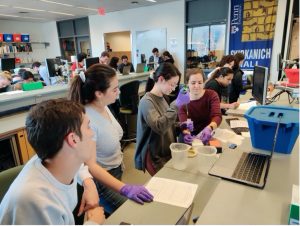
Penn Bioengineering’s BioMakerSpace in action (photo taken pre-pandemic)

Writing for the Penn Health-Tech blog, Hannah Spector profiled the George H. Stephenson Foundation Educational Laboratory and Bio-MakerSpace, the primary teaching lab for the Department of Bioengineering at Penn Engineering. This interdisciplinary Bio-MakerSpace (aka BioMakerSpace) is open to the entire Penn community for independent research and has become a hub for student startups in recent years:

One example is Strella Biotechnology, founded in 2019 by Katherine Sizov (Biology 2019 & President’s Innovation Prize winner). Strella is developing sensors with the ability to reduce the amount of food waste due to going bad in storage. “Having a Bio-MakerSpace that gives you the functionalities of both a wet lab and a traditional electronics lab is extremely helpful in developing novel technologies” says Sizov on the BE Labs Youtube channel.

The Bio-MakerSpace provides students of all academic backgrounds the resources to turn their ideas into realities, including highly knowledgeable lab staff. Seth Fein (BSE ’20, MSE ’21) has worked at the lab since Fall 2020. “Because bioengineering spans many fields, we encourage interdisciplinary work. Students from Mechanical, Electrical, and Chemical Engineering have all found valuable resources in the lab,” says Fein.

The article also discusses the many resources the BioMakerSpace provides to Penn students and their efforts to keep the lab functional, safe, and open for research and education during the current semester.

Penn Health-Tech is an interdisciplinary center launched in 2017 to advance medical device innovation across the Perelman School of Medicine and the School of Engineering and Applied Sciences by forging collaborative connections among Penn researchers and providing seed funding to incubate novel ideas to advance health care.

Continue reading “The Bio-MakerSpace — Fostering Learning and Innovation Across Many Disciplines” at the Penn Health-Tech blog.

Read more BE blog posts featuring the BioMakerSpace.

2020 Awards Season for Bioengineering Students

Each spring, the School of Engineering and Applied Science at the University of Pennsylvania gives out awards of special recognition to honor exceptional work by undergraduate and graduate students. The Department of Bioengineering is proud to announce the following awards given to students in the Class of 2020.

Bioengineering Professor and Chair Ravi Radhakrishnan says, “Congratulations to all the winners! I am so incredibly proud of your accomplishments and I thank you for enriching the Bioengineering environment with your invaluable contributions.” Keep reading below for a list of 2020 award recipients.

UNDERGRADUATE AWARDS:

Katharine Cocherl (BAS 2020), who completed a Bachelor of Applied Science degree in Bioengineering along with a second major in Cinema and Media Studies, was awarded the Ben and Bertha Gomberg Kirsch Prize. This competitive award is decided by the SEAS faculty from among the Engineering undergraduate body and distinguishes a member of the B.A.S. senior class who “in applying the flexibility of the program, has created a personal academic experience involving the most creative use of the resources of the University.”

The Hugo Otto Wolf Memorial Prize, awarded to one or more members of each department’s senior class, distinguishes students who meet with great approval of the professors at large through “thoroughness and originality” in their work. This year, BE chose to share the award between Jacqueline Peng (BSE 2020) and Vera Lee (BSE 2020). In addition to their majors, Jacqueline also minored in Computer Science and is pursuing a Master’s degree in Data Science and Vera minored in the Engineering Entrepreneurship program and is pursuing a Master’s degree in Robotics.

The Herman P. Schwan Award is decided by the Bioengineering Department and honors a graduating senior who demonstrates the “highest standards of scholarship and academic achievement.” The 2020 recipient of the Schwan Award is Alexander Silva (BSE 2020) who is also graduating with a minor in Economics.

Every year, several BE students are recognized with Exceptional Service Awards for their outstanding service to the University and their larger communities. Our winners this year are Arielle Stern (BSE 2020 with a Math minor), Lauren McLeod (BSE 2020), and Evan Paregol (BSE 2020 with an Entrepreneurship minor). Arielle and Evan are also currently in the Accelerated Master’s program, in Data Science and Bioengineering, respectively.

The Student Leadership Award is given annually to a student in Bioengineering who has demonstrated, through a combination of academic performance, service, leadership, and personal qualities, that he or she will be a credit to the Department, the School, and the University. The 2020 recipient of this award is Katherine Simms (BSE 2020 with a minor in Chemistry).

BE also distinguishes a single lab group (four students) with the Albert Giandomenico Award which reflects their “teamwork, leadership, creativity, and knowledge applied to discovery-based learning in the laboratory.” This year’s group consists of Alisa Bhakta (2020 dual degree BSE in Bioengineering and BS in Economics from Wharton), Gabriel Desantis (BSE 2020 with a minor in Math), Lauren McLeod (BSE 2020), and Caroline Raquel (2020 dual degree BSE in Bioengineering and BS in Economics from Wharton).

Of this year’s Bioengineering Senior Design teams, three groups were chosen for special recognition:

  • RelieVR with Nicole Chiou (BSE 2020 with a minor in Computer Science), Gabe Desantis (BSE 2020 with a minor in Math), Ben Habermeyer BSE 2020 with a minor in Computer Science), and Vera Lee (BSE 2020 with an Engineering Entrepreneurship minor). RelieVR also won second place at the 2020 Johns Hopkins Healthcare Design Competition and took home the Berkman Prize this past fall semester.
  • Relila with Alisa Bhakta (dual degree BSE and BS 2020), Alexander Connor (BSE 2020), Lauren McLeod (BSE 2020), Alexa Murray (BSE 2020 in Systems Science and Engineering), and Caroline Raquel (dual degree BSE and BS 2020). Relila also won second place at the annual M&T Program Lab Integration Awards summit.
  • SchistoSpot with Alec Bayliff (dual degree BAS and BS in Economics 2020), Bram Bruno (BAS 2020), Justin Swirbul (BSE 2020 in Computer Science), and Vishal Tien (BSE 2020). SchistoSpot also won the Pioneer Award at the annual Rothberg Catalyzer Makerthon.

Research for these projects was conducted in the George H. Stephenson Foundation Education Laboratory & Bio-Maker Space. The abstracts and presentation videos for each of the 2020 Senior Design Competition winners can be viewed on the BE Labs website.

Additionally, two graduating BAS seniors were awarded prizes for Best Senior Thesis:

  • Katharine Cocherl (BAS 2020 in Bioengineering and Cinema and Media Studies) for her paper “Bioethical Assessments of Film Portrayals of the Opioid Epidemic and Its Relationship with Public Discourse and Policy from the 1990s to Present.” “Insightful, original, and wide-reaching, her study of films related to the opioid epidemic in the U.S. the past 25 years was one of the best senior theses I have advised at Penn the past 15 years, ” says Katharine’s advisor Lance Wahlert, Assistant Professor of Medical Ethics & Health Policy, Program Director of the Master of Bioethics
  • Gayatri Maria Schur (BAS 2020 with a minor in Music) for her paper titled “In Vivo Assessment of OXPHOS Capacity Using 3T CrCEST MRI in Adults and Children with Friedrich’s Ataxia.” Her advisor, Shana McCormack, Assistant Professor of Pediatrics in the Perelman School of Medicine, says that Gayatri’s “work has required that she communicate with collaborators across a variety of disciplines, and has also included interaction with the community of patients we study, and she has excelled here.”

GRADUATE AWARDS:

Master’s student Kayla Prezelski was awarded an Outstanding Teaching Award for students. Kayla served as a TA for the Department of Bioengineering’s two-semester Senior Design courses (BE 495/496).

The following Master’s students were awarded recognition for their Outstanding Research:

  • Linghan Mei – advisor Andrew Tsourkas, Ph.D., Professor of Bioengineering
  • Ayush Aditya Pal – advisor Lukasz Bugaj, Ph.D., Assistant Professor of Bioengineering
  • Robert Pierson – Independent Study advisor Brian Litt, M.D., Professor of Neurology, Neurosurgery, and Bioengineering, and Thesis advisors Insup Lee, Ph.D., Cecilia Fitler Moore of Computer and Information Science and Electrical and Systems Engineering, and James Weimer, Ph.D. Research Assistant Professor of Computer and Information Science
  • Tianjia Zhu – advisor Hao Huang, Ph.D., Research Associate Professor of Radiology in the Perelman School of Medicine

And finally, The Solomon R. Pollack Award for Excellence in Graduate Bioengineering is given annually to the most deserving Bioengineering graduate student who has successfully completed research that is original and recognized as being at the forefront of its field. This year, that award goes to Jonathan Beagan, Ph.D. who recently defended his thesis. Jon conducted his research in the 3D Epigenomics and Systems Neurobiology Lab overseen by Jennifer Phillips-Cremins, Ph.D., Associate Professor of Bioengineering. Research related to Jon’s award-winning doctoral thesis was recently published in the journal Nature Neuroscience. In addition to this prestigious award, Jon was also named a National Science Foundation Graduate Research Fellow during his time at Penn. Jon’s collaborations with Dr. Cremins have been covered several times on the BE blog. “Jon is an excellent researcher — simultaneously rigorous and creative,” says Dr. Cremins. “He has been a force in the lab — reading the literature voraciously, teaching other students, and executing/designing experiments meticulously. Beyond his natural talent, it is Jon’s personal qualities that make him stand out. He is a true leader, a team player, and one of the rare people that raises the bar for everyone around him.”

A full list of SEAS award descriptions and recipients can be found here.

Congratulations once again to the award winners and to all graduating students on an outstanding year of scholarship and service!

Bioengineering Round-Up (October 2019)

by Sophie Burkholder

Innovations in Advancing a Cure for Diabetes

The blue circle is the global symbol for diabetes. Wikimedia Commons.

Diabetes is one of the more common diseases among Americans today, with the American Diabetes Association estimating that approximately 9.5 percent of the population battles the condition today. Though symptoms and causes may vary across types and patients, diabetes generally results from the body’s inability to produce enough insulin to keep blood sugar levels in check. A new experimental treatment from the lab of Sha Jin, Ph.D., a biomedical engineering professor at Binghamton University, aims to use about $1.2 million in recent federal grants to develop a method for pancreatic islet cell transplantation, as those are the cells responsible for producing insulin.

But the catch to this new approach is that relying on healthy donors of these islet cells won’t easily meet the vast need for them in diabetic patients. Sha Jin wants to use her grants to consider the molecular mechanisms that can lead pluripotent stem cells to become islet-like organoids. Because pluripotent stem cells have the capability to evolve into nearly any kind of cell in the human body, the key to Jin’s research is learning how to control their mechanisms and signaling pathways so that they only become islet cells. Jin also wants to improve the eventual culture of these islet cells into three-dimensional scaffolds by finding ways of circulating appropriate levels of oxygen to all parts of the scaffold, particularly those at the center, which are notoriously difficult to accommodate. If successful in her tissue engineering efforts, Jin will not only be able to help diabetic patients, but also open the door to new methods of evolving pluripotent stem cells into mini-organ models for clinical testing of other diseases as well.

A Treatment to Help Others See Better

Permanently crossed eyes, a medical condition called strabismus, affects almost 18 million people in the United States, and is particularly common among children. For a person with strabismus, the eyes don’t line up to look at the same place at the same time, which can cause blurriness, double vision, and eye strain, among other symptoms. Associate professor of bioengineering at George Mason University, Qi Wei, Ph.D., hopes to use almost $2 million in recent funding from the National Institute of Health to treat and diagnose strabismus with a data-driven computer model of the condition. Currently, the most common method of treating strabismus is through surgery on one of the extraocular muscles that contribute to it, but Wei wants her model to eventually offer a noninvasive approach. Using data from patient MRIs, current surgical procedures, and the outcomes of those procedures, Wei hopes to advance and innovate knowledge on treating strabismus.

A Newly Analyzed Brain Mechanism Could be the Key to Stopping Seizures

Among neurological disorders, epilepsy is one of the most common. An umbrella term for a lot of different seizure-inducing conditions, many versions of epilepsy can be treated pharmaceutically. Some, however, are resistant to the drugs used for treatment, and require surgical intervention. Bin He, Ph. D., the Head of the Department of Biomedical Engineering at Carnegie Mellon University, recently published a paper in collaboration with researchers at Mayo Clinic that describes the way that seizures originating at a single point in the brain can be regulated by what he calls “push-pull” dynamics within the brain. This means that the propagation of a seizure through the brain relies on the impact of surrounding tissue. The “pull” he refers to is of the surrounding tissue towards the seizure onset zone, while the “push” is what propagates from the seizure onset zone. Thus, the strength of the “pull” largely dictates whether or not a seizure will spread. He and his lab looked at different speeds of brain rhythms to perform analysis of functional networks for each rhythm band. They found that this “push-pull” mechanism dictated the propagation of seizures in the brain, and suggest future pathways of treatment options for epilepsy focused on this mechanism.

Hyperspectral Imaging Might Provide New Ways of Finding Cancer

A new method of imaging called hyperspectral imaging could help improve the prediction of cancerous cells in tissue specimens. A recent study from a University of Texas Dallas team of researchers led by professor of bioengineering Baowei Fei, Ph.D., found that a combination of hyperspectral imaging and artificial intelligence led to an 80% to 90% level of accuracy in identifying the presence of cancer cells in a sample of 293 tissue specimens from 102 patients. With a $1.6 million grant from the Cancer Prevention and Research Institute of Texas, Fei wants to develop a smart surgical microscope that will help surgeons better detect cancer during surgery.

Fei’s use of hyperspectral imaging allows him to see the unique cellular reflections and absorptions of light across the electromagnetic spectrum, giving each cell its own specific marker and mode of identification. When paired with artificial intelligence algorithms, the microscope Fei has in mind can be trained to specifically recognize cancerous cells based on their hyperspectral imaging patterns. If successful, Fei’s innovations will speed the process of diagnosis, and potentially improve cancer treatments.

People and Places

A group of Penn engineering seniors won the Pioneer Award at the Rothberg Catalyzer Makerthon led be Penn Health-Tech that took place from October 19-20, 2019. SchistoSpot is a senior design project created by students Vishal Tien (BE ‘20), Justin Swirbul (CIS ‘20), Alec Bayliff (BE ‘20), and Bram Bruno (CIS ‘20) in which the group will design a low-cost microscopy dianostic tool that uses computer vision capabilities to automate the diagnosis of schistosomiasis, which is a common parasitic disease. Read about all the winners here.

Virginia Tech University will launch a new Cancer Research Initiative with the hope of creating an intellectual community across engineers, veterinarians, biomedical researchers, and other relevant scientists. The initiative will focus not only on building better connections throughout departments at the university, but also in working with local hospitals like the Carilion Clinic and the Children’s National Hospital in Washington, D.C. Through these new connections, people from all different areas of science and engineering and come together to share ideas.

Associate Professor of Penn Bioengineering Dani Bassett, Ph.D., recently sat down with the Penn Integrates Knowledge University Professor Duncan Watts, Ph.D., for an interview published in Penn Engineering. Bassett discusses the origins of network science, her research in small-world brain networks, academic teamwork, and the pedagogy of science and engineering. You can read the full interview here.

An all-female group of researchers from Northern Illinois University developed a device for use by occupational therapists that can capture three-dimensional images of a patient’s hand, helping to more accurately measure the hand or wrist’s range of motion. The group presented the abstract for their design at this year’s meeting of the Biomedical Engineering Society here in Philadelphia, where Penn students and researchers presented as well.

Students’ Innovative Orthotic Device Wins Rothberg Catalyzer

NB: Penn Bioengineering would like to congratulate one of its current Senior Design teams (Alec Bayliff, Bram Bruno, Justin Swirbul, and Vishal Then) which took home the $500 Pioneer Award at this year’s Rothberg Catalyzer competition this past weekend! Keep reading for more information on the competition, awards, and winners.

Penn Health-Tech’s Rothberg Catalyzer is a two-day makerthon that challenges interdisciplinary student teams to prototype and pitch medical devices that aim to address an unmet clinical need.

The Catalyzer’s third competition was held last weekend and was won by MAR Designs, a team of Mechanical Engineering and Applied Mechanics graduate students: Rebecca Li, Ariella Mansfield and Michael Sobrepera.

MAR Designs took home the top prize of $10,000 for their project, an orthotic device that children with cerebral palsy can more comfortably wear as they sleep.

According to the team’s presentation, existing wrist orthoses “improve function and treat/prevent spasticity. However, patients report that these devices are uncomfortable which leads to lack of compliance and may also prevent patient’s eligibility for surgeries.” MAR Designs’ device initially allows full range of motion, but gradually straightens the wrist as the child is falling asleep.

In second place was Splash Throne. Team members Greg Chen, Nik Evitt, Jake Crawford and Meghan Lockwood proposed a toilet safety frame intended for elderly users. Embedded sensors track basic health information, like weight and heart-rate, as part of a preventative health routine.

Integrated Product Design students Jonah Arheim, Laura Ceccacci, Julia Lin and Alex Wan took third place with ONESCOPE, an untethered, hands-free laproscope designed to make minimally-invasive surgeries faster and safer.

Finally, SchistoSpot took home the Catalyzer’s Pioneer Award. Bioengineering and Computer and Information Science seniors Alec Bayliff, Bram Bruno, Justin Swirbul and Vishal Then designed a low-cost microscopy system that can aid in the diagnosis of the parasitic disease schistosomiasis by detecting eggs in urine samples, eliminating the need for a hospital visit.

The event was made possible by a three-year donation by scientist and entrepreneur Jonathan Rothberg, with the intent of inspiring the next generation of healthcare innovators.

Originally posted on the Penn Engineering Medium blog.

Week in BioE (May 31, 2019)

by Sophie Burkholder

Vector Flow Imaging Helps Visualize Blood Flow in Pediatric Hearts

A group of biomedical engineers at the University of Arkansas used a new ultrasound-based imaging technique called vector flow imaging to help improve the diagnosis of congenital heart disease in pediatric patients. The study, led by associate professor of biomedical engineering Morten Jensen, Ph.D., collaborated with cardiologists at the local Children’s Hospital in Little Rock to produce images of the heart in infants to help potentially diagnose congenital heart defects. Though the use of vector flow imaging has yet to be developed for adult patients, this type of imaging could possibly provide more detail about the direction of blood flow through the heart than traditional techniques like echocardiography do. In the future, the use of both techniques could provide information about both the causes and larger effects of heart defects in patients.

Using Stem Cells to Improve Fertility in Leukemia Survivors

One of the more common side effects of leukemia treatment in female patients is infertility, but researchers at the University of Michigan want to change that. Led by associate professor of biomedical engineering Ariella Shikanov, Ph.D., researchers in her lab found ways of increasing ovarian follicle productivity in mice, which directly relates to the development of mature eggs. The project involves the use of adipose-derived stem cells, that can be found in human fat tissue, to surround the follicles in an ovary-like, three-dimensional scaffold.  Because the radiation treatments for leukemia and some other cancers are harmful to follicles, increasing their survival rate with this stem cell method could reduce the rate of infertility in patients undergoing these treatments. Furthermore, this new approach is innovative in its use of a three-dimensional scaffold as opposed to a two-dimensional one, as it stimulates follicle growth in all directions and thus helps to increase the follicle survival rate.

Penn Engineers Look at How Stretching & Alignment of Collagen Fibers Help Cancer Cells Spread

Cancer has such a massive impact on people’s lives that it might be easy to forget that the disease originates at the cellular level. To spread and cause significant damage, individual cancer cells must navigate the fibrous extracellular environment that cells live in, an environment that Penn Engineer Vivek Shenoy has been investigating for years.

Shenoy is the Eduardo D. Glandt President’s Distinguished Professor with appointments in Materials Science and Engineering, Mechanical Engineering and Applied Mechanics, and Bioengineering. He is also the Director of the Center for Engineering MechanoBiology (CEMB), one of the NSF’s twelve Science and Technology Centers.

Shenoy’s most recent study on cancer’s mechanical environment was led by a postdoctoral researcher in his lab, Ehsan Ban. Paul Janmey, professor in Physiology and Bioengineering, and colleagues at Stanford University also contributed to the study. Shenoy also received the Heilmeier Award this March and delivered the Heilmeier Award Lecture in April.

Read the rest of this story on Penn Engineering’s Medium Blog.

Controlled Electrical Stimulation Can Prevent Joint Replacement Infections

Joint replacements are one of the most common kinds of surgery today, but they still require intense post-operative therapy and have a risk of infection from the replacement implant. These infections are usually due to the inflammatory response that the body has to any foreign object, and can become serious and life-threatening if left untreated. Researchers at the University of Buffalo Jacobs School of Medicine and Biomedical Sciences hope to offer a solution to preventing infections through the use of controlled electrical stimulation. Led by Mark Ehrensberger, Ph.D., Kenneth A. Krackow, M.D., and Anthony A. Campagnari, Ph.D., the treatment system uses the electrical signal to create an antibacterial environment at the interface of the body and the implant. While the signal does not prevent infections completely, these antibacterial properties will prevent infections from worsening to a more serious level. Patented as the Biofilm Disruption Device TM, the final product uses two electrode skin patches and a minimally invasive probe that delivers the electrical signal directly to the joint-body interface. The researchers behind the design hope that it can help create a more standard way of effectively treating joint replacement infections.

People and Places

TBx: Gabriel Koo, Ethan Zhao, Daphne Cheung, and Shelly Teng

For their senior design project, four bioengineering seniors Gabriel Koo, Ethan Zhao, Daphne Cheung, and Shelly Teng created a low-cost tuberculosis diagnostic that they called TBx. Using their knowledge of the photoacoustic effect of certain dyes, the platform the group created can detect the presence of lipoarabinomannan in patient urine. The four seniors presented TBx at the Rice360 Design Competition in Houston, Texas this spring, which annually features student-designed low-cost global health technologies.

Graduating BE Seniors Present Their Senior Design Projects

At the end of April, the graduating seniors in the Penn Department of Bioengineering‘s B.S.E. (Bachelor of Science in Engineering) program presented their Senior Design projects. Developed over the course of their final two semesters in the undergraduate program, these projects are developed in teams of three or four as the students are guided through choosing and understanding an impactful biomedical problem, defining the characteristics of a successful design solution to eliminate or mitigate a problem or fulfill a need, identifying constraints, and creatively developing potential design solutions. Over the course of two days, the students then present their projects to faculty and students from across the Penn Engineering community.

Additionally, three groups (BreatheSmart, RIPT, and Proscopy) were selected to represent BE in the 2019 Penn Engineering Senior Design Project Competition.

Congrats to all of our graduating students on their innovative projects. Check out some photos from the 2019 BE Senior Design presentations and read the full list of this year’s abstracts below.

Group A: BreatheSmart

Caroline Atkinson, Sarah Cai, Rebecca Kellner, Harrison Troché

In the United States, more than 51 million people have been diagnosed with either asthma or chronic obstructive pulmonary disorder (COPD) as of 2016. These conditions result in swelling of the airways, making it difficult to breathe, but the symptoms are commonly managed through the use of a pressurized metered dose inhaler (pMDI), which dispenses aerosolized medication to the lungs, and are currently used by 78% of asthma patients in the US. However, a recent study showed up to 90% of pMDI users incorrectly use their inhalers, reducing the efficacy of medication . Our solution guides patients through the proper technique for using inhalers while providing real-time feedback so patients can correct their technique. This was accomplished by tracking flow rate data converted from a pressure sensor and a smartphone app that provides real-time visual feedback to the user. We were successful in our four design goals as the final device is: (1) lightweight and compact (78 grams with inhaler and 2.5”x1.5”x3.5”), (2) accurate in flow rate measurements (±8.7% error), (3) easy-to-use, with 95% of users surveyed able to use the app successfully without assistance (n=20), and (4) low cost, with the total cost being $76. To improve the design, we will improve accuracy and compactness, and reduce cost. Some ways this could be achieved are through the use of a smaller, custom microcontroller, and a more sensitive pressure sensor. Next steps would include a clinical trial to demonstrate the effectiveness of our device.

Group B: RIPT

Toren Arginteanu, Anna Mujica, Justin Mills, Kayla Prezelski

The tourniquet is the gold standard for treating traumatic extremity bleeding in pre-hospital emergency scenarios. Existing pneumatic tourniquets are safer than non-pneumatic alternatives, minimizing nerve damage and loss of limb function. However, current emergency pneumatic tourniquets are costly and delicate and prone to leaking, punture, and abrasion. Therefore, there is need for an emergency pneumatic tourniquet that is low-cost, highly durable, and easily applied by laypeople. Our design, the Rapidly Inflating Pneumatic Tourniquet (RIPT), consists of an internal bladder made of tough rubber, a durable outer covering made of ballistic nylon, and a pneumatic inflation mechanism involving an inflator valve, pressurized CO 2 cartridge, and pressure relief and regulator valves that maintain a safe and sufficient internal pressure of 250 mmHg. RIPT is tightened around the injured limb and secured with a spring-loaded, automatically locking PLA and aluminum clasp. RIPT’s average application time was 20 ± 0.4 s, which is significantly shorter than that of the Combat Application Tourniquet (CAT): 32 ± 2.9 s. RIPT’s internal bladder pressure reached 250 ± 4.8 mmHg. RIPT packs up to 87.5 in 3 and weighs 18.8 oz. RIPT has comparable circumferential pressure distribution to CAT and superior axial pressure gradient. RIPT’s raw material cost is $65.62. In the future, the clasp mechanism will be modified to improve ease-of-use and minimize size and weight. RIPT will be tested with trauma surgeons at Penn Presbyterian Hospital, and user feedback will indicate ease-of-use and pain of application relative to the CAT.

Group C: Proscopy

Abigail Anmuth, John Forde, Sarah Raizen, Rohit Shinde

Robotically-assisted surgery is a rapidly evolving technology that is expanding the capabilities of surgeons and improving patient outcomes in fields ranging from cardiothoracic surgery to urology. However, as this technology becomes more widely used, there are distinct limitations that impact the safety of surgical maneuvers, largely because of the lack of haptic feedback in the robotic arms of these surgical systems. In the case of prostatectomies, this can lead to the unintentional penetration of the rectal wall and cause pervasive infection, a condition known as rectal injury (RI). Proscopy is a novel real-time proximity sensor that monitors the distance between robotic surgical tools and a patient’s rectal wall to reduce the incidence of RI. This two-part preventative solution consists of a Hall Effect sensor-embedded brace at the tip of the surgical arm and a magnetized flexible insert. The Hall Effect sensors convert magnetic field strength to distance from the rectal wall. In lieu of haptic feedback, Proscopy provides visual feedback within the robot control console as the surgical robot arm nears the rectal wall, displaying information that cannot be perceived from the laparoscopic camera itself. Proscopy gives surgeons the ability to detect the rectal wall with a precision of 0.1±.05mm, rendering it a highly effective and streamlined mechanism for RI prevention during robotically-assisted prostatectomies.

Group D: CerviAid

Dana Abulez, Dayo Adetu, Lamis Elsawah, Yueqi Ren

Cervical insufficiency is premature dilation of the cervix in pregnant women without active labor, and if not addressed, can lead to the loss of pregnancy in the second trimester. This condition results in a protrusion of the amniotic sac out of the uterus and into the cervix, which must be pushed back to extend the pregnancy. In the cervical cerclage procedure, the only treatment for this condition, the cervix is sutured shut to prolong pregnancy. If the cerclage is completed after 14 weeks of pregnancy or if the cervix is more than 3 cm dilated, the procedure is termed an emergency cerclage, which has a higher rate of pregnancy loss due to amniotic sac puncture than regular cerclage procedures. Cerclage procedures are dependent on surgeon experience and utilize no standard method to push back the amniotic sac, which makes this procedure even more high risk. To combat this issue, CerviAid is a device that aims to increase the success rate of emergency cerclages, for which no standardized method exists. The device pushes up the amniotic sac gently with a silicone head and can accommodate a variety of cervix diameters found in patients. The head of the device is controlled by a rack and pinion with a locking mechanism to secure the device and firmly support the sac while the surgeon places the suture. Evaluations of CerviAid with a realistic model of the cervix and amniotic sac produced a 96.15% success rate, in which no membranes were ruptured after pushing through a model cervix. Future steps include further stakeholder evaluations and decreasing manufacturing costs.

Group E: ArcAlert

Kelsey Friberg, Weiwei Meng, Vanessa Moody, Malika Shukurova

Risk of surgical fire increases with use of surgical energy like electrocautery, which presents a source of ignition in an operating room setting with elevated percent oxygen saturation in the air and abundant flammable materials. To address this problem, the FDA recommends human factors strategies, such as heightening awareness, and training on use of surgical energy, which is available yet not standardized across the field. ArcAlert offers a real-time risk management solution for data-driven prevention of surgical fires during use of da Vinci robot electrocautery. The system includes an oxygen sensor device that detects percent oxygen saturation at the surgical field, a machine vision algorithm that detects electrical arcing, and a user-friendly web application. ArcAlert consolidates and assesses risk factor information so that if the oxygen sensor device’s reading exceeds a dangerous threshold or the machine vision algorithm detects arcing, an alert appears on the web application describing the risk, which facilitates improvements to equipment usage by operating room personnel. The oxygen sensor device acquires reliable readings over the timespan of a procedure with an accuracy within +/- 1% for 97% of readings. The machine vision algorithm detects approximately 80% of arcing incidences with only 20% falsely labeled, achieves spatial localization on the order of under 1 centimeter, and executes full pipeline implementation in under half a second. The current design is indicated for transoral surgeries, so future generations will diversify the types of procedures with which the system is compatible, in addition to going fully wireless.

Group F: Grip Glove

Kathryn Khaw, Matthew Rosenwasser, Vidula Kopli

Soft robotics offers many advantages over traditional robotics as an assistive and rehabilitative technology due to its cost-effectiveness and ability to mimic physiological movements. Because of the importance of grip on a patient’s quality of life, we leveraged soft robotic technology to create a flexible, exoskeleton glove for both therapy and assisting daily living for patients with hand impairments. Our device targets muscular dystrophy patients because these patients have a limited selection of devices for their needs. Our design consists of soft robotic actuators attached to a glove that mimic finger movement by bending upon inflation and surface electromyography (SEMG) sensors on the arm to predict the user’s intention to grip or release. Our device effectively recapitulates the forces generated during the grip of a healthy individual using three soft robotic actuators, with each actuator generating 2.83 ± 0.44 N (n = 6). The actuation frequency is 0.2 Hz (12 grips/minute) due to delays in deflation. The EMG sensors, MyoWare and OYMotion Analog EMG Sensor, were not sensitive enough to pick up graded changes in muscle activity, meaning we could only replicate an on-off response through a thresholded spike detection of processed EMG signals in two muscle groups that corresponded to grip and release. Going forward, we will use more sensitive EMG sensors, such as the Biosignalsplux Electromyography Muscle Sensor, increase actuation frequency by actively deflating actuators and include a pressure – solenoid feedback system to have controllable grips.

Group G: SpotOn

Julian Mark, Chase Rapine, Jared Rifkin

In this paper, we discuss SpotOn: a novel bioengineering solution to improving anterior cruciate ligament (ACL) tear recovery. ACL tears are very prevalent injuries and typically take 6-9 months to heal [1]. Physical therapy during recovery is costly and time-intensive, and current knee braces for patients do not provide active support to protect the patient. With SpotOn’s dual-faceted smart mirror and dynamic knee brace technology, patients can begin strengthening the ligament and joint sooner after surgery with less risk of re-injury. When the patient exercises with SpotOn, the smart mirror provides feedback on the patient’s exercise form. If the mirror detects an error, it notifies the user and sends a signal to activate the knee brace and straighten the user’s knee. Key specifications for success include latency of knee brace activation, maximum supported load by the brace, and total cost. Latency and cost met specification goals with a delay less than 0.1s (target under 3s) and total cost of $343.25 (target under $600), but maximum torque output of 18.26ft-lbs was short of the 101.25ft-lbs load goal. Future directions for our design include tracking additional metrics besides squat form, increasing maximum torque output of the knee brace, and lastly adding an on-board battery to the brace to improve portability. Our product was not tested clinically, so the next step to implement our solution would be to reach out to clinicians who are treating injured patients and move forward with gathering clinical data.

Group H: TBx

Daphne Cheung, Gabriel Koo, Shelly Teng, Ethan Zhao

Caused by bacterial strain Mycobacterium tuberculosis, tuberculosis (TB) remains one of the world’s deadliest diseases, with over 10 million new cases and 1.3 million deaths in 2017. Currently, the gold standard for TB diagnosis is to sequence a patient sputum sample using a PCR machine called the GeneXpert. Although this method demonstrates high sensitivity and specificity, it is not ideal in low-income developing countries because it is too expensive and inaccessible, priced at $32,000 for equipment, and $17/test. Thus, diagnosis is most widely determined through smear microscopy. However, this method shows low sensitivity and specificity at only 60% and 81%, respectively. Our solution is TBx, a urine-based diagnostic protocol that offers greater diagnostic power than smear microscopy, while maintaining affordability at only a fraction of the price of the GeneXpert. TBx detects the presence of lipoarabinomannan (LAM) in urine, a glycolipid that is specific to active TB cases. Under TBx protocol, LAM is tagged using photoacoustic dye (IR Dye 800CW) and immunoprecipitation beads via anti-LAM antibodies. Samples are then spun down using a centrifuge to wash out excess dye, concentrated by resuspension in only 5mL 1x PBS, and imaged in a 96-well plate. The photoacoustic dye thermally expands and produces photoacoustic signal in response to photoexcitation at λ = 800nm. Compared to smear microscopy, TBx has higher sensitivity (67%, n = 31) and specificity (92%, n = 31). TBx also only costs $1,500 in equipment, and around $12/test. With further refinements to the TBx protocol and point-of-care packaging, TBx shows promise in developing countries to improve the TB diagnosis landscape, especially with its combination of increased sensitivity and specificity, as well as affordability.

Group I: Sensei “The Cast Master”

Carolina Ferrari, Kristen Ho, Blake Thomas, Alfredo Tovar

Acute compartment syndrome (ACS) occurs in 26,500 people in the US each year and is a result of capillary blood flow becoming compromised when tissue pressure exceeds 30 mmHg. The consequences of ACS are extremely severe if it is not immediately diagnosed. ACS can result in permanent muscle damage, nerve damage and/or amputation, and 70% of cases can be traced back to fractures. The current diagnostic method requires invasive pressure measurements if a patient’s primary symptom assessment is inconclusive. Thus, we have designed SENSEI, a non-invasive device that constantly monitors pressure and can diagnose ACS underneath a cast post-fracture. The device interacts with an Android application via bluetooth to let the user know if they are at risk in real time. SENSEI currently measures compartmental pressure of the forearm within a range of 20-40 mmHg with 91% accuracy. In the future, we plan on improving our pressure sensors so that SENSEI can diagnose ACS with 100% confidence. Other potential additions to SENSEI include designing a sleeve for the lower leg and developing an iOS application to capture more market share.

Group J: UrineLuck: Artificial Urinary Sphincter

Jason Grosz, Richard Adamovich-Zeitlin, Teddy Wang, Sally Pennacchi

Urinary incontinence (UI) is a debilitating ailment that impacts the lives of millions of men. Caused by urinary sphincter damage, prostate issues, or overactive bladder muscles, UI can render men homebound and ruin their social and professional lives. The current gold standard intervention for severe UI is the AMS 800, which consists of a pressure regulated cuff surrounding the urethra and a manual pump in the scrotum. The AMS 800 requires an invasive surgery for implantation and is extremely expensive – around $37,000 for the device, surgery, and hospital stay. It also suffers from a high failure rate, around 33% within three years, due to infections, mechanical failures, and tissue atrophy from the cuff blocking perfusion. In this paper, we fabricate and validate a silicone endourethral valve to treat UI in a less expensive, less invasive, and user-friendly way. These valves are inserted into the penile urethra and mimic the mechanics of bite valves commonly found in water bottles, whereby force applied perpendicularly to a slit enables flow. They are cheap, around $0.08 per device in material costs, do not occlude blood perfusion, and can be inserted at home or in an outpatient clinic with a catheter. Through validation in a PDMS model that recapitulates penile mechanics and geometry, we demonstrated that these valves enable strong flow, 4.99 mL/s, minimal leakage, 0.43 drops, and resistance to cyclic stress. With further cytotoxicity and biofilm testing and insertion protocol development, we hope that this design can improve quality-of-life and prognoses for incontinent men.

Group K: Automated Pathology

Olivia Lang, Joseph Maggiore, Prithvi Pendekanti, Olivia Teter

Clinical and laboratory staining protocols are typically an inefficient use of money, resources and manhours. The highly repetitive and standardized nature of this work poses an opportunity for automation. An automated system would allow a physician or clinician the ability to dedicate their time elsewhere, use less staining fluid, and potentially save on thousands of dollars when compared to similar alternatives. Hemauto is an automated diagnostic device as a proof of concept for a lowcost instrument that combines staining, image acquisition, and image analysis capabilities. Hemauto’s first use case is the detection of Malaria. Malaria, despite being highly treatable, is currently an epidemic in African lowresource regions, disproportionately affecting children. The high incidence rate can be attributed to a lack of standardized and effective earlydetection methods. Providing a lowcost, highthroughput device capable of giving a quantitative diagnosis with limited userinput is critical for these regions because current detection methods are either too hightech, not accurate enough, or are too expensive to be administered frequently. Hemauto’s solution synchronizes the actions of motors and computer technology to deliver a diagnosis. It uses two axes of motion controlled by independent stepper motors to follow the standard Giemsa staining protocol. The stained slide is then automatically focused and imaged in front of a 100x objective lens, where the captured images are analyzed to provide a quantified diagnosis. This proof of concept offers incredible promise for future automated pathology devices across many disease areas.

Group L: Preventear

Matt Riley, Jasmine Wang, Mary Zhuo Ke

Adolescent female athletes are at a high risk of Anterior Cruciate Ligament (ACL) tears as a result of improper form and training techniques. ACL tear prevention training programs are designed to reduce one’s future risk of a tear, but these programs are expensive and not available everywhere. PREVENTEAR is our proposed solution. PREVENTEAR is a self-guided jump training system involving a wearable sock and companion mobile app that like a personal trainer, provides instantaneous feedback on your technique, but is inexpensive and portable. The three main parameters important to jump training are knee angle, knee-to-knee distance, and foot landing technique. The ideal form is to have a knee angle between 30 and 90 degrees with respect to the vertical, have a knee-to-knee distance of greater than 60% with respect to hip width, and land on the ball of the foot before the heel. PREVENTEAR uses bluetooth to communicate with the sensors woven into the sock to a phone app and aims to give feedback on the quality of an athlete’s jump as accurately as ACL tear prevention training programs like Sportsmetrics. The app-reported feedback was compared to true values measured by an observer, phone application, and measuring tools to evaluate performance. PREVENTEAR overall provides a means for ACL tear prevention training by being portable and low cost in comparison to Sportsmetrics, and flexible for adolescent female athletes of varying sizes. PREVENTEAR hopes to further improve accuracy and expand to other forms of training important in reducing risk of ACL injury such as strength, flexibility, and plyometric training in the future.

Group M: Autonomous Home Urinalysis

Saumeel Desai, Anand Prabhu, Eshwar Inapuri

Urinalysis is a crucial diagnostic component of managing heart failure, kidney failure, pre-eclampsia and several other medical conditions. Urinalysis currently requires patient willingness, effort, time and money as patients must travel to a lab, wait for results and rely on caregiver support in order to comply with required follow-ups. Current at-home alternatives, such as dipsticks, lack proper controls and cannot reliably be used by physicians to guide therapy. Moreover, patients are opposed to handling urine, leading to low adherence rates. The goal of this design project was to create an autonomous home urinalysis device that greatly reduces the barriers to diagnostic data collection, enabling detection of deterioration and guidance of therapy with higher efficiency. To meet this goal, the proposed design needed to be affordable, fit on top of a toilet tank, take three or fewer simple steps to use and be capable of rapidly and quantitatively measuring urine volume as well as creatinine, sodium, glucose and albumin concentrations in published clinical reference ranges with a coefficient of variance less than 5%. The solution developed can perform urinalysis within two minutes with only three user steps and is quantitative, controlling assay conditions such as light, volume and time. Each of the initial design requirements have been met, but there is still room for improvement in reducing variation, improving usability and design for manufacturing. This work is a successful starting point upon which future iterations can build to successfully meet patient and provider needs in the urinalysis space.

Group N: BubbleBed

Eden Harris, Candace Jasper, Faith Taliaferro, Sandy Tang

Each year, the U.S. healthcare system spends over $11 billion towards the treatment of pressure ulcer. While pressure ulcers commonly occur in long-term care facilities, they are also prevalent in hospitals, with 700,000 patients admitted to U.S. acute care hospitals developing pressure ulcers each year [1]. The current Braden diagnostic questionnaire assesses risk of ulcers development but fails quantify their locations without nurses having to inspect and rotate patients every two hours. Expensive sensing mats exist, but they offer no pressure relief, and smart-feedback beds offer no information about patient bodily pressure. Thus, we combine pressure sensing capabilities with smart feedback technology [2]. We have developed Bubblebed: a mattress-like device capable of visualizing the user’s bodily pressure as well as responding to areas in need of pressure alleviation, as a means of preventing pressure ulcer development. This device increases or decreases pressure in a specified air cushion (or air cell) based on pressure readings received every ten minutes, actuating until all air cells are restored to safe values, the latter of which was drawn from empirical studies [3]. Our final prototype consists of four air cells controlled by an Arduino algorithm that reads in pressure values within the air cell and opens the corresponding valve to either inflate or deflate a given cell. This prototype serves as a proof-of-concept, demonstrating the efficacy of our pressure-sensing feedback loop and workflow. Future clinical applications would necessitate expanding this to a full hospital bed-size mattress overlay with an associated monitor to visualize pressure ulcer risk.

Week in BioE (February 13, 2019)

by Sophie Burkholder

Bioengineers Tackle Heart Disease

Heart disease is currently the leading cause of death in the United States, resulting in about 630,000 deaths every year according to the Center for Disease Control. One of the most common side effects of heart disease is damage to blood vessels and cardiac tissue, which can ultimately lead to conditions like high blood pressure, arrhythmia, and even cardiac arrest. In serious cases of irreversible heart damage, often the only option for patients is a full heart transplant, and efforts to engineer vascularized cardiac tissue grafts have proved challenging in research so far.

But researchers Ying Zheng, Ph.D., and Charles Murry, M.D., Ph.D., both of whom have joint appointments in Bioengineering at the University of Washington, have found success in using human microvascular grafts to create working blood vessels in vitro to treat infarcted rat hearts. The new heart muscle, developed from human embryonic stem cell-derived endothelial cells in petri dishes, was grown with a focus on not only being able to easily integrate it in vivo, but also in creating a patch of vasculature that closely mirrored that of the heart. In concentrating more on the mechanical aspects of the blood vessel network, Zheng and Murry were able to better restore normal blood flow to the damaged rat hearts after integration of the grafts. The study appears in a recent edition of Nature Communications.

Another team of bioengineers, led by Michael Sacks, Ph.D. at the University of Texas at Austin, recently invented a software-based method for repairing mitral valves in the heart. Their work, published in the International Journal for Numerical Methods in Biomedical Engineering, uses computational modeling techniques to create a noninvasive way of simulating repairs to the mitral valve, which will allow for a better prediction of surgical procedures and postoperative side effects on a more patient-specific basis. This ability to know which treatment plan may be best-suited for a given patient is important especially for valve repair, as heart valves are notoriously difficult to model or image due to the complexity of their functions. But through the use of advanced technology in 3D echocardiography, Sacks and his team say that their new model is accurate enough to rely on in clinical settings.

Virtual Reality Assists in the Evaluation of Surgery

Any form of surgery is always a high risk procedure, as it is subject to a wide variety of sources of human error and irregularity, even with the best surgeons. Certainly, there should be a system in place to not only continually assess the knowledge of surgeons throughout their careers, but also to evaluate their practices and techniques during operation. Such an evaluation, however, would put patients at risk during the assessment of the surgeon.

But now a team of researchers from Rensselaer Polytechnic Institute has developed a way of simulating colorectal surgical procedures using virtual reality technology. Suvranu De, Sc.D. — the J. Erik Jonsson ‘22 Distinguished Professor of Engineering and Head of the Department of Mechanical, Aerospace and Nuclear Engineering with joint appointments in Biomedical Engineering and Information Technology and Web Science —leads the project which incorporates both visual and tactile feedback for users to employ as a tool for both training and evaluating colorectal surgeons. While virtual reality simulators have been used for similar applications related to procedures like the colonoscopy, they have yet to be fully developed for open surgical procedures, because of the difficulties in creating a fully engaged and immersive environment. Nonetheless, De and his team hope that their work will lead to the creation of the first “Virtual Intelligent Preceptor,” which will allow for more advanced technological innovations in aspects of surgical education that have so far been difficult to standardize. Support for the project comes from the National Institute of Biomedical Imaging and Bioengineering (NBIB).

Penn BE’s Dr. Bassett on Understanding Knowledge Networks in the Brain

Dr. Danielle Bassett, Ph.D., Eduardo D. Glandt Faculty Fellow and Associate Professor of Bioengineering

As a network neuroscientist, Danielle Bassett, Ph.D., Eduardo D. Glandt Faculty Fellow and Associate Professor in the Department Bioengineering, brings together insights from a variety of fields to understand how the brain’s connections form and change: mathematics, physics, electrical engineering and developmental biology, to name a few. Bassett’s recent work on the learning process also draws from linguistics, educational theory and other domains even further afield.

The intersection and interaction of knowledge from multiple sources doesn’t just describe Bassett’s methodology; it’s at the heart of her research itself. At the Society for Industrial and Applied MathematicsAnnual Meeting last year, Bassett provided an address on how the structure of knowledge networks can influence what our brains can do when it comes to learning new things.

Read the full story on Penn Engineering’s Medium blog.

People & Places:

Tammy Dorsey, a graduate student at Wichita State University, created a non-invasive in utero tool to help read the oxygen levels of unborn babies as part of her senior design project. Dorsey says the inspiration for the project came from complications during the birth of her middle child, who despite having a normal heart rate throughout the entire pregnancy, was born blue. The device Dorsey created uses measurements of the baby’s pH to read fetal oxygen levels. She hopes that the design will help doctors better detect when a fetus is in distress during pregnancy and childbirth.

The field of bioengineering is constantly growing, and new programs are always in development. Boise State University has announced the launch of a new doctoral program in bioengineering that will begin in the fall of 2019. Developed through the collaboration of the university’s College of Health Sciences, College of Engineering, Graduate College, and College of Arts and Sciences, this new opportunity to do research in the field of bioengineering will have three study tracks available in biomechanics, mechanobiology, and human performance.

The new biomedical engineering department at the University of Massachusetts Amherst has announced the department’s first faculty appointments. The founding department head will be Professor Tammy L. Haut Donahue, Ph.D., whose research focus is on the biomechanics of the musculoskeletal system. Another professor joining the department’s new faculty is Seth W. Donahue, Ph.D., who has also done research in the field of biomechanics, and specifically how it pertains to tissue regeneration.

Since we last posted, there have also been several significant academic appointments in the field of Bioengineering. This week, we would like to congratulate Bruce Tromberg, Ph.D., on his appointment as the director of the National Institute of Biomedical Imaging and Bioengineering (NIBIB). Dr. Tromberg is currently a Professor with appointments in Biomedical Engineering and Surgery at the University of California at Irvine, where he leads research in bioimaging and biophotonics. He has also served on the External Advisory Board of NIH P41 Center for Magnetic Resonance and Optical Imaging here at Penn since 2009, and has also given several lectures here on his work in bioimaging.

Secondly, we congratulate the University of Toronto’s Professor Warren Chan, Ph.D., who was recently named as a Tier 1 Canada Research Chair in Nanobioengineering. Professor Chan, who is also the director of the Institute of Biomaterials and Biomedical Engineering at the University of Toronto, conducts research in the field of nanotechnology for applications in the treatment and diagnosis of cancer and viral diseases.

And finally, we also want to congratulate Frank Pintar, Ph.D., on his appointment as the Founding Chair of the Marquette University and Medical College of Wisconsin. Dr. Pintar’s research in bioengineering involves the study of the biomechanics involved with brain and spinal cord injury, with a focus on motor vehicle crash trauma.

SockRocker Aims to Rehab Sprained Ankles

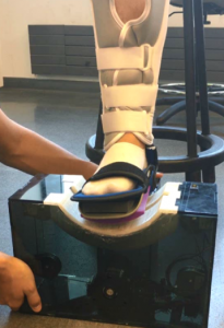
Ankle sprains are among the most common injuries suffered. Not only do 23,000 sprains occur annually, but nearly two-thirds of people with sprained ankles don’t finish their rehabilitation programs, and more than one-third will sprain the same ankle again. A senior design project team that addressed this topic was one of this year’s three winners: the SockRocker

SockRocker
The SockRocker

Among the problems with the currently available rehab technologies are issues of effectiveness, lack of personalization, and poor accessibility. The team — which consisted of Aras Fanuscu, Andrea Frank, David Hernandez, and Angel Xiao — sought to address these issues, coming up with the SockRocker (right). The device, which cost approximately $350 to produce, combines targeted muscle therapy, individualized physician input, and a universal design. The patient places his/her foot into the SockRocker and is then able to move the ankle 30° in either direction, thus strengthening the injured joint. In a pilot study, the design team found that the SockRocker rated 4.8 out of 5 for comfort. In addition, the device is fully portable and can run on 24-volt battery for one month.

Going forward, the team hopes that the SockRocker can be tested clinically to determine its long-term efficacy. According to Timothy Dillingham, MD, MS, chair of the Department of Physical Medicine and Rehabilitation in the Perelman School of Medicine, the device has potential to close “an unfortunate gap in our clinical rehabilitation and management” of patients with ankle sprains.

Pressure Sores Targeted by Flysole

Among the myriad medical complications caused by diabetes, pressure sores of the feet are among the most troubling. Because of the common  complication of peripheral neuropathy, people with diabetes are often unable to determine how much pressure is being exerted on their feet. As a result, they cause foot ulcers, which can become infected, leading in the worst cases to amputation.

pressure sores
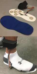
The Flysole combines an insole with five sensors (top) and an ankle band (bottom) to house the electrical components, including the circuit for the pressure sensors as well as the microcontroller and SD card to log the pressure data.

One of the senior design teams from the Department of Bioengineering at the University of Pennsylvania developed a project to address this problem. Their solution was Flysole (right), a prognostic implant that diabetic patients can wear to collect data on foot pressure so that the doctor can prescribe an optimal orthotic to prevent sores from developing. The team was named one of the three winners of this year’s competition.

The team, which consisted of Parag Bapna, Karthik Ramesh, Jane Shmushkis, and Amey Vrudhula, designed the Flysole as a lightweight insole with ankle band paired with software that generates a profile of the pressure on the sole of the patient’s foot. The insole has five sensors to collect these data. The cost is approximately $75 per pair.

In addition, the team made the Flysole to be reusable by including a polyurethane laminate sleeve for the individual patient. Future improvements envisioned by the students include improving the software to include recommendations for orthotics and alternate arrangements for the sole sensors.