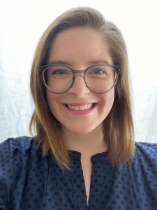
Congratulations to Alison Pouch, Assistant Professor in Bioengineering in the School of Engineering and Applied Science, and in Radiology in the Perelman School of Medicine, on winning a 2024 Cardiac Center Innovation Award for scientific research from the Children’s Hospital of Philadelphia (CHOP)’s Philly Spin-In. Pouch’s study, titled “Systemic Semilunar Valve Mechanics and Simulated Repair in Congenital Heart Disease,” is a collaboration with Matthew Jolley, Assistant Professor of Anesthesiology and Critical Care at CHOP:
“Through biomechanical assessment, Drs. Matthew Jolley and Alison Pouch are leading an interdisciplinary CHOP-Penn team that plans to determine why current approaches to systemic semilunar valve (SSV) repair fail. They will also investigate methods to design improved repairs before going to the operating room by using computational simulation to iteratively optimize repair.
‘We believe that understanding biomechanics of abnormal SSVs and explorations of simulated repair will markedly improve our ability to characterize, risk stratify, and surgically treat SSV dysfunction, thereby improving long-term outcomes and quality of life in patients with SSV dysfunction,’ Dr. Jolley said.”
Pouch’s lab focuses on 3D/4D segmentation and modeling of heart valves in echocardiographic images with applications to surgical treatment of valvular regurgitation as part of the Penn Image Computing and Science Laboratory.
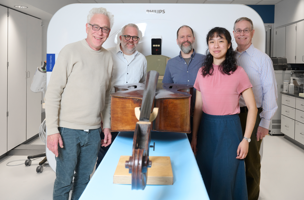
The instrument imaging team, from left: Philadelphia Orchestra bassist Duane Rosengard; Peter Noël, PhD, director of CT Research at the Perelman School of Medicine; luthier Zachary S. Martin; Leening Liu, a PhD student in Noël’s Laboratory of Advanced Computed Tomography Imaging; and Mark Kindig.
When you’re an expert in medical CT imaging, two things are bound to happen, says Peter Noël, PhD, associate professor of Radiology and director of CT Research at the Perelman School of Medicine. One: You develop an insatiable curiosity about the inner workings of all kinds of objects, including those unrelated to your research. And two: Both colleagues and complete strangers will ask for your help in imaging a wide variety of unexpected items.
Over the course of his career, in between managing his own research projects, Noël has imaged diverse objects ranging from animal skulls to tree samples from a German forest, all in the name of furthering scientific knowledge. But none has intrigued him as much as his current extracurricular project: the first known attempt to perform CT imaging of some of the world’s finest string basses.
The goal is to crack the code on what makes a world-class instrument. This knowledge could both increase the ability to better care for masterworks built between the 17th and 19th centuries, as well as providing insights into refining the building of new ones, including possibly shifting from older, scarcer European wood to the use of sustainably harvested U.S. wood.
That’s why Noël and Leening Liu, a PhD student in Noël’s Laboratory of Advanced Computed Tomography Imaging, have found themselves volunteering to run the basses through a Penn CT scanner occasionally, when they’re not developing next-generation CT technology.
“We always learn something out of projects like this … the more appealing part is that medical research can also be applied to non-medical things,” Noël said. “We have the opportunity to take what we learn in medicine and use it for something else—in this case, moving the arts forward.”
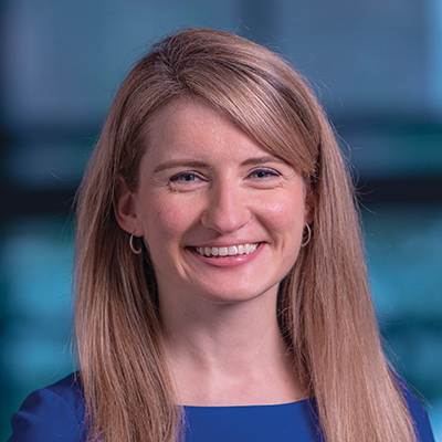
Leening Liu is a Ph.D. student in Bioengineering. She is a member of the Laboratory for Advanced Tomography Imaging (LACTI) with research interests including clinical applications of spectral CT and spectral CT thermometry.
The first few waves of COVID-19 slowed life across the United States, affecting everything from attending school to eating out for dinner and going on vacation. Segments of health care were also affected: Services that were not considered immediately crucial to fighting the virus were slowed or stopped during the pandemic’s first wave.
But once Penn Medicine invited patients back to resume normal health care—including preventive care, like screenings for disease—there was some lag in numbers.
“As we opened up to routine outpatient care, screening rates for situations when patients didn’t have symptoms were not returning back to normal,” said Mitchell Schnall, MD, PhD, FACR, a professor of Radiology, now the senior vice president for Data and Technology Solutions at Penn Medicine, and then the head of a team focused on the “resurgence” efforts to ease patients back into outpatient care. “Although a short delay in health screening is likely not going to cause long-term health problems, we were concerned whether screening rates would stay lower and lead to a long-term impact.”
Congratulations to the members of the Penn Bioengineering community who were awarded 2023 Accelerating from Lab to Market Pre-Seed Grants from the University of Pennsylvania Office of the Vice Provost for Research (OVPR).
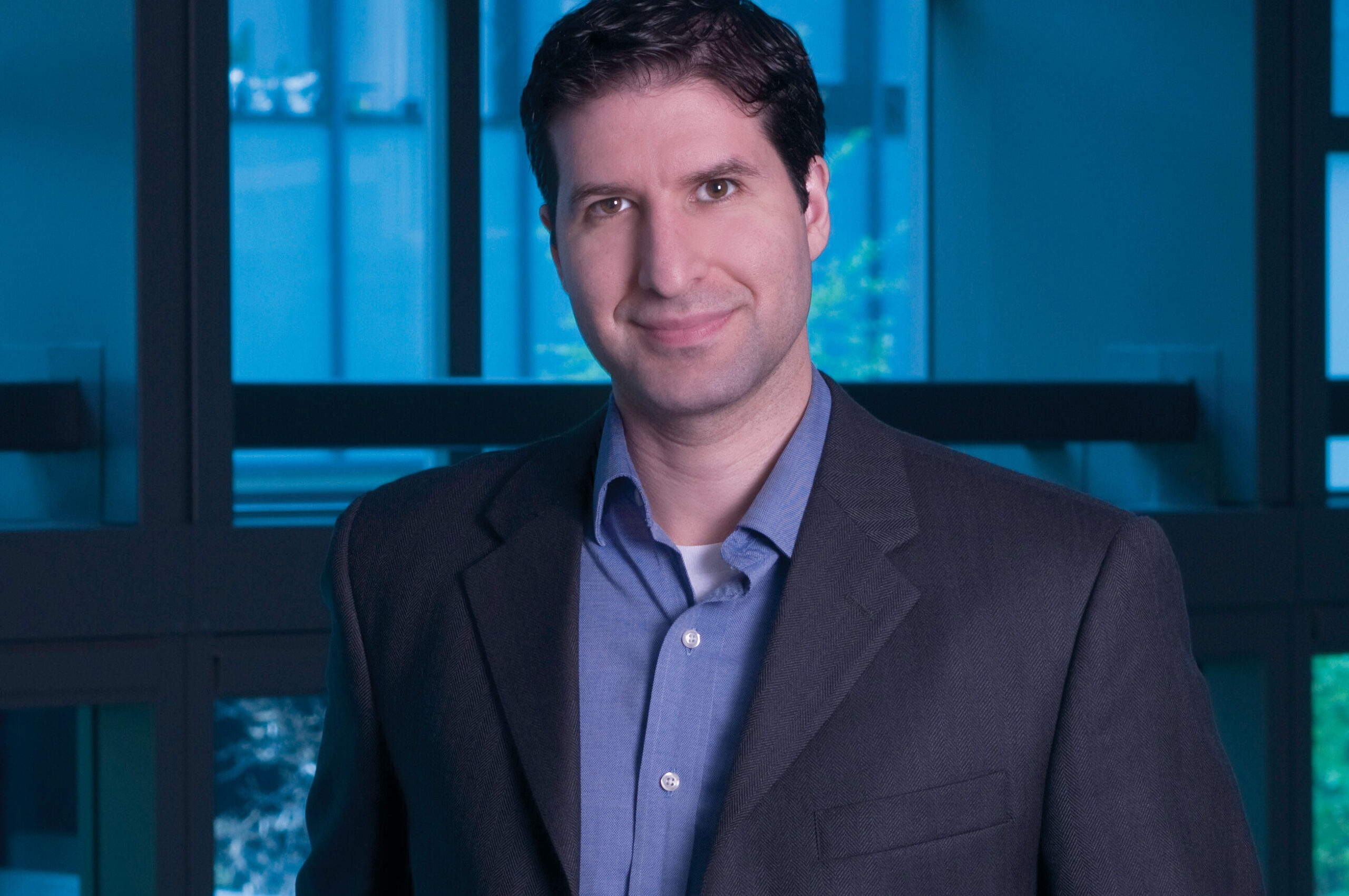
Andrew Tsourkas, Ph.D.
Three faculty affiliated with Bioengineering were included among the four winners. Andrew Tsourkas, Professor in Bioengineering and Co-Director of the Center for Targeted Therapeutics and Translational Nanomedicine (CT3N), was awarded for his project titled “Precise labeling of protein scaffolds with fluorescent dyes for use in biomedical applications.” Tsourkas’s team created protein scaffold that can better control the location and orientation of fluorescent dyes, commonly used for a variety of biomedical applications, such as labeling antibodies or fluorescence-guided surgery. The Tsourkas Lab specializes in “creating novel targeted imaging and therapeutic agents for the detection and/or treatment of diverse diseases.”
Also awarded were Penn Bioengineering Graduate Group members Mark Anthony Sellmeyer, Assistant Professor in Radiology in the Perelman School of Medicine, and Rahul M. Kohli, Associate Professor of Medicine in the Division of Infectious Diseases in the Perelman School of Medicine.
From the OVPR website:
“Penn makes significant commitments to academic research as one of its core missions, including investment in faculty research programs. In some disciplines, the path by which discovery makes an impact on society is through commercialization. Pre-seed grants are often the limiting step for new ideas to cross the ‘valley of death’ between federal research funding and commercial success. Accelerating from Lab to Market Pre-Seed Grant program aims to help to bridge this gap.”
Read the full list of winning projects and abstracts at the OVPR website.
Cosette Tomita, a master’s student in Bioengineering, spoke with Penn Engineering Graduate Admissions about her research in cellular therapy and her path to Penn Engineering.
“What were you doing before you came to Penn Engineering?
After college I wanted to get some industry experience before going to graduate school, so I spent a year working for a pharmaceutical company in New Jersey. I learned a lot—but mostly I learned that I wanted to go back into academia. So I was looking for a more research-oriented position to boost my graduate school applications, and I found a position at Penn’s cyclotron facility. Shortly after that, I applied to the master’s program. I’m still working at the cyclotron, so I’m doing the program part time.
How has your experience in the program been so far?
I love the research I’m doing here. I love the collaboration we have and the fact that I’m able to work with whoever I want to. And I can only say good things about my PI, Robert Mach. He’s a very busy man, but he makes time for his people. And he recognizes when somebody has a lot on their plate and he will go to bat for that person.
What’s your research all about?
The focus of my PI’s lab is on neurodegenerative diseases and opiate use, so we’re looking to make imaging agents and antagonists that can help with the opioid crisis.
For my project, I wanted to look at treating neurodegenerative disease from the perspective of cellular therapy. My PI doesn’t have that expertise, so when I came to him with this idea, he said I should talk to Mark Sellmyer in the bioengineering department. He does a lot of cellular therapies, cell engineering, protein engineering and things of that nature. So his lab is more biological.
I don’t have a grant for my research, so my advisors are supporting it out of their own pockets. They could have said, no, you need to work on this project that’s already going on in the lab. But they gave me the intellectual freedom to do what I wanted to do.”
Congratulations to the fourteen Bioengineering students to receive 2023 National Science Foundation Graduate Research Fellowship Program (NSF GRFP) fellowships. The prestigious NSF GRFP program recognizes and supports outstanding graduate students in NSF-supported fields. The recipients honorees were selected from a highly-competitive, nationwide pool. Further information about the program can be found on the NSF website.
Carlos Armando Aguila, Ph.D. student in Bioengineering, is a member of the Center of Neuroengineering and Therapeutics, advised by Erin Conrad, Assistant Professor in Neurology, and Brian Litt, Professor in Bioengineering and Neurology. His research focuses on analyzing electroencephalogram (EEG) signals to better understand epilepsy.
Joseph Lance Victoria Casila is a Ph.D. student in Bioengineering in the lab of Riccardo Gottardi, Assistant Professor in Pediatrics and Bioengineering. His research focuses on probing environmental factors that influence stem cell differentiation towards chondrogenesis for cartilage engineering and regeneration.
Trevor Chan is a Ph.D. student in Bioengineering in the lab of Felix Wehrli, Professor of Radiologic Science. His research is in developing computational methods for medical image refinement and analysis. Two ongoing projects are: self-supervised methods for CT super-resolution and assessment of osteoporosis, and semi-supervised segmentation of 3D and 4D echocardiograms for surgical correction of congenital heart-valve defects.
Rakan El-Mayta is an incoming Ph.D. student in the lab of Drew Weissman, Roberts Family Professor in Vaccine Research. Rakan studies messenger RNA-lipid nanoparticle vaccines for the treatment and prevention of infectious diseases. Prior to starting in the Bioengineering graduate program, he worked as a Research Assistant in Weissman lab and in the lab of Michael Mitchell, Associate Professor in Bioengineering.
Austin Jenk is a Ph.D. student in the lab of Robert Mauck, Mary Black Ralston Professor in Orthopaedic Surgery and Bioengineering. Austin aims to develop early intervention, intra-articular therapeutics to combat the onset of post-traumatic osteoarthritis following acute joint injuries. His work focuses on developing a therapeutic that can be employed not only in conventional healthcare settings, but also emergency and battlefield medicine.
Jiageng Liu is a Ph.D. student in the lab of Alex Hughes, Assistant Professor in Bioengineering. His work aims to precisely control the bio-physical/chemical properties of iPSC-derived organoids with advanced synthetic biology approaches to create functional replacement renal tissues.
Alexandra Neeser is a Ph.D. student in the lab of Leyuan Ma, Assistant Professor of Pathology and Laboratory Medicine. Her research focuses on solid tumor microenvironment delivery of therapeutics.
William Karl Selboe Ojemann, a Ph.D. Student in Bioengineering, is a member of the Center for Neuroengineering and Therapeutics directed by Brian Litt, Professor in Bioengineering and Neurology. His research is focused on developing improved neurostimulation therapies for epilepsy and other neurological disorders.
Savan Patel (BSE Class of 2023) conducted research in the lab of Michael Mitchell, Associate Professor in Bioengineering, where he worked to develop lipid nanoparticle formulations for immunotherapy and extrahepatic delivery of mRNA. He will be joining the Harvard-MIT HST MEMP Ph.D. program in the fall of 2023.
David E. Reynolds, a Ph.D. student in Bioengineering, is a member of the lab of Jina Ko, Assistant Professor in Bioengineering and Pathology and Laboratory Medicine. His research focuses on developing novel and translatable technologies to address currently intractable diagnostic challenges for precision medicine.
Andre Roots is a Ph.D. student in the lab of Christopher Madl, Assistant Professor in Materials Science and Engineering. His research focuses on the use of protein engineering techniques and an optimized 3D human skeletal muscle microtissue platform to study the effects of biophysical material properties on cells.
Emily Sharp, a second year Ph.D. student in Bioengineering, is a member of the lab of Robert Mauck, Mary Black Ralston Professor in Orthopaedic Surgery and Bioengineering, part of the McKay Orthopaedic Research Laboratories. Her research focuses on designing multi-functional biomaterials to enhance tissue repair, specifically intervertebral disc repair following herniation and discectomy.
Nat Thurlow is a Ph.D. student in the lab of Louis J. Soslowsky, Fairhill Professor in Orthopedic Surgery and Bioengineering. Their current work focuses on delineating the roles of collagens V and XI in tendon mechanics, fibril structure, and gene expression during tendon development and healing.
Maggie Wagner, Ph.D. student in Bioengineering, is a member in the labs of Josh Baxter, Assistant Professor of Orthopaedic Surgery, and Flavia Vitale, Assistant Professor in Neurology and Bioengineering. Her research focuses on the development of novel sensors to record and monitor muscle neuromechanics.
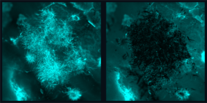
Candida albicans is a species of yeast that is a normal part of the human microbiota but can also cause severe infections that pose a significant global health risk due to their resistance to existing treatments, so much so that the World Health Organization has highlighted this as a priority issue. The picture above shows a before (left) and after (right) fluorescence image of fungal biofilms being precisely targeted by nanozyme microrobots without bonding to or disturbing the tissue sample. (Image: Min Jun Oh and Seokyoung Yoon)
Infections caused by fungi, such as Candida albicans, pose a significant global health risk due to their resistance to existing treatments, so much so that the World Health Organization has highlighted this as a priority issue.
Although nanomaterials show promise as antifungal agents, current iterations lack the potency and specificity needed for quick and targeted treatment, leading to prolonged treatment times and potential off-target effects and drug resistance.
“Candida forms tenacious biofilm infections that are particularly hard to treat,” Koo says. “Current antifungal therapies lack the potency and specificity required to quickly and effectively eliminate these pathogens, so this collaboration draws from our clinical knowledge and combines Ed’s team and their robotic expertise to offer a new approach.”
The team of researchers is a part of Penn Dental’s Center for Innovation & Precision Dentistry, an initiative that leverages engineering and computational approaches to uncover new knowledge for disease mitigation and advance oral and craniofacial health care innovation.
For this paper, published in Advanced Materials, the researchers capitalized on recent advancements in catalytic nanoparticles, known as nanozymes, and they built miniature robotic systems that could accurately target and quickly destroy fungal cells. They achieved this by using electromagnetic fields to control the shape and movements of these nanozyme microrobots with great precision.
“The methods we use to control the nanoparticles in this study are magnetic, which allows us to direct them to the exact infection location,” Steager says. “We use iron oxide nanoparticles, which have another important property, namely that they’re catalytic.”
Other authors include Min Jun Oh, Alaa Babeer, Yuan Liu, Zhi Ren, Zhenting Xiang, Yilan Miao, and Chider Chen of Penn Dental; and David P. Cormode and Seokyoung Yoon of the Perelman School of Medicine. Cormode also holds a secondary appointment in Bioengineering.
This research was supported in part by the National Institute for Dental and Craniofacial Research (R01 DE025848, R56 DE029985, R90DE031532 and; the Basic Science Research Program through the National Research Foundation of Korea of the Ministry of Education (NRF-2021R1A6A3A03044553).
The Solomon R. Pollack Award for Excellence in Graduate Bioengineering Research is given annually to the most deserving Bioengineering graduate students who have successfully completed research that is original and recognized as being at the forefront of their field. This year, the Department of Bioengineering at the University of Pennsylvania recognizes the stellar work of four graduate students in Bioengineering.
Margaret Billingsley
Dissertation: “Ionizable Lipid Nanoparticles for mRNA CAR T Cell Engineering”
Maggie Billingsley
Margaret earned a bachelor’s degree in Biomedical Engineering from the University of Delaware where she conducted research in the Day Lab on the use of antibody-coated gold nanoparticles for the detection of circulating tumor cells. She conducted doctoral research in the lab of Michael J. Mitchell, J. and Peter Skirkanich Assistant Professor in Bioengineering. After defending her thesis at Penn in 2022, Margaret began postdoctoral training at the Massachusetts Institute of Technology (MIT) in the Hammond Lab where she is investigating the design and application of polymeric nanoparticles for combination therapies in ovarian cancer. She plans to use these experiences to continue a research career focused on drug delivery systems.
“Maggie was an absolutely prolific Ph.D. student in my lab, who pioneered the development of new mRNA lipid nanoparticle technology to engineer the immune system to target and kill tumor cells,” says Mitchell. “Maggie is incredibly well deserving of this honor, and I am so excited to see what she accomplishes next as a Postdoctoral Fellow at MIT and ultimately as a professor running her own independent laboratory at a top academic institution.”
Victoria Muir
Dissertation: “Designing Hyaluronic Acid Granular Hydrogels for Biomaterials Applications”
Victoria Muir
Victoria is currently a Princeton University Presidential Postdoctoral Research Fellow in the lab of Sujit S. Datta, where she studies microbial community behavior in 3D environments. She obtained her Ph.D. in 2022 as an NSF Graduate Research Fellow at Penn Bioengineering under the advisement of Jason A. Burdick, Adjunct Professor in Bioengineering at Penn and Bowman Endowed Professor in Chemical and Biological Engineering at the University of Colorado, Boulder. She received a B.ChE. in Chemical Engineering from the University of Delaware in 2018 as a Eugene DuPont Scholar. Outside of research, Victoria is highly active in volunteer and leadership roles within the American Institute of Chemical Engineers (AIChE), currently serving as Past Chair of the Young Professionals Community and a member of the Career and Education Operating Council (CEOC). Victoria’s career aspiration is to become a professor of chemical engineering and to lead a research program at the interaction of biomaterials, soft matter, and microbiology.
“Victoria was a fantastic Ph.D. student,” says Burdick. “She worked on important projects related to granular materials from the fundamentals to applications in tissue repair. She was also a leader in outreach activities, a great mentor to numerous undergraduates, and is already interviewing towards an independent academic position.”
Sadhana Ravikumar
Dissertation: “Characterizing Medial Temporal Lobe Neurodegeneration Due to Tau Pathology in Alzheimer’s Disease Using Postmortem Imaging”
Sadhana Ravikumar
Sadhana completed her B.S. in Electrical Engineering at the University of Cape Town, South Africa in 2014 and her M.S. in Biomedical Engineering from Carnegie Mellon University in 2017. Outside of the lab, she enjoys spending time in nature and exploring restaurants in Philadelphia with friends. She focused her doctoral work on the development of computational image analysis techniques applied to ex vivo human brain imaging data in the Penn Image Computing and Science Laboratory of Paul Yushkevich, Professor of Radiology at the Perelman School of Medicine and member of the Penn Bioengineering Graduate Group. She hopes to continue working at the intersection of machine learning and biomedical imaging to advance personalized healthcare and drug development.
“Dr. Sadhana Ravikumar’s Ph.D. work is a tour de force that combines novel methodological contributions crafted to address the challenge of anatomical variability in ultra-high resolution ex vivo human brain MRI with new clinical knowledge on the contributions of molecular pathology to neurodegeneration in Alzheimer’s disease,” says Yushkevich. “I am thrilled that this excellent contribution, as well as Sadhana’s professionalism and commitment to mentorship, have been recognized through the Sol Pollack award.”
Hannah Zlotnick
Dissertation: “Remote Force Guided Assembly of Complex Orthopaedic Tissues”
Hannah Zlotnick
Hannah was a Ph.D. candidate in the lab of Robert Mauck, Mary Black Ralston Professor in Orthopaedic Surgery and in Bioengineering. She successfully defended her thesis and graduated in August 2022. During her Ph.D., Hannah advanced the state-of-the-art in articular cartilage repair by harnessing remote fields, such as magnetism and gravity. Using these non-invasive forces, she was able to control cell positioning within engineered tissues, similar to the cell patterns within native cartilage, and enhance the integration between cartilage and bone. Her work could be used in many tissue engineering applications to recreate complex tissues and tissue interfaces. Hannah earned a B.S. in Biological Engineering from the Massachusetts Institute of Technology (MIT) in 2017 during which time she was also a member of the women’s varsity soccer team. At Penn, Hannah was also involved in the Graduate Association of Bioengineers (GABE) intramurals & leadership, and helped jumpstart the McKay DEI committee. Since completing her Ph.D., Hannah has begun her postdoctoral research as a Schmidt Science Fellow in Jason Burdick’s lab at the University of Colorado Boulder where she looks to improve in vitro disease models for osteoarthritis.
“Hannah was an outstanding graduate student, embodying all that is amazing about Penn BE – smart, driven, inventive and outstanding in every way,” says Mauck. “ I can’t wait to see where she goes and what she accomplishes!”
Congratulations to our four amazing 2023 Sol Pollack Award winners!
Penn Engineering’s newly established ASSET Center aims to make AI-enabled systems more “safe, explainable and trustworthy” by studying the fundamentals of the artificial neural networks that organize and interpret data to solve problems.
ASSET’s first funding collaboration is with Penn’s Perelman School of Medicine (PSOM) and the Penn Institute for Biomedical Informatics (IBI). Together, they have launched a series of seed grants that will fund research at the intersection of AI and healthcare.
Teams featuring faculty members from Penn Engineering, Penn Medicine and the Wharton School applied for these grants, to be funded annually at $100,000. A committee consisting of faculty from both Penn Engineering and PSOM evaluated 18 applications and judged the proposals based on clinical relevance, AI foundations and potential for impact.
Artificial intelligence and machine learning promise to revolutionize nearly every field, sifting through massive amounts of data to find insights that humans would miss, making faster and more accurate decisions and predictions as a result.
Applying those insights to healthcare could yield life-saving benefits. For example, AI-enabled systems could analyze medical imaging for hard-to-spot tumors, collate multiple streams of disparate patient information for faster diagnoses or more accurately predict the course of disease.
Given the stakes, however, understanding exactly how these technologies arrive at their conclusions is critical. Doctors, nurses and other healthcare providers won’t use such technologies if they don’t trust that their internal logic is sound.
“We are developing techniques that will allow AI-based decision systems to provide both quantifiable guarantees and explanations of their predictions,” says Rajeev Alur, Zisman Family Professor in Computer and Information Science and Director of the ASSET Center. “Transparency and accuracy are key.”
“Development of explainable and trustworthy AI is critical for adoption in the practice of medicine,” adds Marylyn Ritchie, Professor of Genetics and Director of the Penn Institute for Biomedical Informatics. “We are thrilled about this partnership between ASSET and IBI to fund these innovative and exciting projects.”
Seven projects were selected in the inaugural class, including projects from Dani S. Bassett, J. Peter Skirkanich Professor in the Departments of Bioengineering, Electrical and Systems Engineering, Physics & Astronomy, Neurology, and Psychiatry, and several members of the Penn Bioengineering Graduate Group: Despina Kontos, Matthew J. Wilson Professor of Research Radiology II, Department of Radiology, Penn Medicine and Lyle Ungar, Professor, Department of Computer and Information Science, Penn Engineering; Spyridon Bakas, Assistant Professor, Departments of Pathology and Laboratory Medicine and Radiology, Penn Medicine; and Walter R. Witschey, Associate Professor, Department of Radiology, Penn Medicine.
Optimizing clinical monitoring for delivery room resuscitation using novel interpretable AI
Elizabeth Foglia, Associate Professor, Department of Pediatrics, Penn Medicine and the Children’s Hospital of Philadelphia
Dani S. Bassett, J. Peter Skirkanich Professor, Departments of Bioengineering and Electrical and Systems Engineering, Penn Engineering
This project will apply a novel interpretable machine learning approach, known as the Distributed Information Bottleneck, to solve pressing problems in identifying and displaying critical information during time-sensitive clinical encounters. This project will develop a framework for the optimal integration of information from multiple physiologic measures that are continuously monitored during delivery room resuscitation. The team’s immediate goal is to detect and display key target respiratory parameters during delivery room resuscitation to prevent acute and chronic lung injury for preterm infants. Because this approach is generalizable to any setting in which complex relations between information-rich variables are predictive of health outcomes, the project will lay the groundwork for future applications to other clinical scenarios.
The Solomon R. Pollack Award for Excellence in Graduate Bioengineering Research is given annually to the most deserving Bioengineering graduate students who have successfully completed research that is original and recognized as being at the forefront of their field. This year Penn Bioengineering recognizes the outstanding work of two graduate students in Bioengineering: Erin Berlew and Rhea Chitalia.
Erin Berlew, Ph.D. candidate in Bioengineering
Erin Berlew is a Ph.D. candidate in the lab of Brian Chow, Associate Professor in Bioengineering. She successfully defended her thesis, titled “Single-component optogenetic tools for cytoskeletal rearrangements,” in December 2021. In her research, she used the BcLOV4 optogenetic platform discovered/developed in the Chow lab to control RhoGTPase signaling. Erin earned a B.S. in Chemistry from Haverford College in 2015 and was an Americorps member with City Year Philadelphia from 2015-2016. “Erin is a world-class bioengineering with an uncommon record of productivity gained through her complementary expertise in molecular, cellular, and computational biology,” says Chow. “She embodies everything wonderful, both academically and culturally, about our graduate program and its distinguished history.” Erin’s hobbies outside the lab include spending time with family, reading mystery novels, enjoying Philadelphia, and crossword puzzles. In the future, she hopes to continue to teach for the BE department (she has already taught ENGR 105 and served as a TA for undergraduate and graduate courses) and to conduct further research at Penn.
Rhea Chitalia, Ph.D. candidate in Bioengineering
Rhea Chitalia is a Ph.D. candidate in Bioengineering and a member of the Computational Biomarker Imaging Group (CBIG), advised by Despina Kontos, Matthew J. Wilson Associate Professor of Research Radiology II in the Perelman School of Medicine. Rhea completed her B.S.E. in Biomedical Engineering at Duke University in 2015. Her doctoral research concerns leveraging machine learning, bioinformatics, and computer vision to develop computational imaging biomarkers for improved precision cancer care. In December 2021 she successfully defended her thesis titled “Computational imaging biomarkers for precision medicine: characterizing intratumor heterogeneity in breast cancer.” “It has been such a privilege to mentor Rhea on her dissertation research,” says Kontos. “Rhea has been a star graduate student. Her work has made fundamental contributions in developing computational methods that will allow us to gain important insight into tumor heterogeneity by utilizing a multi-modality imaging approach.” David Mankoff, Matthew J. Wilson Professor of Research Radiology in the Perelman School of Medicine, served as Rhea’s second thesis advisor. “It was a true pleasure for me to work with Rhea and to Chair her BE Thesis Committee,” Mankoff adds. “Rhea’s Ph.D. thesis and thesis presentation was one of the best I have had the chance to be involved with in my graduate mentoring career.” After graduation, Rhea hopes to further precision medicine initiatives through the use of real world, multi-omic data in translational industry settings. She will be joining Invicro as an Imaging Scientist. In her spare time, Rhea enjoys trying new restaurants, reading, and spending time with friends and family.


 Cosette Tomita, a master’s student in Bioengineering, spoke with Penn Engineering Graduate Admissions about her research in cellular therapy and her path to Penn Engineering.
 Carlos Armando Aguila, Ph.D. student in Bioengineering, is a member of the Center of Neuroengineering and Therapeutics, advised by
  Joseph Lance Victoria Casila is a Ph.D. student in Bioengineering in the lab of
  Trevor Chan is a Ph.D. student in Bioengineering in the lab of
  Rakan El-Mayta is an incoming Ph.D. student in the lab of
  Austin Jenk is a Ph.D. student in the lab of
  Jiageng Liu is a Ph.D. student in the lab of
  Alexandra Neeser is a Ph.D. student in the lab of
  William Karl Selboe Ojemann, a Ph.D. Student in Bioengineering, is a member of the Center for Neuroengineering and Therapeutics directed by
  Savan Patel (BSE Class of 2023) conducted research in the lab of
  David E. Reynolds, a Ph.D. student in Bioengineering, is a member of the lab of
  Andre Roots is a Ph.D. student in the lab of
  Emily Sharp, a second year Ph.D. student in Bioengineering, is a member of the lab of
  Nat Thurlow is a Ph.D. student in the lab of
  Maggie Wagner, Ph.D. student in Bioengineering, is a member in the labs of