Understanding the Cellular Mechanisms Driving Solid Tumors’ Robust Defense System

by Nathi Magubane

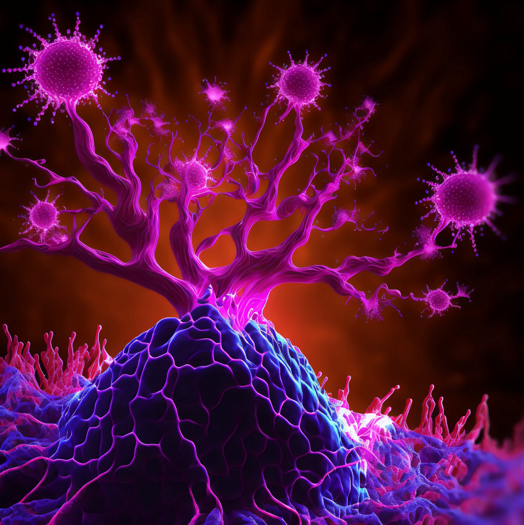
In a collaborative interdisciplinary study, Michael Mitchell of the School of Engineering and Applied Science, Wei Guo of the School of Arts & Sciences, and Drew Weissman of the Perelman School of Medicine show that solid tumors can block drug-delivery mechanisms with a “forcefield-like” effect but certain genetic elements that can effectively “shut down” the forcefield. Their findings hint at new targets for delivering cancer treatments that use the body’s immune system to fight tumors. (Image: iStock / CIPhotos)

The tumor microenvironment—an ad hoc, messy amalgamation of signaling molecules, immune cells, fibroblasts, blood vessels, and the extracellular matrix—acts like a “powerful security system that protects solid tumors from invaders seeking to destroy them,” says Michael Mitchell, a bioengineer at the University of Pennsylvania working on nanoscale therapeutics aimed at targeting cancers.

“A lot like the Death Star with its surrounding fleet of fighter ships and protective shields, solid tumors can use features like immune cells and vasculature to exert force, acting as a physical barrier to rebel forces (nanoparticles) coming in to deliver the payload that destroys it,” Mitchell says.

Now, researchers in the Mitchell lab have teamed up with Wei Guo’s group in the School of Arts & Sciences at Penn and Drew Weissman of the Perelman School of Medicine to figure out the molecular mechanisms that make tumor microenvironments seemingly impenetrable and found that small extracellular vesicles (sEVs) are secreted by tumor cells and act as a “forcefield,” blocking therapeutics. Their findings are published in Nature Materials.

“This discovery reveals how tumors create a robust defense system, making it challenging for nanoparticle-based therapies to reach and effectively target cancer cells,” Guo says. “By understanding the cellular mechanisms driving these responses, we can potentially develop strategies to disable this defense, allowing therapeutics to penetrate and attack the tumor more efficiently.”

The research builds on a prior collaboration between Guo and Mitchell’s labs, wherein the teams focused on how tumor-associated immune cells, known as macrophages, contribute to the suppression of anti-tumor immunity by secreting extracellular vesicles.

Read the full story in Penn Today.

Michael Mitchell is an associate professor in the Department of Bioengineering in the School of Engineering and Applied Science and director of the Lipid Nanoparticle Synthesis Core at the Penn Institute for RNA Innovation at the University of Pennsylvania.

Wei Guo is the Hirsch Family President’s Distinguished Professor in the Department of Biology in Penn’s School of Arts & Sciences.

Ningqiang Gong, a former postdoctoral researcher in the Mitchell lab at Penn Engineering, is an assistant professor at the University of Science and Technology of China.

Wenqun Zhong is a reseearch associate in the Guo Laboratory in Penn Arts & Sciences.

Other authors include: Alex G Hamilton, Dongyoon Kim, Junchao Xu, and Lulu Xue of Penn Engineering; Junhyong Kim, Zhiyuan Qin, and Fengyuan Xu of Penn Arts & Sciences; Mohamad-Gabriel Alameh and Drew Weissman of the Perelman School of Medicine; Andrew E. Vaughn and Gan Zhao of the Penn School of Veterinary Medicine; Jinghong Li and Xucong Teng of the University of Beijing; and Xing-Jie Liang of the Chinese Academy of Sciences.

This research received support from the U.S. National Institutes of Health (DP2 TR002776, R35 GM141832, and NCI P50 CA261608), Burroughs Wellcome Fund, U.S. National Science Foundation CAREER Award (CBET-2145491), and an American Cancer Society Research Scholar Grant (RGS-22-1122-01-ET.)

Researchers Breathe New Life into Lung Repair

by Nathi Magubane

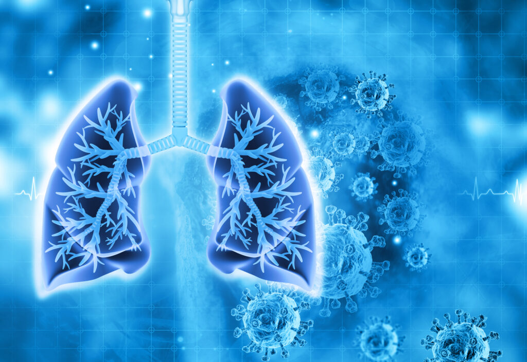
Image: iStock/Mohammed Haneefa Nizamudeen

In the human body, the lungs and their vasculature can be likened to a building with an intricate plumbing system. The lungs’ blood vessels are the pipes essential for transporting blood and nutrients for oxygen delivery and carbon dioxide removal. Much like how pipes can get rusty or clogged, disrupting normal water flow, damage from respiratory viruses, like SARS-CoV-2 or influenza, can interfere with this “plumbing system.”

In a recent study, researchers looked at the critical role of vascular endothelial cells in lung repair. Their work, published in Science Translational Medicine, was led by Andrew Vaughan of the University of Pennsylvania’s School of Veterinary Medicine and shows that, by using techniques that deliver vascular endothelial growth factor alpha (VEGFA) via lipid nanoparticles (LNPs), that they were able to greatly enhance modes of repair for these damaged blood vessels, much like how plumbers patch sections of broken pipes and add new ones.

“While our lab and others have previously shown that endothelial cells are among the unsung heroes in repairing the lungs after viral infections like the flu, this tells us more about the story and sheds light on the molecular mechanisms at play,” says Vaughan, assistant professor of biomedical sciences at Penn Vet. “Here we’ve identified and isolated pathways involved in repairing this tissue, delivered mRNA to endothelial cells, and consequently observed enhanced recovery of the damaged tissue. These findings hint at a more efficient way to promote lung recovery after diseases like COVID-19.”

They found VEGFA’s involvement in this recovery, while building on work in which they used single cell RNA sequencing to identify transforming growth factor beta receptor 2 (TGFBR2) as a major signaling pathway. The researchers saw that when TGFBR2 was missing it stopped the activation of VEGFA. This lack of signal made the blood vessel cells less able to multiply and renew themselves, which is vital for the exchange of oxygen and carbon dioxide in the tiny air sacs of the lungs.

“We’d known there was a link between these two pathways, but this motivated us to see if delivering VEGFA mRNA into endothelial cells could improve lung recovery after disease-related injury,” says first author Gan Zhao, a postdoctoral researcher in the Vaughan Lab.

The Vaughan Lab then reached out to Michael Mitchell of the School of Engineering and Applied Science, whose lab specializes in LNPs, to see if delivery of this mRNA cargo would be feasible.

“LNPs have been great for vaccine delivery and have proven incredibly effective delivery vehicles for genetic information. But the challenge here was to get the LNPs into the bloodstream without them heading to the liver, which is where they tend to congregate as its porous structure lends favor to substances passing from the blood into hepatic cells for filtration,” says Mitchell, an associate professor of bioengineering at Penn Engineering and a coauthor of the paper. “So, we had to devise a way to specifically target the endothelial cells in the lungs.”

Lulu Xue, a postdoctoral researcher in the Mitchell Lab and a co-first author of the paper, explains that they engineered the LNP to have an affinity for lung endothelial cells, this is known as extra hepatic delivery, going beyond the liver.

Read the full story in Penn Today.

How Penn Medicine Is Changing the World with mRNA

by Rachel Ewing

Vaccines for COVID-19 were the first time that mRNA technology was used to address a worldwide health challenge. The Penn Medicine scientists behind that technology were awarded the 2023 Nobel Prize in Physiology or Medicine. Next come all the rest of the potential new treatments made possible by their discoveries.

Starting in the late 1990s, working together at Penn Medicine, Katalin Karikó, PhD, and Drew Weissman, MD, PhD, discovered how to safely use messenger RNA (mRNA) as a whole new type of vaccine or therapy for diseases. When the COVID-19 pandemic hit in 2020, these discoveries made Pfizer/BioNTech and Moderna’s new vaccines possible—saving millions of lives. 

But curbing the pandemic was only the beginning of the potential for this Nobel Prize-winning technology. 

These biomedical innovations from Penn Medicine in using mRNA represent a multi-use tool, not just a treatment for a single disease. The technology’s potential is virtually unlimited; if researchers know the sequence of a particular protein they want to create or replace, it should be possible to target a specific disease. Through the Penn Institute for RNA Innovation led by Weissman, who is the Roberts Family Professor of Vaccine Research in Penn’s Perelman School of Medicine, researchers are working to ensure this limitless potential meets the world’s most challenging and important needs.

Infectious Diseases and Beyond

Just consider some of the many projects Weissman’s lab is partnering in: “We’re working on malaria with people across the U.S. and in Africa,” Weissman said. “We’re working on leptospirosis with people in Southeast Asia. We’re working on vaccines for peanut allergies. We’re working on vaccines for autoimmunity. And all of this is through collaboration.”

Clinical trials are underway for the new malaria vaccine, as well as for a Penn-developed mRNA vaccine for genital herpes and one that aims to protect against all varieties of coronaviruses. Trials should begin soon for vaccines for norovirus and the bacterium C. difficile.

Single-Injection Gene Therapies for Sickle Cell and Heart Disease

Drew Weissman, MD, PhD, is a co-winner of the 2023 Nobel Prize in Physiology or Medicine for discoveries with mRNA.

The Weissman lab is working to deploy mRNA technology as an accessible gene therapy for sickle cell anemia, a devastating and painful genetic disease that affects about 20 million people around the world. About 300,000 babies are born each year with the condition, mainly in sub-Saharan Africa. Weissman’s team has developed technology to efficiently deliver modified mRNA to bone marrow stem cells, instructing red blood cells to produce normal hemoglobin instead of the malformed “sickle” version that causes the illness. Conventional gene therapies are complex and expensive treatments, but the mRNA gene therapy could be a simple, one-time intravenous injection to cure the disease. Such a treatment would have applications to many other congenital gene defects in blood and stem cells.

In another new program, Penn Medicine researchers have found a way to target the muscle cells of the heart. This gene therapy method developed by Weissman’s team, together with Vlad Muzykantov, MD, PhD, the Founders Professor in Nanoparticle Research could potentially repair the heart or increase blood flow to the heart, noninvasively, after a heart attack or to correct a genetic deficiency in the heart. “That is important because heart disease is the number one killer in the U.S. and in the world,” Weissman said. “Drugs for heart disease aren’t specific for the heart. And when you’re trying to treat a myocardial infarction or cardiomyopathy or other genetic deficiencies in the heart, it’s very difficult, because you can’t deliver to the heart.”

Weissman’s team also is partnering on programs for neurodevelopmental diseases and for neurodegenerative diseases, to replace genes or deliver therapeutic proteins that will treat and potentially cure these diseases.

“The potential is unbelievable,” Weissman said. “We haven’t thought of everything that can be done.”

Read the full story in Penn Medicine News.

Vladimir R. Muzykantov is Founders Professor in Nanoparticle Research in the Department of Systems Pharmacology and Translational Therapeutics in the Perelman School of Medicine. He is a member of the Penn Bioengineering Graduate Group.