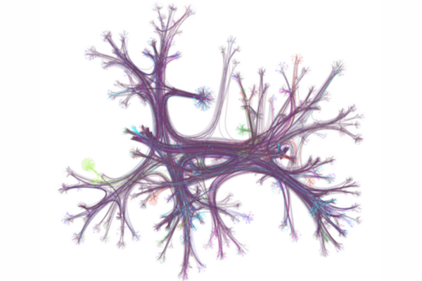
A hyperlink network from English Wikipedia, with only 0.1% of articles (nodes) and their connections (edges) visualized. Seven different reader journeys through this network are highlighted in various colors. The network is organized by topic and displayed using a layout that groups related articles together. (Image: Dale Zhou)
At one point or another, you may have gone online looking for a specific bit of information and found yourself “going down the Wiki rabbit hole” as you discover wholly new, ever-more fascinating related topics — some trivial, some relevant — and you may have gone so far down the hole it’s difficult to piece together what brought you there to begin with.
According to the University of Pennsylvania’s Dani Bassett, who recently worked with a collaborative team of researcher to examine the browsing habits of 482,760 Wikipedia readers from 50 different countries, this style of information acquisition is called the “busybody.” This is someone who goes from one idea or piece of information to another, and the two pieces may not relate to each other much.
“The busybody loves any and all kinds of newness, they’re happy to jump from here to there, with seemingly no rhyme or reason, and this is contrasted by the ‘hunter,’ which is a more goal-oriented, focused person who seeks to solve a problem, find a missing factor, or fill out a model of the world,” says Bassett.
In the research, published in the journal Science Advances, Bassett and colleagues discovered stark differences in browsing habits between countries with more education and gender equality versus less equality, raising key questions about the impact of culture on curiosity and learning.
Dani S. Bassett is the J. Peter Skirkanich Professor at the University of Pennsylvania with a primary appointment in the School of Engineering and Applied Science’s Department of Bioengineering and secondary appointments in the School of Arts & Sciences’ Department of Physics & Astronomy, Penn Engineering’s Department of Electrical and Systems Engineering, and the Perelman School of Medicine’s Departments of Neurology and Psychiatry.
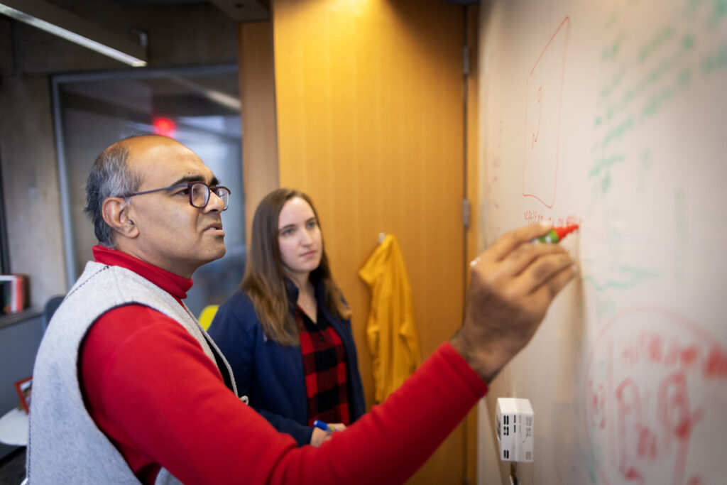
Younger scientists often ask him about exploring multiple fields, Balasubramanian says. The advice he offers is to “have a central line where you have credibility, where you’ve established that you’re really, really good at what you do, and you can be trusted.” (Image: Eric Sucar)
Academia is a long journey of specialization and behind any professor’s CV are long hours of research and study. While the path can be direct for some, for others there’s a pivot, a moment or experience that changes the course of that journey.
Penn Today spoke with four professors whose academic paths diverged, to learn about the trajectory of their interdisciplinary work. Vijay Balasubramanian traverses the boundaries of physics and neuroscience. Tukufu Zuberi is a demographer-turned-curator. Brittany Watson integrates education, research, and veterinary medicine. Amy Hillier began her career studying historical mortgage redlining and moved into supporting trans youth.
Vijay Balasubramanian The Cathy and Marc Lasry Professor of Physics in the School of Arts & Sciences
Wandering through Kolkata’s markets in India stimulates the mind. Hawkers’ cries pass through the inner ear as electrical signals; the pungent, earthy smell of turmeric enters the brain through olfactory sensory neurons. In 1976, a 7-year-old Vijay Balasubramanian had his own market revelation through a bookseller’s portico, where the cover of a slim volume showed a man peering through a microscope lens and a smattering of white paint scattered like stars across the firmament of man and machine.
“What is a scientist?” the book asked, running through a series of exciting adventure shots: archeologic discovery, venom extraction, missile control. In that moment, Balasubramanian knew he would be a scientist. It looked, he says, “amazingly cool.”
When he arrived at the Massachusetts Institute of Technology, Balasubramanian wanted to study the fundamental laws of nature. “So that’s physics,” he says. While earning his doctoral degree at Princeton University, a mentor suggested Balasubramanian read papers in the burgeoning field of neuroscience. It immediately resonated. “Oh my god, this stuff is so cool,” Balasubramanian thought. “But the final year of a Ph.D. is not the time to switch.”
He earned his degree and took a position as a junior fellow of the Harvard Society of Fellows. During the day, he worked on string theory and the information loss paradox for black holes. But in the evening, he would moonlight in a neuroscience lab.
As a young theoretical physicist at Penn, Balasubramanian met Peter Sterling. A former Freedom Rider and professor of neuroscience at the Perelman School of Medicine, Sterling was “a true intellectual,” Balasubramanian says. He knew everything, was interested in everything, and would talk with anybody.
The pair wrote a series of papers together regarding information processing and transmission. “He’s so quick and so much fun and so lively,” Sterling says of Balasubramanian. “He’s fearless; there’s nothing he won’t try.”
While in Cairo with wife Heather J. Sharkey, professor of modern Middle Eastern and North African history at Penn, Balasubramanian prepared a neuroscience grant and submitted it to the National Science Foundation, “sort of on a whim,” he says. “I put it in from an internet café on an island in the middle of the Nile.” He got the grant and started a research group.
After that, Balasubramanian says, “I was off and running.”
“I was certainly told,” Balasubramanian says of his work in neuroscience, “do not do this before tenure.” But, if he waited, “then I’d be too set my ways,” he says. “I just wouldn’t know enough; it would be too hard to learn; I wouldn’t have the time.”
Younger scientists often ask him about exploring multiple fields, Balasubramanian says. The advice he offers is to “have a central line where you have credibility, where you’ve established that you’re really, really good at what you do, and you can be trusted.” That gives you more latitude, he says.
After that, it’s just sheer discipline. “You’re going to have to wake up earlier than everybody else. You’re going to have to work longer days,” he says. “Otherwise, you know, everybody else is working hard too, and you’ll never be able to achieve the level of expertise and knowledge to be able to do things at that world-class level.”
Balasubramanian wants to see more interdisciplinary collaboration. “Each field trains its students with a certain body of techniques that has been found historically useful in that field,’ he says. “Very often, those techniques also have uses elsewhere, but they don’t know to apply it.”
Traversing borders can be helpful in producing new insights, Balasubramanian says. You can ask questions that people in the field won’t. You might experiment with new ideas or put two disjointed ideas together, he says. “If you’re coming from outside, you have the leeway to do all kinds of silly things. Sometimes, they’re not silly.”
Why not ask new questions and propose new answers? In the end, the data will tell you what’s true. “It gives me comfort to know how things tick.”
This post is adapted from a longer story in Penn Today. Read the full story here.
Balasubramanian is Cathy and Marc Lasry Professor in the Department of Physics and Astronomy in the Penn School of Arts and Sciences and is a member of the Penn Bioengineering Graduate Group. Read more stories featuring his research here.
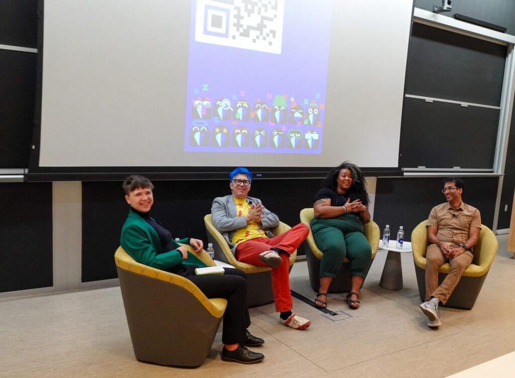
Jamie Moffa, host of In Plain English; Konrad Kording, Kaela Singleton and Arjun Raj
One of the pillars of science is the idea that experimental results can be replicated. If they cannot be reproduced, what if the findings of an experiment were due just to chance? Over the last two decades, a growing chorus of scientists has raised concerns about the “reproducibility crisis,” in which many published research findings can’t be independently validated, calling into question the rigor of contemporary science.
Two years ago, a group led by Konrad Kording, a Penn Integrates Knowledge Professor in Bioengineering and Neuroscience, founded the Community For Rigor (C4R) to build a grassroots movement to improve the rigor of scientific research.
Supported by a grant from the National Institutes of Health (NIH) and partners at Harvard, Duquesne, Smith College and Johns Hopkins, among other institutions, C4R creates educational materials that teach the principles of rigorous research, from data collection to pre-registration of results. “Everyone has done wrong things,” says Kording. “We’re all making these mistakes and we need to be able to talk about it.”
Last month, Kording appeared on In Plain English, a podcast devoted to making science more accessible, alongside Kaela Singleton, the co-founder and President of Black in Neuro; Arjun Raj, Professor in Bioengineering in Penn Engineering and in Genetics in Penn Medicine; and Jamie Moffa, a physician-scientist in training at Washington University in St. Louis, to discuss scientific rigor, including actionable strategies for students and faculty alike.
The conversation touched on everything from successfully managing the reams of data produced by experiments to the power of community to drive cultural change, as well as the difficulty of filtering useful feedback from the noise of social media. “I hope we can get to a point where people feel comfortable sharing what’s working and what’s not working,” says Raj.
Lasya Sreepada has always been fascinated by the brain and the underlying biology that shapes how people develop and age. “My curiosity traces back to observing differences between myself and my sister,” says Sreepada, a Ph.D. candidate in Bioengineering whose research unites efforts across Penn Medicine and Penn Engineering. “We grew up in the same environment but had remarkably different personalities, which led me to question what drove these differences and which brought me to the brain.”
Her academic journey began by applying medical imaging to understand how brain injuries sustained by professional athletes or military veterans impact their brain structure and chemistry over time. She became curious about how neurotrauma impacts aging and degeneration in the long term. Now, she leverages large, multimodal datasets to investigate neurodegenerative disease, with a particular focus on Alzheimer’s.
TDP-43 may be one of the most dangerous proteins in the human body, implicated in neurodegenerative conditions like ALS and Alzheimer’s disease. But the protein remains mysterious: how TDP-43 interacts with the immune system, for instance, is still unclear.
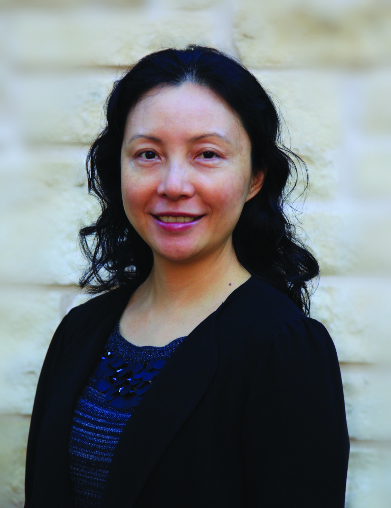
Now, Ning Jenny Jiang, J. Peter and Geri Skirkanich Associate Professor of Innovation in Bioengineering, has been selected for the Collaborative Pairs Pilot Project Awards, sponsored by the Chan Zuckerberg Initiative (CZI), to investigate the relationship between TDP-43 and the immune system.
Launched in 2018, the Collaborative Pairs Pilot Project Awards support pairs of investigators to explore “innovative, interdisciplinary approaches to address critical challenges in the fields of neurodegenerative disease and fundamental neuroscience.” Professor Jiang will partner with Pietro Fratta, MRC Senior Clinical Fellow and MNDA Lady Edith Wolfson Fellow at the University College London Queen Square Institute of Neurology.
The TDP-43 protein is associated with neurodegenerative diseases affecting the central nervous system, including ALS and Alzehimer’s disease. While the loss of neurons and muscle degeneration cause the progressive symptoms, the diseases themselves may be a previously unidentified trigger for abnormal immune system activity.
One possible link is the intracellular mislocalization of TDP-43 (known as TDP-43 proteinopathy), when the protein winds up in the wrong location, which the Jiang and Fratta Labs will investigate. Successfully proving this link could result in potentially game-changing new therapies for these neurodegenerative diseases.
The Jiang Lab at Penn Engineering specializes in systems immunology, using high-throughput sequencing and single-cell and quantitative analysis to understand how the immune system develops and ages, as well as the molecular signatures of immune related diseases. Jiang joined Penn Bioengineering in 2021.
Since arriving on campus, Jiang has teamed with the recently formed Penn Anti-Cancer Engineering Center (PACE), which seeks to understand the forces that determine how cancer grows and spreads, and Engineers in the Center for Precision Engineering (CPE4H), which focuses on innovations in diagnostics and delivery in the development of customizable biomaterials and implantable devices for individualized care.
Jiang was elected a member of the American Institute for Medical and Biological Engineering (AIMBE) College of Fellows in 2021, and has previously won multiple prestigious awards including the NSF CAREER, a Cancer Research Institute Lloyd J. Old STAR Award, and a CZI Neurodegeneration Challenge Network Ben Barres Early Career Acceleration Award.
Jiang is a leader in high-throughput and high-dimensional analysis of T cells, a type of white blood cell crucial to the functioning of a healthy immune system. A recent study in Nature Immunology described the Jiang Lab’s TetTCR-SeqHD technology, the first approach to provide a multifaceted analysis of antigen-specific T cells in a high-throughput manner.
The CZI Collaborative Pairs Pilot Project Awards will provide $200,000 of funding over 18 months with a chance to advance to the second phase of $3.2 million in funding over a four-year period.
Read the full list of grantees on the CZI’s Neurodegeneration Challenge Network (NDCN) Projects website here.
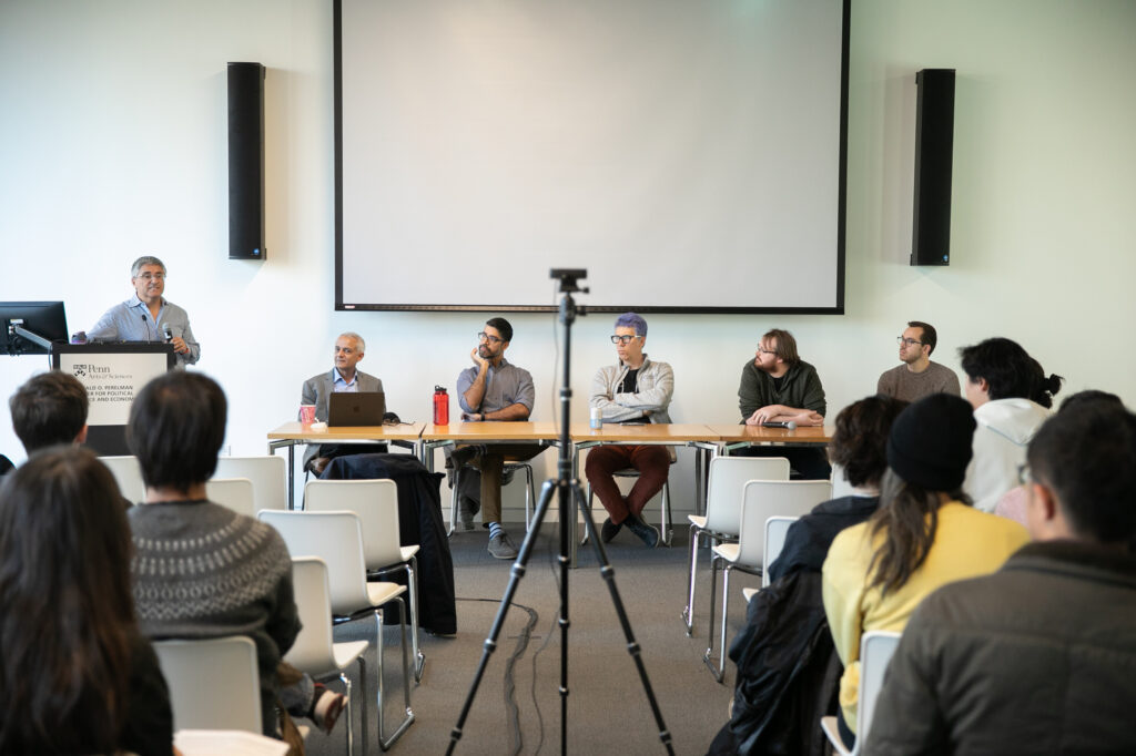
René Vidal, at the podium, introduces the event “ChatGPT turns one: How is generative AI reshaping science?” Bhuvnesh Jain, left at the table, moderated the discussion with Sudeep Bhatia, Konrad Kording, Andrew Zahrt, and Nick Pangakis.
As a neuroscientist surveying the landscape of generative AI—artificial intelligence capable of generating text, images, or other media—Konrad Kording cites two potential directions forward: One is the “weird future” of political use and manipulation, and the other is the “power tool direction,” where people use ChatGPT to get information as they would use a drill to build furniture.
“I’m not sure which of those two directions we’re going but I think a lot of the AI people are working to move us into the power tool direction,” says Kording, a Penn Integrates Knowledge (PIK) University professor with appointments in the Perelman School of Medicine and School of Engineering and Applied Science. Reflecting on how generative AI is shifting the paradigm of science as a discipline, Kording said he thinks “it will push science as a whole into a much more collaborative direction,” though he has concerns about ChatGPT’s blind spots.
Kording joined three University of Pennsylvania researchers from the chemistry, political science, and psychology departments sharing their perspectives in the recent panel “ChatGPT turns one: How is generative AI reshaping science?” PIK Professor René Vidal opened the event, which was hosted by the School of Arts & Sciences’ Data Driven Discovery Initiative (DDDI), and Bhuvnesh Jain, physics and astronomy professor and co-faculty director of DDDI, moderated the discussion.
“Generative AI is moving so rapidly that even if it’s a snapshot, it will be very interesting for all of us to get that snapshot from these wonderful experts,” Jain said. OpenAI launched ChatGPT, a large language model (LLM)-based chatbot, on Nov. 30, 2022, and it rapidly ascended to ubiquity in news reports, faculty discussions, and research papers. Colin Twomey, interim executive director of DDDI, told Penn Today that it’s an open question as to how it will change the landscape of scientific research, and the` idea of the event was to solicit colleagues’ opinions on interesting directions in their fields.
Konrad Paul Kording is Nathan Francis Mossell University Professor in Bioengineering and Computer and Information Science in Penn Engineering and in Neuroscience in the Perelman School of Medicine.
Let’s say you typically eat eggs for breakfast but were running late and ate cereal. As you crunched on a spoonful of Raisin Bran, other contextual similarities remained: You ate at the same table, at the same time, preparing to go to the same job. When someone asks later what you had for breakfast, you incorrectly remember eating eggs.
This would be a real-world example of a false memory. But what happens in your brain before recalling eggs, compared to what would happen if you correctly recalled cereal?
In a paper published in Proceedings of the National Academy of Sciences, University of Pennsylvania neuroscientists show for the first time that electrical signals in the human hippocampus differ immediately before recollection of true and false memories. They also found that low-frequency activity in the hippocampus decreases as a function of contextual similarity between a falsely recalled word and the target word.
“Whereas prior studies established the role of the hippocampus in event memory, we did not know that electrical signals generated in this region would distinguish the imminent recall of true from false memories,” says psychology professor Michael Jacob Kahana, director of the Computational Memory Lab and the study’s senior author. He says this shows that the hippocampus stores information about an item with the context in which it was presented.
Researchers also found that, relative to correct recalls, the brain exhibited lower theta and high-frequency oscillations and higher alpha/beta oscillations ahead of false memories. The findings came from recording neural activity in epilepsy patients who were already undergoing invasive monitoring to pinpoint the source of their seizures.
Noa Herz, lead author and a postdoctoral fellow in Kahana’s lab at the time of the research, explains that the monitoring was done through intracranial electrodes, the methodology researchers wanted to use for this study. She says that, compared to scalp electrodes, this method “allowed us to more precisely, and directly, measure the neural signals that were generated in deep brain structures, so the activity we are getting is much more localized.”
Michael Kahana is the Edmund J. and Louise W. Kahn Term Professor of Psychology in the School of Arts & Sciences and director of the Computational Memory Lab at the University of Pennsylvania. He is a member of the Penn Bioengineering Graduate Group.
Ongoing clinical trials have demonstrated that psychedelics like psilocybin and LSD can have rapid and long-lived antidepressant and anti-anxiety effects. A related clinical problem is chronic pain, which is notoriously difficult to treat and often associated with depression and anxiety.
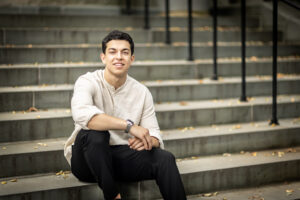
This summer, Ahmad Hammo, a rising third-year student in bioengineering in the School of Engineering and Applied Science, is conducting a pilot study to explore psilocybin’s potential as a therapy for chronic pain and the depression that often accompanies it.
“There’s a strong correlation between chronic pain and depression, so I’m looking at how a psychedelic might be used for treating both of these things simultaneously,” says Hammo, who is originally from Amman, Jordan.
Hammo’s project focuses on neuropathic pain, pain associated with nerve damage. Like other forms of chronic pain, most experts believe that chronic neuropathic pain is stored in the brain.
“Neuropathic pain can lead to a centralized pain syndrome where the pain is still being processed in the brain,” Cichon says. “It’s as if there’s a loop that keeps playing over and over again, and this chronic form is completely divorced from that initial injury.”
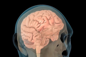
Traumatic brain injury (TBI) has disabled 1 to 2% of the population, and one of their most common disabilities is problems with short-term memory. Electrical stimulation has emerged as a viable tool to improve brain function in people with other neurological disorders.
Now, a new study in the journal Brain Stimulation shows that targeted electrical stimulation in patients with traumatic brain injury led to an average 19% boost in recalling words.
Led by University of Pennsylvania psychology professor Michael Jacob Kahana, a team of neuroscientists studied TBI patients with implanted electrodes, analyzed neural data as patients studied words, and used a machine learning algorithm to predict momentary memory lapses. Other lead authors included Wesleyan University psychology professor Youssef Ezzyat and Penn research scientist Paul Wanda.
“The last decade has seen tremendous advances in the use of brain stimulation as a therapy for several neurological and psychiatric disorders including epilepsy, Parkinson’s disease, and depression,” Kahana says. “Memory loss, however, represents a huge burden on society. We lack effective therapies for the 27 million Americans suffering.”
Michael Kahana is the Edmund J. and Louise W. Kahn Term Professor of Psychology at the University of Pennsylvania. He is a member of the Penn Bioengineering Graduate Group.
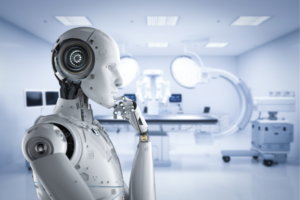
Machine learning (ML) programs computers to learn the way we do – through the continual assessment of data and identification of patterns based on past outcomes. ML can quickly pick out trends in big datasets, operate with little to no human interaction and improve its predictions over time. Due to these abilities, it is rapidly finding its way into medical research.
People with breast cancer may soon be diagnosed through ML faster than through a biopsy. Those suffering from depression might be able to predict mood changes through smart phone recordings of daily activities such as the time they wake up and amount of time they spend exercising. ML may also help paralyzed people regain autonomy using prosthetics controlled by patterns identified in brain scan data. ML research promises these and many other possibilities to help people lead healthier lives.
But while the number of ML studies grow, the actual use of it in doctors’ offices has not expanded much past simple functions such as converting voice to text for notetaking.
The limitations lie in medical research’s small sample sizes and unique datasets. This small data makes it hard for machines to identify meaningful patterns. The more data, the more accuracy in ML diagnoses and predictions. For many diagnostic uses, massive numbers of subjects in the thousands would be needed, but most studies use smaller numbers in the dozens of subjects.
But there are ways to find significant results from small datasets if you know how to manipulate the numbers. Running statistical tests over and over again with different subsets of your data can indicate significance in a dataset that in reality may be just random outliers.
This tactic, known as P-hacking or feature hacking in ML, leads to the creation of predictive models that are too limited to be useful in the real world. What looks good on paper doesn’t translate to a doctor’s ability to diagnose or treat us.
These statistical mistakes, oftentimes done unknowingly, can lead to dangerous conclusions.
To help scientists avoid these mistakes and push ML applications forward, Konrad Kording, Nathan Francis Mossell University Professor with appointments in the Departments of Bioengineering and Computer and Information Science in Penn Engineering and the Department of Neuroscience at Penn’s Perelman School of Medicine, is leading an aspect of a large, NIH-funded program known as CENTER – Creating an Educational Nexus for Training in Experimental Rigor. Kording will lead Penn’s cohort by creating the Community for Rigor which will provide open-access resources on conducting sound science. Members of this inclusive scientific community will be able to engage with ML simulations and discussion-based courses.
“The reason for the lack of ML in real-world scenarios is due to statistical misuse rather than the limitations of the tool itself,” says Kording. “If a study publishes a claim that seems too good to be true, it usually is, and many times we can track that back to their use of statistics.”
Such studies that make their way into peer-reviewed journals contribute to misinformation and mistrust in science and are more common than one might expect.


 Machine learning (ML) programs computers to learn the way we do – through the continual assessment of data and identification of patterns based on past outcomes. ML can quickly pick out trends in big datasets, operate with little to no human interaction and improve its predictions over time. Due to these abilities, it is rapidly finding its way into medical research.