Nader Engheta (left) and Firooz Aflatouni swap ideas over cups of tea.
According to Chinese legend, the first cup of tea was an accident. Shennong, a mythical emperor, boiled a pot of water, only for the wind to add a handful of leaves.
Nader Engheta, H. Nedwill Ramsey Professor, regularly joins his colleague Firooz Aflatouni, associate professor and undergraduate chair in ESE, for a cup of tea in the latter’s office. “We talk about academic life,” says Engheta. “We talk about history, politics.” And, of course, science.
Engheta, who won the Benjamin Franklin Medal last year, is known for his groundbreaking contributions to the design of materials that interact with electromagnetic waves at tiny scales with unprecedented functionalities. More than a decade ago, the Department recruited Aflatouni, who specializes in the design of electronic and photonic chips, and Engheta became his mentor. “We come from different angles to the field of optics,” says Engheta.
Over tea, the two brew up new ideas. While perhaps not as directly inspired by teatime as James Watt, who famously experimented with kettles en route to inventing the steam engine, the pair nonetheless finds that ideas rise like the steam from their teacups. “It’s a pleasure to collaborate with Firooz,” says Engheta. “We love to see how we can bring our ideas together.”
Nader Engheta is H. Nedwill Ramsey Professor of Electrical and Systems Engineering at Penn Engineering, with secondary appointments in the departments of Bioengineering, Materials Science and Engineering, and Physics and Astronomy in the School of Arts & Sciences. Read more stories featuring Engheta in the BE Blog.
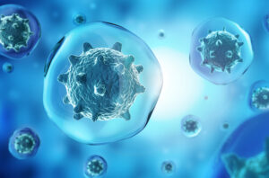
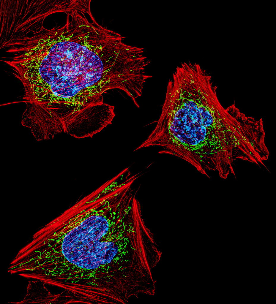
nucleus and membrane of pathogen micro organisms in blue background
Up to 50 percent of cancer-signaling proteins once believed to be immune to drug treatments due to a lack of targetable protein regions may actually be treatable, according to a new study from the Perelman School of Medicine at the University of Pennsylvania. The findings, published this month in Nature Communications, suggest there may be new opportunities to treat cancer with new or existing drugs.
Researchers, clinicians, and pharmacologists looking to identify new ways to treat medical conditions—from cancer to autoimmune diseases—often focus on protein pockets, areas within protein structures to which certain proteins or molecules can bind. While some pockets are easily identifiable within a protein structure, others are not. Those hidden pockets, referred to as cryptic pockets, can provide new opportunities for drugs to bind to. The more pockets scientists and clinicians have to target with drugs, the more opportunities they have to control disease.
The research team identified new pockets using a Penn-designed neural network, called PocketMiner, which is artificial intelligence that predicts where cryptic pockets are likely to form from a single protein structure and learns from itself. Using PocketMiner—which was trained on simulations run on the world’s largest super computer—researchers simulated single protein structures and successfully predicted the locations of cryptic pockets in 35 cancer-related protein structures in thousands of areas of the body. These once-hidden targets, now identified, open up new approaches for potentially treating existing cancer.
What’s more, while successfully predicting the cryptic pockets, the method scientists used in this study was much faster than previous simulation or machine-learning methods. The network allows researchers to nearly instantaneously decide if a protein is likely to have cryptic pockets before investing in more expensive simulations or experiments to pursue a predicted pocket further.
“More than half of human proteins are considered undruggable due to an apparent lack of binding proteins in the snapshots we have,” said Gregory R. Bowman, PhD, a professor of Biochemistry and Biophysics and Bioengineering at Penn and the lead author of the study. “This PocketMiner research and other research like it not only predict druggable pockets in critical protein structures related to cancer but suggest most human proteins likely have druggable pockets, too. It’s a finding that offers hope to those with currently untreatable diseases.”
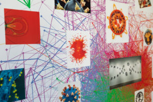
Artist-in-residence and visiting scholar Rebecca Kamen has blended AI and art to produce animated illustrations representing how a dyslexic brain interprets information.
A work that Penn artist-in-residence Rebecca Kamen produced for the show, “Dyslexic Dictionary” at Arion Press in San Francisco. Here, she reinterprets Ph.D. candidate Dale Zhou’s network visualization. (Image: Cat Fennell)
Communicating thoughts with words is considered a uniquely human evolutionary adaptation known as language processing. Fundamentally, it is an information exchange, a lot like data transfer between devices, but one riddled with discrete layers of complexity, as the ways in which our brains interpret and express ideas differ from person to person.
Learning challenges such as dyslexia are underpinned by these differences in language processing and can be characterized by difficulty learning and decoding information from written text.
Artist-in-residence in Penn’s Department of Physics and Astronomy Rebecca Kamen has explored her personal relationship with dyslexia and information exchange to produce works that reflect elements of both her creative process and understanding of language. Kamen unveiled her latest exhibit at Arion Press Gallery in San Francisco, where nine artists with dyslexia were invited to produce imaginative interpretations of learning and experiencing language.
The artists were presented with several prompts in varying formats, including books, words, poems, quotes, articles, and even a single letter, and tasked with creating a dyslexic dictionary: an exploration of the ways in which their dyslexia empowered them to engage in information exchange in unique ways.
Undiagnosed dyslexia
“[For the exhibit], each artist selected a word representing the way they learn, and mine was ‘lens,’” explains Kamen. “It’s a word that captures how being dyslexic provides me with a unique perspective for viewing and interacting with the world.”
From an early age, Kamen enjoyed learning about the natural sciences and was excited about the process of discovery. She struggled, however, with reading at school, which initially presented an obstacle to achieving her dreams of becoming a teacher. “I had a difficult time getting into college,” says Kamen. “When I graduated high school, the word ‘dyslexia’ didn’t really exist, so I assumed everyone struggled with reading.”
Kamen was diagnosed with dyslexia well into her tenure as a professor. “Most dyslexic people face challenges that may go unnoticed by others,” she says, “but they usually find creative ways to overcome them.”
This perspective on seeing and experiencing the world through the lens of dyslexia not only informed Kamen’s latest work for the exhibition “Dyslexic Dictionary,” but also showcased her background in merging art and science. For decades, Kamen’s work has investigated the intersection of the two, creating distinct ways of exploring new relationships and similarities.
“Artists and scientists are curious creatures always looking for patterns,” explains Kamen. “And that’s because patterns communicate larger insights about the world around us.”
The researchers studied different information-seeking approaches by monitoring how participants explore Wikipedia pages and categorically related these to two ideas rooted in philosophical understandings of learning: a “busybody,” who typically jumps between diverse ideas and collects loosely connected information; and a more purpose-driven “hunter,” who systematically ties in closely related concepts to fill their knowledge gaps.
They used these classifications to inform their computational model, the knowledge network. This uses text and context to determine the degree of relatedness between the Wikipedia pages and their content—represented by dots connected with lines of varying thickness to illustrate the strength of association.
In an adaption of the knowledge network, Kamen was classified as a dancer, an archetype elaborated on in an accompanying review paper by Dale Zhou, a Ph.D. candidate in Bassett’s Complex Systems Lab, who had also collaborated with Kamen on “Reveal.”
“The dancer can be described as an individual that breaks away from the traditional pathways of investigation,” says Zhou. “Someone who takes leaps of creative imagination and in the process, produces new concepts and radically remodels knowledge networks.”
Dani Smith Bassett is J. Peter Skirkanich Professor in Bioengineering with secondary appointments in the Departments of Physics & Astronomy, Electrical & Systems Engineering, Neurology, and Psychiatry.
David Lydon-Staley is an Assistant Professor in the Annenberg School for Communications and Bioengineering and is an alumnus of the Bassett Lab.
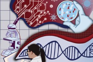
Penn Engineering’s newly established ASSET Center aims to make AI-enabled systems more “safe, explainable and trustworthy” by studying the fundamentals of the artificial neural networks that organize and interpret data to solve problems.
ASSET’s first funding collaboration is with Penn’s Perelman School of Medicine (PSOM) and the Penn Institute for Biomedical Informatics (IBI). Together, they have launched a series of seed grants that will fund research at the intersection of AI and healthcare.
Teams featuring faculty members from Penn Engineering, Penn Medicine and the Wharton School applied for these grants, to be funded annually at $100,000. A committee consisting of faculty from both Penn Engineering and PSOM evaluated 18 applications and judged the proposals based on clinical relevance, AI foundations and potential for impact.
Artificial intelligence and machine learning promise to revolutionize nearly every field, sifting through massive amounts of data to find insights that humans would miss, making faster and more accurate decisions and predictions as a result.
Applying those insights to healthcare could yield life-saving benefits. For example, AI-enabled systems could analyze medical imaging for hard-to-spot tumors, collate multiple streams of disparate patient information for faster diagnoses or more accurately predict the course of disease.
Given the stakes, however, understanding exactly how these technologies arrive at their conclusions is critical. Doctors, nurses and other healthcare providers won’t use such technologies if they don’t trust that their internal logic is sound.
“We are developing techniques that will allow AI-based decision systems to provide both quantifiable guarantees and explanations of their predictions,” says Rajeev Alur, Zisman Family Professor in Computer and Information Science and Director of the ASSET Center. “Transparency and accuracy are key.”
“Development of explainable and trustworthy AI is critical for adoption in the practice of medicine,” adds Marylyn Ritchie, Professor of Genetics and Director of the Penn Institute for Biomedical Informatics. “We are thrilled about this partnership between ASSET and IBI to fund these innovative and exciting projects.”
Seven projects were selected in the inaugural class, including projects from Dani S. Bassett, J. Peter Skirkanich Professor in the Departments of Bioengineering, Electrical and Systems Engineering, Physics & Astronomy, Neurology, and Psychiatry, and several members of the Penn Bioengineering Graduate Group: Despina Kontos, Matthew J. Wilson Professor of Research Radiology II, Department of Radiology, Penn Medicine and Lyle Ungar, Professor, Department of Computer and Information Science, Penn Engineering; Spyridon Bakas, Assistant Professor, Departments of Pathology and Laboratory Medicine and Radiology, Penn Medicine; and Walter R. Witschey, Associate Professor, Department of Radiology, Penn Medicine.
Optimizing clinical monitoring for delivery room resuscitation using novel interpretable AI
Elizabeth Foglia, Associate Professor, Department of Pediatrics, Penn Medicine and the Children’s Hospital of Philadelphia
Dani S. Bassett, J. Peter Skirkanich Professor, Departments of Bioengineering and Electrical and Systems Engineering, Penn Engineering
This project will apply a novel interpretable machine learning approach, known as the Distributed Information Bottleneck, to solve pressing problems in identifying and displaying critical information during time-sensitive clinical encounters. This project will develop a framework for the optimal integration of information from multiple physiologic measures that are continuously monitored during delivery room resuscitation. The team’s immediate goal is to detect and display key target respiratory parameters during delivery room resuscitation to prevent acute and chronic lung injury for preterm infants. Because this approach is generalizable to any setting in which complex relations between information-rich variables are predictive of health outcomes, the project will lay the groundwork for future applications to other clinical scenarios.
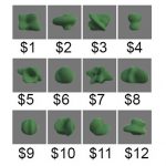
These 12 object-number value pairs were taught to the participants, who had to properly learn the associations to succeed in value judgement tests. The researchers investigated the differences in their brain activity patterns to see why some were faster learners than others.
Why do some people naturally excel at learning instruments, languages or technology while others take longer to pick up new knowledge? Learning requires the brain to encode information, changing its neural “wiring” and creating networks between brain regions.
Earlier research has suggested that part of what might slow down learners is over-thinking. A 2015 study led by Danielle Bassett, Eduardo D. Glandt Faculty Fellow and associate professor in the Department of Bioengineering, showed a correlation between slow learning and cognitive control: the brain’s ability to regulate itself by activating the necessary networks and inhibiting unnecessary activity. In that study, when people unnecessarily engaged parts of the brain linked to cognitive control, they were more likely to take longer to learn a simple task.
But beyond what might make an individual learn more slowly, the researchers want to know what sort of geometric patterns of brain activity make for better learning.
Evelyn Tang and Danielle Bassett
Their new study was led by Bassett and Evelyn Tang, who was an Africk Family Postdoctoral Fellow in Bassett’s Complex Systems Lab before starting at the Max Planck Institute this fall. Sharon Thompson-Schill, Christopher H. Browne Distinguished Professor and chair of Psychology, also contributed to the study.
We’ve known for many years that exercise is good for you, but it was less clear how muscle strength and stamina were assembled at the molecular level. Based the principle that the health of mitochondria – a key organelle within the muscle cell – regulates muscle health, recent work identifies some of the key signaling pathways in vivo that can switch a cell between degrading damaged mitochondria or creating new mitochondria. Zhen Yan, M.D., Ph.D., of the University of Virginia, used a fluorescent reporter gene (MitoTimer) to “report back” the information for individual mitochondria in muscle cells prior to and following exercise. The results reported in a recent issue of Nature Communications show very clearly that mitochondria can switch a muscle cell’s fate. Dr. Yan’s research team identified a new signaling pathway within skeletal muscle that is essential to mitophagy. Knowledge of this pathway could help to develop a variety of therapies for diseases of the muscles or damage to the muscles due to injury.
Understanding How the Brain Processes Visual Data
As a model for how the brain “computes” the information surrounding all of us, researchers have studied how visual information is processed by the brain. One method for investigating this question is the use of artificial neural networks to recognize visual information that they have previously “seen.” A recent article in Cerebral Cortex details how a team at Purdue University, led by Zhongming Liu, Ph.D., Assistant Professor of Electrical and Computer Engineering and Biomedical Engineering, used an artificial neural network to predict and decode information obtained with functional magnetic resonance imaging (fMRI). By collecting fMRI brain activation data when people watch movies, the artificial neural network could generate feature maps that strongly resembled the objects depicted by the initial stimuli. Available now in open access format, the team at Purdue intends to repeat these experiments with more complex networks and more detailed imaging modalities
Preventing Prosthesis-related Infection
Prostheses have improved by leaps and bounds over the years, with the development of osseointegrated prostheses — which are fused directly to the existing bones — a major step in this evolution. However, these prostheses can lead to severe infections that would require the removal of the prosthesis. These problems have been seen more commonly over the last decade or so in the military, where wounded soldiers have received prostheses but suffered subsequent infections.
In a major step forward to address this issue, Mark Ehrensberger, PhD, assistant professor of biomedical engineering at SUNY Buffalo, is the principal investigator on a two-year $1.1 million grant from the Office of Naval Research in the U.S. Department of Defense, awarded for the purpose of investigating implant-related infections. Initial research by Dr. Ehrensberger, who shares the grant award with scientists from the departments of orthopaedics and microbiology and immunology, showed that delivering electrical stimulation to the site of the prosthesis could be effective. One method the team will investigate is using titanium from within the implants themselves to conduct the current to the site.
Success with this grant could mean that patients receiving prostheses show better recovery rates and much lower rates of rejection. It could also reduce the antibiotics used by such patients, which would be a welcome outcome given the increasing rates of antibiotic resistance in health care.
Bioengineering Treatments for Depression
Depression is a largely invisible illness, but it brings with it a massive burden on both the patient and society, with health care costs exceeding $200 billion per year in the U.S. alone. Different drugs are used to treat depression, but all have significant side effects. Psychotherapy also has some effectiveness, but not all patients are helped with therapy.
One promising alternative to treat depression uses transcranial magnetic stimulation, but the devices used in this treatment are often cumbersome. In response to calls to develop more accessible forms of therapy for depression, a startup company in Sweden called Flow Neuroscience has developed a wearable device that uses transcranial direct-current stimulation targeted at the left frontal lobe. The device is noninvasive and is smaller than a sun visor, and the company claims it will be relatively inexpensive (estimated at $750). Flow Neuroscience is in the process of applying for regulatory approval in the European Union.
People and Places
United Kingdom Chancellor of the Exchequer Philip Hammond has announced that the British government will provide £7 million (approximately $9.2 million) in funding to create the UK Centre for Engineering Biology, Metrology and Standards. The government is collaborating with the the Francis Crick Institute in London, with the goal of supporting startup companies in Great Britain dedicated to using engineering and the biological sciences to develop new products.
Closer to home, the Universities of Shady Grove — a partnership of nine Maryland public universities where each university provides its most heavily demanded program — have begun construction on a $162 million biomedical sciences building. The building is slated for completion in 2019 and is expected to nearly double the enrollment at Shady Grove.
Here at Penn, Adam Pardes, a current Ph.D. candidate in our own Department of Engineering, is one of the cofounders of NeuroFlow, a company developing a mobile platform to track and record biometric information obtained from wearables. NeuroFlow recently received $1.25 million in investments to continue developing its technology and ultimately bring it to market. Congratulations, Adam!
Finally, California State University, Long Beach, is our newest national BME program this fall. Burkhard Englert, Ph.D., professor and chair of the Department of Computer Engineering and Computer Science at CSULB, heads the new program as interim chair until a permanent chair is hired.