Shedding Light on Cellular Metabolism to Fight Disease

by

Enamored by the chemical processes of life, Yihui Shen, J. Peter and Geri Skirkanich Assistant Professor of Innovation in Bioengineering, started her research career as a chemist studying the way that proteins fold and the intricate dynamics underlying life processes.

“As an undergraduate, I studied physical chemistry, thinking that one day I’d be addressing challenges in hardcore STEM fields,” she says. “It wasn’t until I observed the dynamics of a single protein molecule that I fell in love with microscopy. I realized that this imaging tool could not only help us observe biological processes on a small scale, but it could also provide new insight at the interface of engineering, chemistry and physics and solve problems on a large scale.”

When Shen turned her attention to microscopy, the field itself was advancing quickly, with improvements being made and new techniques being released every month. Without missing a beat, Shen dove deeper into the most current tools available when she joined Dr. Wei Min’s lab at Columbia University as a doctoral student.

“Professor Wei Min is a pioneer in a new imaging technique called coherent Raman imaging,” says Shen. “In this type of microscopy, we focus light on a very specific point in the cell and measure the amount of scattered light that comes back after exchanging energy with the molecular vibration. This approach allows us to visualize the spatial distribution of different molecules, the very chemistry of life I had studied as an undergraduate, at a high enough resolution to gain insights into biological processes, such as tissue organization, drug distribution and cellular metabolism.”

With this new tool under her belt, Shen was able to ask the kinds of questions that could connect the use of this observation tool to practical applications for real-world challenges.

“I started thinking outside the box,” says Shen. “What if we could observe the chemical exchanges involved in metabolism as they are happening on the scale of a single cell, and then use that insight to pinpoint the exact metabolic pathways and molecules that facilitate tumor growth and disease?”

Read the full story in Penn Engineering Today.

Researchers Develop Technology to Keep Track of Living Cells and Tissues

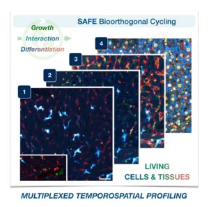
SAFE Bioorthogonal Cycling

Cells in complex organisms undergo frequent changes, and researchers have struggled to monitor these changes and create a comprehensive profile for living cells and tissues. Historically researchers have been limited to only 3-5 markers due to spectral overlaps in fluorescence microscopy, an essential tool required for imaging cells. With only this small handful of markers, it is difficult to monitor protein expressions of live cells and a comprehensive profile of cellular dynamics cannot be created. However, a new study in Nature Biotechnology addresses these limitations by demonstrating a new method for comprehensive profiling of living cells.

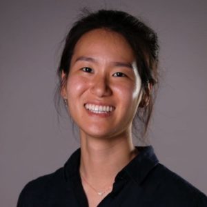
Jina Ko, PhD

Jina Ko, Assistant Professor in Bioengineering in the School of Engineering and Applied Science and in Pathology and Laboratory Medicine in the Perelman School of Medicine, conducted postdoctoral research at Massachusetts General Hospital (MGH) and the Wyss Institute at Harvard University, and the work for this study was done under the supervision of Jonathan Carlson M.D., Ph.D. and Ralph Weissleder M.D., Ph.D. of MGH. Ko’s lab at Penn develops novel technologies using bioengineering, molecular biology, and chemistry to address diagnostic challenges for precision medicine.

To address these limitations in microscopy, the team developed a new chemistry tool which was highly gentle to cells. This “scission-accelerated fluorophore exchange (or SAFE)” method utilizes “click” chemistry, a type of chemistry that follows examples found in nature to create fast and simple reactions. This new SAFE method functions with non-toxic conditions to living cells and tissues, whereas previous methods have used harsh chemicals that would strip off fluorophores and consequently would not work with living cells and tissues.

With the development of SAFE, the authors demonstrated that researchers can now effectively perform multiple cycles of cell profiling and can monitor cellular changes over the course of their observations. Instead of the previous limitation of 3-5 markers total, SAFE allows for many more cycles and can keep track of almost as many markers as the researcher wants. One can now stain cells and quench/release fluorophores and repeat the cycle multiple times for multiplexing on living cells. Each cycle can profile 3 markers, and so someone interested in profiling 15 markers could easily perform 5 cycles to achieve this much more comprehensive cell profile. With this breakthrough in more detailed imaging of cells, SAFE demonstrates broad applicability for allowing researchers to better investigate the physiologic dynamics in living systems.

Read the paper, “Spatiotemporal multiplexed immunofluorescence imaging of living cells and tissues with bioorthogonal cycling of fluorescent probes,” in Nature Biotechnology.

This study was supported by the Schmidt Science Fellows in Partnership with the Rhodes Trust and National Institutes of Health, National Cancer Institute (K99CA256353).

BE Seminar: “Dynamics of 3D Cell Migration and Organ Formation” (Kenneth Yamada)

Our next Penn Bioengineering seminar will be held on zoom next Thursday.

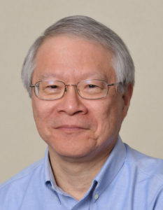
Kenneth Yamada, MD, PhD

Speaker: Kenneth Yamada, M.D., Ph.D.
NIH Distinguished Investigator
Cell Biology Section
National Institute of Dental and Craniofacial Research, National Institutes of Health (NIH)

Date: Thursday, September 9, 2021
Time: 3:30-4:30 PM EDT
Zoom – check email for link or contact ksas@seas.upenn.edu
Location: Moore Room 216, 200 S. 33rd Street

Abstract: Real-time microscopy of the dynamics of cells and tissues in 3D environments is opening new windows to understanding the biophysical mechanisms of complex biological processes. Direct visualization is allowing us to explore fundamental questions in more depth that include: How do cells migrate in 3D? How do cancer cells invade? How is the extracellular matrix assembled? How are organs formed? Visualizing how cells move and organize into tissues is not only providing descriptive insights, but is also leading to the identification of novel, unexpected physical and mechanical mechanisms relevant to tissue engineering. Cells can use varying combinations of cell adhesion to adjacent cells and to the surrounding extracellular matrix with localized cellular contractility to migrate, invade, and produce the complex tissue architecture needed for organ formation.

Kenneth Yamada Bio: Kenneth Yamada has been an NIH Distinguished Investigator since 2011. He received MD and PhD degrees from Stanford. He was a Section Chief at the National Cancer Institute for 10 years and has been a Section Chief at NIDCR since 1990. He is an elected Fellow of the AAAS and American Society for Cell Biology. His research focuses on discovering novel mechanisms and regulators of cell interactions with the extracellular matrix and their roles in embryonic development and cancer. His research group focuses on the mechanisms by which three-dimensional (3D) extracellular matrix mediates key biological events, including cell migration, tissue morphogenesis, and cancer cell invasion. His research places particular emphasis on characterizing the dynamic movements of cells and their extracellular matrix as tissues are remodeled in 3D in real time. The biological systems they study include human primary cells migrating in 3D, human tumor cells and tissues, and mouse organ development. He places particularly high priority on developing future independent research leaders.

BE Seminar: “Imaging and Sequencing Single Cells” (Aaron Streets, UC Berkeley)

The Penn Bioengineering virtual seminar series continues on October 8th.

Aaron Streets, PhD


Speaker: Aaron Streets, Ph.D.
Associate Professor of Bioengineering
University of California, Berkeley

Date: Thursday, October 8, 2020
Time: 2:00-3:00 pm (note the change from our regular seminar time)
Zoom – check email for link or contact ksas@seas.upenn.edu

Title: “Imaging and Sequencing Single Cells”

Abstract:

Recent advances in microfluidics and high-throughput sequencing technology have enabled rapid profiling of genomic material in single cells. Valve- and droplet-based microfluidic platforms can precisely and efficiently manipulate, sort, and process cells to generate indexed sequencing libraries, allowing for high-throughput single-cell analysis of the genome, transcriptome, proteome, and epigenome. Such technology has been instrumental in the global effort to create a human cell atlas, with the ambitious goal of identifying and cataloging all human cell types and cell states in health and disease. However, not all cell phenotypes are directly encoded in the genome and high-throughput sequencing cannot probe the full space of cellular identity. Therefore, microscopy remains one of the most powerful and versatile tools for characterizing cells. Fluorescent imaging and quantitative non-linear optical imaging can reveal morphological characteristics, protein localization, chromatin organization, and chemical composition in single cells. Both single-cell genomics and microscopy can uncover heterogeneity in cellular populations that would otherwise be obscured in ensemble measurement. In this talk, I will discuss a suite of new microfluidic platforms for coupling genomic measurements and optical measurements of the same single cell, and some novel computational approaches to grapple with these new datasets. With a combination of new hardware and software, our goal is to converge on a quantitative and comprehensive understanding of cellular identity.

Bio:

Aaron received a Bachelor of Science in Physics and a Bachelor of Arts in Art at UCLA. He completed his PhD in Applied Physics at Stanford with Dr. Stephen Quake. Aaron then went to Beijing, China as a Whitaker International Postdoctoral Fellow and a Ford postdoctoral fellow and worked with Dr. Yanyi Huang in the Biodynamic Optical Imaging Center (BIOPIC) at Peking University. Aaron joined the faculty of UC Berkeley as an Assistant Professor in Bioengineering in 2016 and is currently a core member of the Biophysics Program and the Center for Computational Biology and he is a Chan Zuckerberg Biohub investigator. Aaron has received the NSF Early Career award and was recently named a Pew Biomedical Scholar.

See the full list of upcoming Penn Bioengineering fall seminars here.

Students’ Innovative Orthotic Device Wins Rothberg Catalyzer

NB: Penn Bioengineering would like to congratulate one of its current Senior Design teams (Alec Bayliff, Bram Bruno, Justin Swirbul, and Vishal Then) which took home the $500 Pioneer Award at this year’s Rothberg Catalyzer competition this past weekend! Keep reading for more information on the competition, awards, and winners.

Penn Health-Tech’s Rothberg Catalyzer is a two-day makerthon that challenges interdisciplinary student teams to prototype and pitch medical devices that aim to address an unmet clinical need.

The Catalyzer’s third competition was held last weekend and was won by MAR Designs, a team of Mechanical Engineering and Applied Mechanics graduate students: Rebecca Li, Ariella Mansfield and Michael Sobrepera.

MAR Designs took home the top prize of $10,000 for their project, an orthotic device that children with cerebral palsy can more comfortably wear as they sleep.

According to the team’s presentation, existing wrist orthoses “improve function and treat/prevent spasticity. However, patients report that these devices are uncomfortable which leads to lack of compliance and may also prevent patient’s eligibility for surgeries.” MAR Designs’ device initially allows full range of motion, but gradually straightens the wrist as the child is falling asleep.

In second place was Splash Throne. Team members Greg Chen, Nik Evitt, Jake Crawford and Meghan Lockwood proposed a toilet safety frame intended for elderly users. Embedded sensors track basic health information, like weight and heart-rate, as part of a preventative health routine.

Integrated Product Design students Jonah Arheim, Laura Ceccacci, Julia Lin and Alex Wan took third place with ONESCOPE, an untethered, hands-free laproscope designed to make minimally-invasive surgeries faster and safer.

Finally, SchistoSpot took home the Catalyzer’s Pioneer Award. Bioengineering and Computer and Information Science seniors Alec Bayliff, Bram Bruno, Justin Swirbul and Vishal Then designed a low-cost microscopy system that can aid in the diagnosis of the parasitic disease schistosomiasis by detecting eggs in urine samples, eliminating the need for a hospital visit.

The event was made possible by a three-year donation by scientist and entrepreneur Jonathan Rothberg, with the intent of inspiring the next generation of healthcare innovators.

Originally posted on the Penn Engineering Medium blog.

Week in BioE (July 12, 2019)

by Sophie Burkholder

DNA Microscopy Gives a Better Look at Cell and Tissue Organization

A new technique that researchers from the Broad Institute of MIT and Harvard University are calling DNA microscopy could help map cells for better understanding of genetic and molecular complexities. Joshua Weinstein, Ph.D., a postdoctoral associate at the Broad Institute, who is also an alumnus of Penn’s Physics and Biophysics department and former student in Penn Bioengineering Professor Ravi Radhakrishnan’s lab, is the first author of this paper on optics-free imaging published in Cell.

The primary goal of the study was to find a way of improving analysis of the spatial organization of cells and tissues in terms of their molecules like DNA and RNA. The DNA microscopy method that Weinstein and his team designed involves first tagging DNA, and allowing the DNA to replicate with those tags, which eventually creates a cloud of sorts that diffuses throughout the cell. The DNA tags subsequent interactions with molecules throughout the cell allowed Weinstein and his team to calculate the locations of those molecules within the cell using basic lab equipment. While the researchers on this project focused their application of DNA microscopy on tracking human cancer cells through RNA tags, this new method opens the door to future study of any condition in which the organization of cells is important.

Read more on Weinstein’s research in a recent New York Times profile piece.

Penn Engineers Demonstrate Superstrong, Reversible Adhesive that Works like Snail Slime

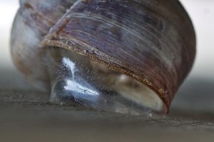
A snail’s epiphragm. (Photo: Beocheck)

If you’ve ever pressed a picture-hanging strip onto the wall only to realize it’s slightly off-center, you know the disappointment behind adhesion as we typically experience it: it may be strong, but it’s mostly irreversible. While you can un-stick the used strip from the wall, you can’t turn its stickiness back on to adjust its placement; you have to start over with a new strip or tolerate your mistake. Beyond its relevance to interior decorating, durable, reversible adhesion could allow for reusable envelopes, gravity-defying boots, and more heavy-duty industrial applications like car assembly.

Such adhesion has eluded scientists for years but is naturally found in snail slime. A snail’s epiphragm — a slimy layer of moisture that can harden to protect its body from dryness — allows the snail to cement itself in place for long periods of time, making it the ultimate model in adhesion that can be switched on and off as needed. In a new study, Penn Engineers demonstrate a strong, reversible adhesive that uses the same mechanisms that snails do.

This study is a collaboration between Penn Engineering, Lehigh University’s Department of Bioengineering, and the Korea Institute of Science and Technology.

Read the full story on Penn Engineering’s Medium blog. 

Low-Dose Radiation CT Scans Could Be Improved by Machine Learning

Machine learning is a type of artificial intelligence growing more and more popular for applications in bioengineering and therapeutics. Based on learning from patterns in a way similar to the way we do as humans, machine learning is the study of statistical models that can perform specific tasks without explicit instructions. Now, researchers at Rensselaer Polytechnic Institute (RPI) want to use these kinds of models in computerized tomography (CT) scanning by lowering radiation dosage and improving imaging techniques.

A recent paper published in Nature Machine Intelligence details the use of modularized neural networks in low-dose CT scans by RPI bioengineering faculty member Ge Wang, Ph.D., and his lab. Since decreasing the amount of radiation used in a scan will also decrease the quality of the final image, Wang and his team focused on a more optimized approach of image reconstruction with machine learning, so that as little data as possible would be altered or lost in the reconstruction. When tested on CT scans from Massachusetts General Hospital and compared to current image reconstruction methods for the scans, Wang and his team’s method performed just as well if not better than scans performed without the use of machine learning, giving promise to future improvements in low-dose CT scans.

A Mind-Controlled Robotic Arm That Requires No Implants

A new mind-controlled robotic arm designed by researchers at Carnegie Mellon University is the first successful noninvasive brain-computer interface (BCI) of its kind. While BCIs have been around for a while now, this new design from the lab of Bin He, Ph.D.,  a Trustee Professor and the Department Head of Biomedical Engineering at CMU, hopes to eliminate the brain implant that most interfaces currently use. The key to doing this isn’t in trying to replace the implants with noninvasive sensors, but in improving noisy EEG signals through machine learning, neural decoding, and neural imaging. Paired with increased user engagement and training for the new device, He and his team demonstrated that their design enhanced continuous tracking of a target on a computer screen by 500% when compared to typical noninvasive BCIs. He and his team hope that their innovation will help make BCIs more accessible to the patients that need them by reducing the cost and risk of a surgical implant while also improving interface performance.

People and Places

Daeyeon Lee, professor in the Department of Chemical and Biomolecular Engineering and member of the Bioengineering Graduate Group Faculty here at Penn, has been selected by the U.S. Chapter of the Korean Institute of Chemical Engineers (KIChE) as the recipient of the 2019 James M. Lee Memorial Award.

KIChE is an organization that aims “to promote constructive and mutually beneficial interactions among Korean Chemical Engineers in the U.S. and facilitate international collaboration between engineers in U.S. and Korea.”

Read the full story on Penn Engineering’s Medium blog.

We would also like to congratulate Natalia Trayanova, Ph.D., of the Department of Biomedical Engineering at Johns Hopkins University on being inducted into the Women in Tech International (WITI) Hall of Fame. Beginning in 1996, the Hall of Fame recognizes significant contributions to science and technology from women. Trayanova’s research specializes in computational cardiology with a focus on virtual heart models for the study of individualized heart irregularities in patients. Her research helps to improve treatment plans for patients with cardiac problems by creating virtual simulations that help reduce uncertainty in either diagnosis or courses of therapy.

Finally, we would like to congratulate Andre Churchwell, M.D., on being named Vanderbilt University’s Chief Diversity Officer and Interim Vice Chancellor for Equity, Diversity, and Inclusion. Churchwell is also a professor of medicine, biomedical engineering, and radiology and radiological sciences at Vanderbilt, with a long career focused in cardiology.

Week in BioE (March 22, 2019)

by Sophie Burkholder

A New Microscopy Technique Could Reduce the Risk of LASIK Surgery
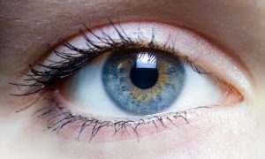
Though over ten million Americans have undergone LASIK vision corrective surgery since the option became available about 20 years ago, the procedure still poses some risk to patients. In addition to the usual risks of any surgery however, LASIK has even more due to the lack of a precise way to measure the refractive properties of the eye, which forces surgeons to make approximations in their measurements during the procedure. In an effort to eliminate this risk, a University of Maryland team of researchers in the Optics Biotech Laboratory led by Giuliano Scarcelli, Ph. D., designed a microscopy technique that would allow for precise measurements of these properties.

Using a form of light-scattering technology called Brillouin spectroscopy, Scarcelli and his lab found a way to directly determine a patient’s refractive index – the quantity surgeons need to know to be able to measure and adjust the way light travels through the eye. Often used as a way to sense mechanical properties of tissues and cells, this technology holds promise for taking three-dimensional spatial observations of these structures around the eye. Scarcelli hopes to keep improving the resolution of the new technique, to further understanding of the eye, and reduce even more of the risks involved with LASIK surgery.

Taking Tissue Models to the Final Frontier

Space flight is likely to cause deleterious changes to the composition of bacterial flora, leading to an increased risk of infection. The environment may also affect the susceptibility of microorganisms within the spacecraft to antibiotics, key components of flown medical kits, and may modify the virulence of bacteria and other microorganisms that contaminate the fabric of the International Space Station and other flight platforms.

“It has been known since the early days of human space flight that astronauts are more prone to infection,” says Dongeun (Dan) Huh, Wilf Family Term Assistant Professor in Bioengineering at Penn Engineering. “Infections can potentially be a serious threat to astronauts, but we don’t have a good fundamental understanding of how the microgravity environment changes the way our immune system reacts to pathogens.”

In collaboration with G. Scott Worthen, a physician-scientist in neonatology at the Children’s Hospital of Philadelphia, Huh will attempt to answer this question by sending tissues-on-chips to space. Last June, the Center for the Advancement of Science in Space (CASIS) and the National Center for Advancing Translational Sciences (NCATS), part of the National Institutes of Health (NIH), announced that the duo had received funding to study lung host defense in microgravity at the International Space Station.

Huh and Worthen aim to model respiratory infection, which accounts for more than 30 percent of all infections reported in astronauts. The project’s goals are to test engineered systems that model the airway and bone marrow, a critical organ in the immune system responsible for generating white blood cells, and to combine the models to emulate and understand the integrated immune responses of the human respiratory system in microgravity.

Read the rest of the article on Penn Engineering’s Medium Blog. Media contacts Evan Lerner and Janelle Weaver.

Sappi Limited Teams Up with the University of Maine to Develop Paper Microfluidics

At the Westbrook Technology Center of Sappi, a global pulp and paper company, researchers found ways to apply innovations in paper texture for medical use. So far, these include endeavors in medical test devices and patches for patient diagnostics. In collaboration with the Caitlin Howell, Ph.D., Assistant Professor of Chemical and Biomedical Engineering at the University of Maine, Sappi hopes to continue advances in these unconventional uses of their paper, especially as the business in paper for publishing purposes declines.

Sappi’s projects with the university focus on the development of paper microfluidics devices as what’s now becoming a widespread solution for obstacles in point-of-care diagnostics. One project in particular, called Sharklet, uses a paper that mimics shark skin as a way to impede unwanted microbial growth on the device – a key characteristic needed for its transition into commercial use. Beyond this example, Sappi’s work in developing paper microfluidics underscores the benefits of these devices in their mass producibility and adaptability.

New Observations of the WNT Pathway Deepen the Understanding of Protein Signaling in Cellular Development

Scientists at Rice University recently found that a protein signaling pathway called WNT, typically associated with its role in early organism development, can both listen for signals from a large amount of triggers and influence cell types throughout embryonic development. These new findings, published in PNAS, add to the already known functions of WNT, deepening our understanding of it and opening the doors to new potential applications of it in stem cell research.

Led by Aryeh Warmflash, Ph. D., researchers discovered that the WNT pathway is different between stem cells and differentiated cells, contrary to prior belief that it was the same for both. Using CRISPR-Cas9 gene editing technology, the Warmflash lab observed that the WNT signaling pathway is actually context-dependent throughout the process of cellular development. This research brings a whole new understanding to the way the WNT pathway operates, and could open the doors to new forms of gene therapy and treatments for diseases like cancers that involve genetic pathway mutations.

People and Places

In a recent article from Technical.ly Philly, named Group K Diagnostics on a list of ten promising startups in Philadelphia. Group K Diagnostics founder Brianna Wronko graduated with a B.S.E. from Penn’s Department of Bioengineering in 2017, and her point-of-care diagnostics company raised over $2 million in funding last year. Congratulations Brianna!

We would also like to congratulate Pamela K. Woodward, M.D., on her being named as the inaugural Hugh Monroe Wilson Professor of Radiology at the Washington University School of Medicine in St. Louis. Also a Professor of Biomedical Engineering at the university, Dr. Woodward leads a research lab with a focus on cardiovascular imaging, including work on new standards for diagnosis of pulmonary blood clots and on an atherosclerosis imaging agent.

Lastly, we would like to congratulate all of the following researchers on their election to the National Academy of Engineering:

  • David Bishop, Ph. D., a professor at the College of Engineering at Boston University whose current research involves the development of personalized heart tissue as an all-encompassing treatment for patients with heart disease.
  • Joanna Aizenberg, Ph. D., a professor of chemistry and chemical biology at Harvard University who leads research in the synthesis of biomimetic inorganic materials
  • Gilda Barabino, Ph. D., the dean of the City College of New York’s Grove School for Engineering whose lab focuses on cartilage tissue engineering and treatments for sickle cell disease.
  • Karl Deisseroth, M.D., Ph. D., a professor of bioengineering at Stanford University whose research involves the re-engineering of brain circuits through novel electromagnetic brain stimulation techniques.
  • Rosalind Picard, Ph.D., the founder and director of the Affective Computing Research Group at the Massachusetts Institute of Technology’s Media Lab whose research focuses on the development of technology that can measure and understand human emotion.
  • And finally, Molly Stevens, Ph. D., the Research Director for Biomedical Material Sciences at the Imperial College of London with research in understanding biomaterial interfaces for biosensing and regenerative medicine.