Enamored by the chemical processes of life, Yihui Shen, J. Peter and Geri Skirkanich Assistant Professor of Innovation in Bioengineering, started her research career as a chemist studying the way that proteins fold and the intricate dynamics underlying life processes.
“As an undergraduate, I studied physical chemistry, thinking that one day I’d be addressing challenges in hardcore STEM fields,” she says. “It wasn’t until I observed the dynamics of a single protein molecule that I fell in love with microscopy. I realized that this imaging tool could not only help us observe biological processes on a small scale, but it could also provide new insight at the interface of engineering, chemistry and physics and solve problems on a large scale.”
When Shen turned her attention to microscopy, the field itself was advancing quickly, with improvements being made and new techniques being released every month. Without missing a beat, Shen dove deeper into the most current tools available when she joined Dr. Wei Min’s lab at Columbia University as a doctoral student.
“Professor Wei Min is a pioneer in a new imaging technique called coherent Raman imaging,” says Shen. “In this type of microscopy, we focus light on a very specific point in the cell and measure the amount of scattered light that comes back after exchanging energy with the molecular vibration. This approach allows us to visualize the spatial distribution of different molecules, the very chemistry of life I had studied as an undergraduate, at a high enough resolution to gain insights into biological processes, such as tissue organization, drug distribution and cellular metabolism.”
With this new tool under her belt, Shen was able to ask the kinds of questions that could connect the use of this observation tool to practical applications for real-world challenges.
“I started thinking outside the box,” says Shen. “What if we could observe the chemical exchanges involved in metabolism as they are happening on the scale of a single cell, and then use that insight to pinpoint the exact metabolic pathways and molecules that facilitate tumor growth and disease?”
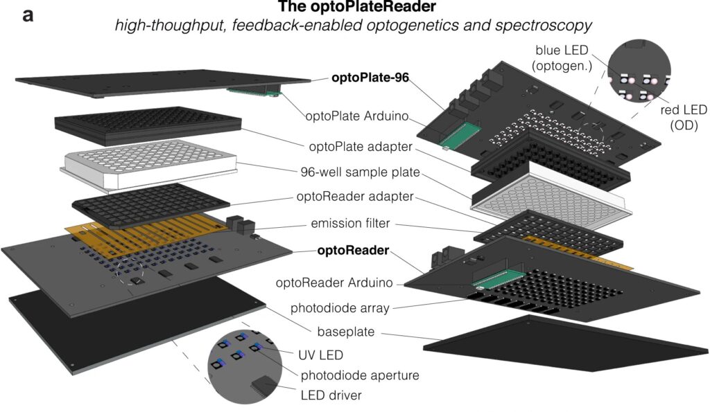
Diagram of the optoPlateReader, a high-throughput, feedback-enabled optogenetics and spectroscopy device initially developed by Penn 2021 iGEM team.
For bioengineers today, light does more than illuminate microscopes. Stimulating cells with light waves, a field known as optogenetics, has opened new doors to understanding the molecular activity within cells, with potential applications in drug discovery and more.
Thanks to recent advances in optogenetic technology, much of which is cheap and open-source, more researchers than ever before can construct arrays capable of running multiple experiments at once, using different wavelengths of light. Computing languages like Python allow researchers to manipulate light sources and precisely control what happens in the many “wells” containing cells in a typical optogenetic experiment.
However, researchers have struggled to simultaneously gather data on all these experiments in real time. Collecting data manually comes with multiple disadvantages: transferring cells to a microscope may expose them to other, non-experimental sources of light. The time it takes to collect the data also makes it difficult to adjust metabolic conditions quickly and precisely in sample cells.
Now, a team of Penn Engineers has published a paper in Communications Biology, an open access journal in the Nature portfolio, outlining the first low-cost solution to this problem. The paper describes the development of optoPlateReader (or oPR), an open-source device that addresses the need for instrumentation to monitor optogenetic experiments in real time. The oPR could make possible features such as automated reading, writing and feedback in microwell plates for optogenetic experiments.
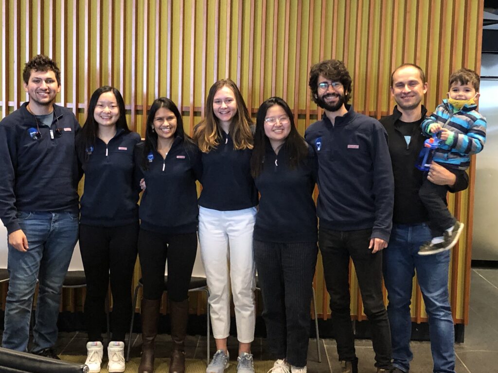
Left to right: Will Benman, Gloria Lee, Saachi Datta, Juliette Hooper, Grace Qian, David Gonzalez-Martinez, and Lukasz Bugaj (with Max).
The paper follows up on the award-winning work of six University of Pennsylvania alumni — Saachi Datta, M.D. Candidate at Stanford School of Medicine; Juliette Hooper, Programmer Analyst in Penn’s Perelman School of Medicine; Gabrielle Leavitt, M.D. Candidate at Temple University; Gloria Lee, graduate student at Oxford University; Grace Qian, Drug Excipient and Residual Analysis Research Co-op at GSK; and Lana Salloum, M.D. Candidate at Albert Einstein College of Medicine — who claimed multiple prizes at the 2021 International Genetically Engineered Machine Competition (iGEM) as Penn undergraduates.
The International Genetically Engineered Machine Competition (or iGEM) is the largest synthetic biology community and the premiere synthetic biology competition for both university and high school students from around the world. Hundreds of interdisciplinary teams of students compete annually, combining molecular biology techniques and engineering concepts to create novel biological systems and compete for prizes and awards through oral presentations and poster sessions.
The optoPlateReader was initially developed by Penn’s 2021 iGEM team, combining a light-stimulation device with a plate reader. At the iGEM competition, the invention took home Best Foundational Advance (best in track), Best Hardware (best from all undergraduate teams), and Best Presentation (best from all undergraduate teams), as well as a Gold Medal Distinction and inclusion in the Top 10 Overall and Top 10 Websites lists. (Read more about the 2021 iGEM team on the BE Blog.)
The original iGEM project focused on the design, construction, and testing of the hardware and software that make up the oPR, the focus of the new paper. After iGEM concluded, the team showed that the oPR could be used with real biological samples, such as cultures of bacteria. This work demonstrated that the oPR could be applied to real research questions, a necessary precursor to publication, and that the device could simultaneously monitor and manipulate living samples.
The main application for the oPR is in metabolic production (such as the creation of pharmaceuticals and bio-fuels). The oPR is able to issue commands to cells via light but can also take live readings about their current state. In the oPR, certain colors of light cause cells to carry out different tasks, and optical measurements give information on growth rates and protein production rates.
In this way, the new device is able to support production processes that can adapt in real time to what cells need, altering their behavior to maximize yield. For example, if an experiment produces a product that is toxic to cells, the oPR could instruct those cells to “turn on” only when the population of cells is dense and “turn off” when the concentration of that product becomes toxic and the cellular population needs to recover. This ability to pivot in real time could assist industries that rely on bioproduction.
The main challenges in developing this device were in incorporating the many light emitting diodes (LEDs) and sensors into a tiny space, as well as insulating the sensors from the nearby LEDs to ensure that the measured light came from the sample and not from the instrument itself. The team also had to create software that could coordinate the function of nearly 100 different sets of LEDs and sensors. Going forward, the team hopes to spread the word about the open-source oPR to other researchers studying metabolic production to enable more efficient research.
Lukasz Bugaj, Assistant Professor in Bioengineering and senior author of the paper, served as the team’s mentor along with Brian Chow, formerly an Associate Professor in Bioengineering and a founding member of the iGEM program at MIT, and Jose Avalos, Associate Professor of Chemical and Biological Engineering at Princeton University.
Key to the project’s development was the guidance of Bioengineering graduate students Will Benman, David Gonzalez Martinez, and Gabrielle Ho, as well as that of Saurabh Malani, a graduate student at Princeton University.
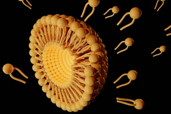
Like space shuttles using booster rockets to breach the atmosphere, lipid nanoparticles (LNPs) equipped with the new molecule more successfully deliver medicinal payloads. (Love Employee via Getty Images)
Inspired by the design of space shuttles, Penn Engineering researchers have invented a new way to synthesize a key component of lipid nanoparticles (LNPs), the revolutionary delivery vehicle for mRNA treatments including the Pfizer-BioNTech and Moderna COVID-19 vaccines, simplifying the manufacture of LNPs while boosting their efficacy at delivering mRNA to cells for medicinal purposes.
In a paper in Nature Communications, Michael J. Mitchell, Associate Professor in the Department of Bioengineering, describes a new way to synthesize ionizable lipidoids, key chemical components of LNPs that help protect and deliver medicinal payloads. For this paper, Mitchell and his co-authors tested delivery of an mRNA drug for treating obesity and gene-editing tools for treating genetic disease.
Previous experiments have shown that lipidoids with branched tails perform better at delivering mRNA to cells, but the methods for creating these molecules are time- and cost-intensive. “We offer a novel construction strategy for rapid and cost-efficient synthesis of these lipidoids,” says Xuexiang Han, a postdoctoral student in the Mitchell Lab and the paper’s co-first author.
Fat is a normal and necessary part of the body. Fat cells store and release energy, as well as play significant roles in hormonal regulation and immunity.
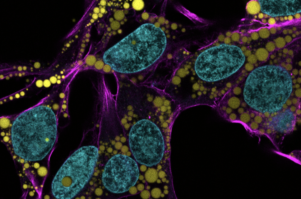
Engineers and scientists at the University of Pennsylvania are the first to discover fat-filled lipid droplets’ (pictured above in green) surprising capability to indent and puncture the nucleus, the organelle which contains and regulates a cell’s DNA.
In recent decades, a concerning rise in metabolic illnesses – such as cardiovascular disease, high blood pressure and diabetes – has focused scientific attention on the biology and chemistry of fat, resulting in a wealth of information about how fat cells work.
But fat cells and their metabolic activities are only part of the story.
Fat-filled lipid droplets, tiny spheres of fat many times smaller than fat cells, are a growing subject of scientific interest. Found inside many different cell types, these lipid particles have long been little understood. Studies have begun to illuminate these droplets’ participation in metabolic functions and cellular protection, but we still know next to nothing about the physical nature of fat.
Now, researchers at the University of Pennsylvania School of Engineering and Applied Science have looked beyond biochemistry to publish groundbreaking work on the physics of these droplets, revealing them to be a potential threat to a cell’s nucleus. In the August issue of the Journal of Cell Biology, they are the first to discover fat-filled lipid droplets’ surprising capability to indent and puncture the nucleus, the organelle which contains and regulates a cell’s DNA.
The stakes of their findings are high: a ruptured nucleus can lead to elevated DNA damage that is characteristic of many diseases, including cancer.
“Intuitively, people think of fat as soft,” says Discher. “And on a cellular level it is. But at this small size of droplet— measuring just a few microns rather than the hundreds of microns of a mature fat cell—it stops being soft. Its shape has a much higher curvature, bending other objects very sharply. This changes its physics in the cell. It can deform. It can damage. It can rupture.”