Through Brain Imaging Analysis in Rats, Penn Researchers Show Potential to Predict Whether Pain Will be Acute or Persistent

Beth Winkelstein, Megan Sperry, and Eric Granquist

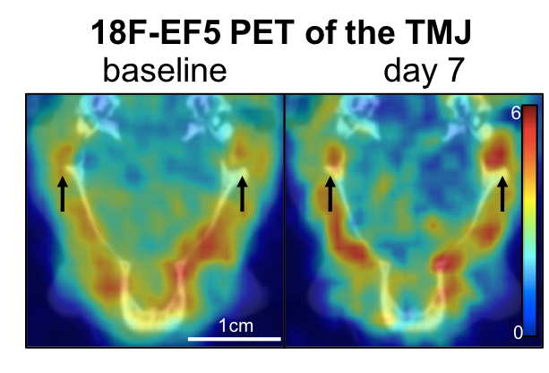
Pain may be a universal experience, but what actually causes that experience within our brains is still poorly understood. Pain often continues long after the relevant receptors in the body have stopped being stimulated and can persist even after those receptors cease to exist, as is the case with “phantom limb” pain.

The exact experience an individual will have after a painful incident comes down to the complex, variable connections formed between several different parts of the brain. The inability to predict how those connections will form and evolve can make pain management a tricky, frustrating endeavor for both healthcare providers and patients.

Now, a team of Penn researchers has shown a way to make such predictions from the pattern of neural connections that begin to take shape soon after the first onset of pain. Though their study was conducted in rats, it suggests that similar brain imaging techniques could be used to guide treatment decisions in humans, such as which individuals are most likely to benefit from different drugs or therapies.

The study, published in the journal Pain, was led by Beth Winkelstein, Eduardo D. Glandt President’s Distinguished Professor in Penn Engineering’s Department of Bioengineering and Deputy Provost of the University of Pennsylvania, along with Megan Sperry, then a graduate student in her lab. Eric Granquist, Director of the Center for Temporomandibular Joint Disease at the Hospital of the University of Pennsylvania in the Department of Oral & Maxillofacial Surgery, and assistant professor of Oral & Maxillofacial Surgery in Penn’s School of Dental Medicine, also contributed to the research.

“Our findings provide the first evidence that brain networks differ between acute and persistent pain states, even before those different groups of rats actually show different pain symptoms,” says Winkelstein.

Read the full story at Penn Engineering Today. Media contact Evan Lerner.

Sperry Wins BMES Design and Research Award

Sperry
Megan Sperry

Megan Sperry, a Ph.D. student in the Department of Bioengineering, is a recipient of a Student Design and Research Award from the Biomedical Engineering Society (BMES). Megan works in the Spine Pain Research Lab of Beth Winkelstein, Ph.D., professor of Bioengineering and Vice Provost for Education at Penn’s School of Engineering and Applied Science, as well as with Eric Granquist, DMD, MD, an oral and maxillofacial surgeon at Penn Dental Medicine.

With Drs. Winkelstein and Granquist, Megan studies temporomandibular joint (TMJ) pain and osteoarthritis, the latter of which can develop as a long-term consequence of untreated TMJ dysfunction.  There’s currently no way to determine which patients will progress to TMJ osteoarthritis, so Megan’s extended abstract, which was submitted to the BMES competition, detailed a study using 18F-EF5 PET, an imaging modality used mainly in oncology. Hypothesizing that hypoxia, or low oxygen, was a key factor in the development of TMJ osteoarthritis, Megan studied the relationship between hypoxia and persistent TMJ pain and found that hypoxia preceded reorganization of the cartilage of the TMJ, part of the process culminating in TMJ osteoarthritis (see image below).

Sperry
An example of 18F-EF5-PET imaging of the TMJ.

“This project has been both fun and challenging because it brings together concepts and techniques from multiple fields, including orthopedics, neuroscience, and, with the use of 18F-EF5, radiation oncology,” Megan said. “I’m excited to have the opportunity to share my work at the BMES Annual Meeting and receive feedback as we continue to move the project forward.”

Each year, BMES awards up to five graduate students the Student Design and Research Award from dozens of submissions. Congratulations to Megan for this elite recognition of her research!