Team Current Care (Andrew Lee, Antranig Baghdassarian, Johnson Liu, Leah Lackey, Brianna Leung, and Justin Liu), took home the $3,000 Grand Prize in the Cornell Hackathon.
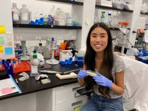
Brianna Leung, a rising senior majoring in Bioengineering and minoring in Neuroscience and Healthcare Management at the University of Pennsylvania, led a diverse team of student scientists and engineers to resounding success at the 2024 Cornell Health Tech Hackathon, where the team won the $3,000 Grand Prize.
Held in March 2024 on Cornell’s campus in New York City, the event brought together students from 29 different universities for a weekend of finding “hacks” to patient wellness and healthcare issues inspired by the theme of “patient safety.”
ADAPT members enjoy a pancake-making marathon in preparation for their pancake sale.
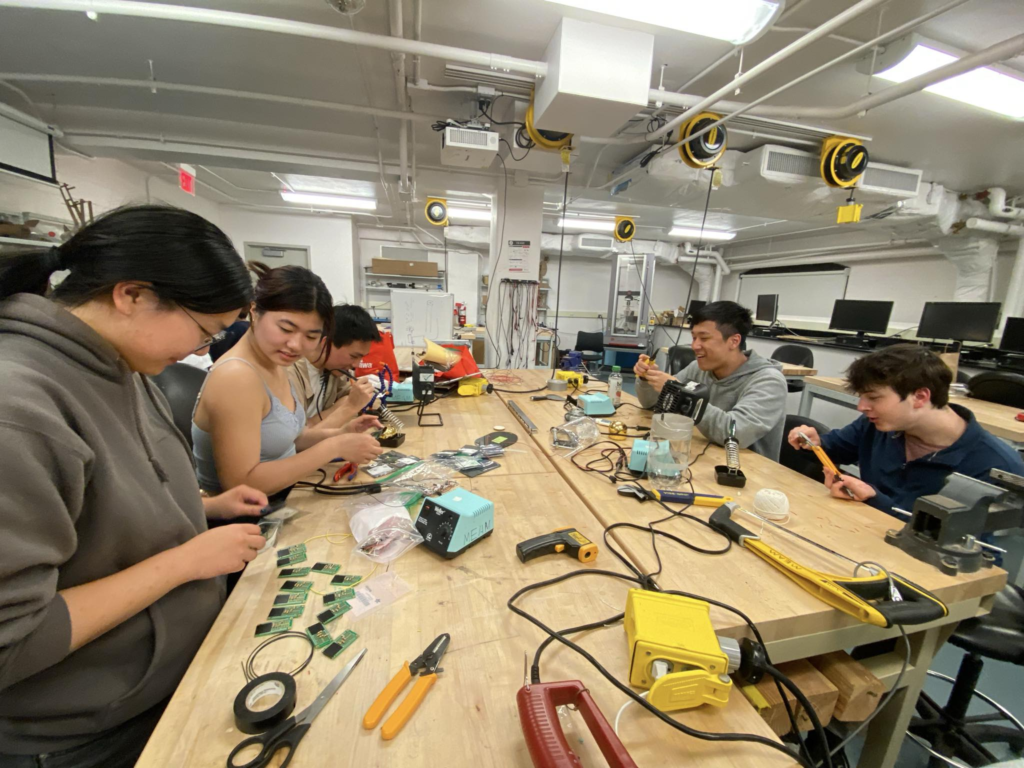
Leung serves as President of Penn Assistive Devices and Prosthetic Technologies (ADAPT), a medical-device project club whose members pursue personal projects, community partnerships and national design competitions. Penn ADAPT’s activities range from designing, building and improving assistive medical devices for conditions such as cerebral palsy and limb loss, to community engagement activities like their semesterly 3D-printed pancake sale.
In her role, Leung has increased the program’s hackathon participation to give club members greater exposure to fast-paced, competition-based design. She also leads the HMS School project, which develops and manufactures switch interfaces for children with cerebral palsy, enabling these students to interact with computers.
Leung’s passion for medical devices extends to her academic research. As a member of the robotics lab of Cynthia Sung, Gabel Family Term Assistant Professor in Mechanical Engineering and Applied Mechanics, Computer and Information Science, and Electrical and Systems Engineering, Leung characterizes origami patterns for energy-saving applications in the heart and in facial reconstruction. Leung has also served as Vice President External for the Penn Lions and Vice President of Member Engagement for the Wharton Undergraduate Healthcare Club, and belongs to the Phi Gamma Nu professional business fraternity.
ADAPT members working on medical devices.
For the Cornell Hackathon, Leung’s team developed a prototype for Current Care, a closed-loop device to prevent pressure ulcers through electrical muscle stimulation. Pressure ulcers, often called bed sores, result from prolonged pressure, which often occurs during extended hospitalization or in patients who are bedridden. This condition is exacerbated by understaffing and strained resources, and can create an extra burden on hospitals, patients and healthcare workers. The U.S. Department of Health and Human Services estimates that pressure ulcers cost the U.S. healthcare system approximately $9.1 billion to $11.6 billion per year.
Current Care is designed to deliver electrical stimulation, which increases blood flow to affected body parts. Conceptualizing and designing complex devices on short notice is the nature of a hackathon, so the team focused their efforts on creating proof-of-concept prototypes for all the different sensors required for the device, as well as providing the judges with on-screen read-outs to demonstrate the logic and hypothetical inputs for the device.
For their design, the team was awarded the $3,000 Grand Prize in the Cornell Hackathon. In addition to Leung, the team consisted of Johnson Liu (Cornell ECE & MSE’26); Antranig Baghdassarian (Cornell BME’27); Andrew Lee (Weill Cornell M.D.’25); Leah Lackey (Cornell ECE Ph.D.’28); and Justin Liu (Northeastern CS’27).
In choosing a project, Leung was inspired by her late grandmother’s experiences. “My role on the team largely consisted of coordinating and leading aspects of its development as needed. I also ultimately presented our idea to the judges,” she says. “This was actually all of my teammates’ first hackathon, so it was really exciting to serve a new role (considering it was actually only my second hackathon!). I had a lot of fun working with them, and we have actually been meeting regularly since the event to continue to work on the project. We had a range of expertise and experience on our team, and I deeply appreciate their hard work and enthusiasm for a project that means so much to me.”
Having found success at the Cornell hackathon, the team is discussing next steps for Current Care. “Our team is still very motivated to continue working on the project, and we’ve been speaking with professors across all of our schools to discuss feasibility and design plans moving forward,” says Leung.
Several other projects developed by Penn ADAPT members were recognized in the Cornell Hackathon:
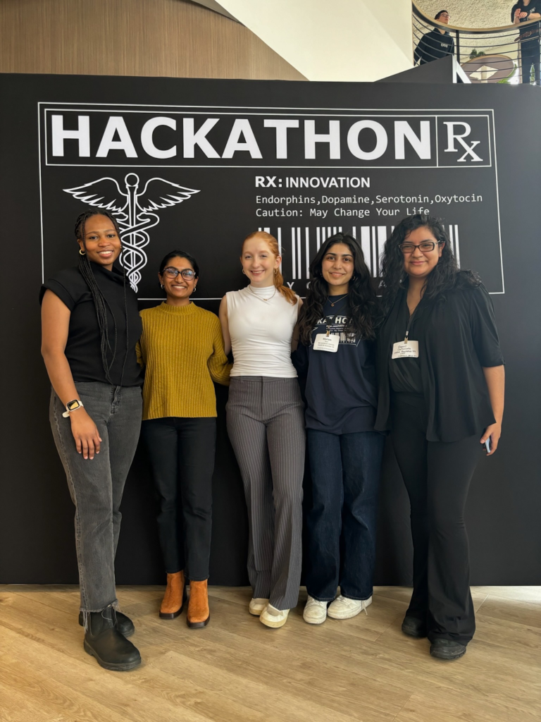
ADAPT members and Hackathon participants, left to right: Brianna Leung, Rebecca Wang, Claire Zhang, Amy Luo, Mariam Rizvi, Natey Kim, Joe Kojima. Also in attendance but not pictured: Suhani Patel, Harita Trivedi, Dwight Koyner.
Claire Zhang, a sophomore studying Bioengineering and Biology in the VIPER program, was a member and presenter for team CEDAR (winner of Most Innovative/2nd Place), a portable ultrasound imaging device used to monitor carotid artery stenosis development in rural areas.
Natey Kim, a sophomore in Bioengineering, was a member and presenter for team HMSS (finalist), a low-cost digital solution for forecasting infections in hospitals.
Rebecca Wang, a sophomore in Bioengineering and Social Chair of Penn ADAPT, was a member of Team Femnostics (winner of Most Market Ready/4th Place) which developed QuickSense, an all-in-one diagnostic tool that streamlines testing for a handful of the most common vaginal disease infections simultaneously.
Mariam Rizvi, a sophomore in Computational Biology, was a member of team IPVision (winner of Most Potential Impact/5th Place), an application programming interface (or API) that integrates into electronic health records such as Epic, leveraging AI to detect intimate partner violence cases and provide personalized treatment in acute-care settings.
Suhani Patel and Dwight Koyner worked with team RealAIs, which developed a full-stack multi-platform application using React Native and Vertex AI on the Google Cloud Platform (GCP). Patel, a sophomore double majoring in Bioengineering and Computer and Information Science in Penn Engineering, serves as ADAPT’s treasurer, while Koyner is a first-year M&T student studying Business and Systems Engineering in Penn Engineering and Wharton.
Team Femnostics from left to right: Antonia Li, Bhavishya Agarwal, Urmila Sehrawat, Rebecca Wang, Justin Xiang, and Edward Kim.Team IPVision from left to right: Elizabeth Madamidola, Keshika Gopinathan, Emily L. Leventhal, Mariam Rizvi, Raquel Castromonte, and Lina ChihoubTeam Cedar from left to right: Claire Zhang, Ethan Tse, Danny Nguyen, Allen Dinh, and Reem UllayTeam RealAIs from left to right: Rishi Shah, Suhani Patel, Saanvi Mehra, Dwight Koyner, Chimdi Ejiogu, and Garrett Breslin
Learn more about Penn ADAPT here and follow their Instagram.
Read more about the 2024 Cornell Tech Hackathon in the Cornell Chronicle.
Wu is among 55second-year and third-year students selected from 406 candidates nominated by 192colleges and universities nationwide. Scholars are recognized for leadership, public service, and commitment to issues related to the environment or to Native American nations. Each scholar will be awarded as much as $7,000.
A Taiwanese-American undergraduate scientist from Woodbury, Minnesota, Wu is the founder and international director of Waterroots, a nonprofit environmental education project that uses climate storytelling to combat water insecurity in more than 20 countries. Wu is a researcher in Penn Engineering’s McBride Lab, where he works as a plant specialist for a project that promotes environmental stability and sustainable agriculture. He is the deputy director of research for the nonprofit Climate Cardinals, a member of Penn’s Student Advisory Group for the Environment, and the North America representative for the Tunza Eco-Generation Ambassador program. Wu is a Clinton Global Initiative Scholar, a Duke Interfaith Climate Fellow, an IEEE Bio-X Scholar, a 2023 Millennium Fellow, and a 2024 UN ECOSOC Youth Delegate. In addition, he is a resident advisor in Penn’s Stouffer College House, as well as a Penn Engineering and a VIPER student ambassador.
Wu is the 10th student from Penn to be named a Udall Scholar since Congress established the foundation in 1992 to honor Morris and Stewart Udall for their impact on the nation’s environment, public lands, and natural resources and for their support of the rights and self-governance of American Indians and Alaska Natives.
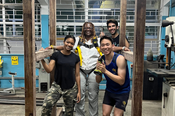
Students test the GaitMate harness and structure as a tool to help recovering patients walk.
Penn students have been building their knowledge and hands-on experience in places all over the world through Penn Global Seminars. Last May, “Robotics and Rehabilitation” brought Penn students back to the tropical island of Jamaica to collaborate with local university students and make an impact on recovery and quality of life for patients in Kingston and beyond.
Course leaders Camillo Jose (CJ) Taylor, Raymond S. Markowitz President’s Distinguished Professor in Computer and Information Science (CIS), and Michelle J. Johnson, Associate Professor of Physical Medicine and Rehabilitation at the Perelman School of Medicine and Associate Professor in Bioengineering (BE) and Mechanical Engineering and Applied Mechanics (MEAM) at Penn Engineering, brought the first cohort of students to the island in 2019.
“CJ and I are both Jamaicans by birth,” says Johnson. “We were both excited to introduce the next generation of engineers to robotics, rehabilitation and the process of culturally sensitive design in a location that we are personally connected to.”
As they built relationships with colleagues at the University of West Indies, Mona (UWI, Mona) and the University of Technology, Jamaica (UTECH), both Johnson and Taylor worked to tie the goals of the course to the location.
“In the initial iteration of the course, our goal was to focus on the applications of robotics to rehabilitation in a developing country where it is necessary to create solutions that are cost effective and will work in under-resourced settings,” says Taylor.
Taylor and Johnson wanted to make the course a regular offering, however, due to COVID-related travel restrictions, it wasn’t until last spring that they were able to bring it back. But when they did, they made up for lost time and expanded the scope of the course to include solving health problems for both people and the environment.
“While we started with a focus on people, we realized that the health and quality of life of a community is also impacted by the health of the environment,” says Taylor. “Jamaica has rich terrestrial and marine ecosystems, but those resources need to be monitored and regulated. We ventured into developing robotics tools to make environmental monitoring more effective and cost-friendly.”
One of those student-invented tools was a climate survey drone called “BioScout.”
“Our aim was to create a drone to monitor the ecosystem and wildlife in Jamaica,” says Rohan Mehta, junior in Systems Science and Engineering. “We wanted to help researchers and rangers who need to monitor wildlife and inspect forest sectors without entering and disturbing territories, but there were no available drones that met all of the following criteria necessary for the specific environment: affordable, modular, water-resistant and easy to repair. So we made our own.”
Another team of students created a smart buoy to reduce overfishing. The buoy was equipped with an alarm that goes off when fishermen get too close to a no-fishing zone.
Five other student teams dove into projects aligned to the original goals of the course. Their devices addressed patients’ decreased mobility due to diabetes, strokes and car accidents. These projects were sponsored by the Sir John Golding Rehabilitation Center.
One of which, the GaitMate, was engineered to help stroke patients who had lost partial muscle control regain their ability to walk.
“We developed a device that supports a patient’s weight and provides sensory feedback to help correct their form and gait as they walk on a treadmill, ultimately enhancing the recovery process and providing some autonomy to the patient,” says Taehwan Kim, senior in BE. “The device is also relatively cheap and simple, making it an option for a wide variety of physical therapy needs in Jamaica and other countries.”
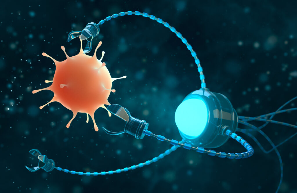
Biofilms—structured communities of microorganisms that create a protective matrix shielding them from external threats, including antibiotics—are responsible for about 80% of human infections and present a significant challenge in medical treatments, often resisting conventional methods.
In a Q&A with Penn Today, Hyun (Michel) Koo of the School of Dental Medicine and Edward Steager of the School of Engineering and Applied Science at Penn discuss an innovative approach they’ve partnered on: the use of small-scale robotics, microrobots, to offer a promising solution to tackle these persistent infections, bringing new capabilities and precision to the field of biomedical engineering.
Q: What is the motivation behind opting for tiny robots to tackle infections?
Koo: Treating biofilms is a broad yet unresolved biomedical problem, and conversely, the strategies for tackling biofilms are limited for a number of reasons. For instance, biofilms typically occur on surfaces that can be tricky to reach, like between the teeth in the oral cavity, the respiratory tract, or even within catheters and implants, so treatments for these are usually restricted to antibiotics (or antimicrobials) and other physical methods reliant on mechanical disruption. However, this touches on the problem of antimicrobial resistance: targeting specific microorganisms present in these structures is difficult, so antibiotics often fail to reach and penetrate the biofilm’s protective layers, leading to persistent infections and increased risk of antibiotic resistance.
We needed a way to circumvent these constraints, so Ed and I teamed up in 2017 to develop new, more precise and effective approaches that leverage the engineers’ ability to generate solutions that we, the clinicians and life science researchers, identify.
Hyun (Michel) Koo is a professor in the Department of Orthodontics and in the divisions of Pediatric Dentistry and Community Oral Health and the co-founder of the Center for Innovation & Precision Dentistry in the School of Dental Medicine at the University of Pennsylvania. He is a member of the Penn Bioengineering Graduate Group.
Edward Steager is a senior research investigator in Penn’s School of Engineering and Applied Science.
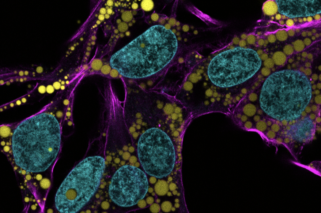
Fat is a normal and necessary part of the body. Fat cells store and release energy, as well as play significant roles in hormonal regulation and immunity.
Engineers and scientists at the University of Pennsylvania are the first to discover fat-filled lipid droplets’ (pictured above in green) surprising capability to indent and puncture the nucleus, the organelle which contains and regulates a cell’s DNA.
In recent decades, a concerning rise in metabolic illnesses – such as cardiovascular disease, high blood pressure and diabetes – has focused scientific attention on the biology and chemistry of fat, resulting in a wealth of information about how fat cells work.
But fat cells and their metabolic activities are only part of the story.
Fat-filled lipid droplets, tiny spheres of fat many times smaller than fat cells, are a growing subject of scientific interest. Found inside many different cell types, these lipid particles have long been little understood. Studies have begun to illuminate these droplets’ participation in metabolic functions and cellular protection, but we still know next to nothing about the physical nature of fat.
Now, researchers at the University of Pennsylvania School of Engineering and Applied Science have looked beyond biochemistry to publish groundbreaking work on the physics of these droplets, revealing them to be a potential threat to a cell’s nucleus. In the August issue of the Journal of Cell Biology, they are the first to discover fat-filled lipid droplets’ surprising capability to indent and puncture the nucleus, the organelle which contains and regulates a cell’s DNA.
The stakes of their findings are high: a ruptured nucleus can lead to elevated DNA damage that is characteristic of many diseases, including cancer.
“Intuitively, people think of fat as soft,” says Discher. “And on a cellular level it is. But at this small size of droplet— measuring just a few microns rather than the hundreds of microns of a mature fat cell—it stops being soft. Its shape has a much higher curvature, bending other objects very sharply. This changes its physics in the cell. It can deform. It can damage. It can rupture.”
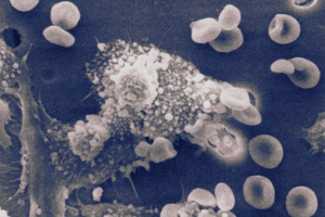
“Macrophages killing cancer cell” photographed by Susan Arnold.
By silencing the molecular pathway that prevents macrophages from attacking our own cells, Penn Engineers have manipulated these white blood cells to eliminate solid tumors.
Cancer remains one of the leading causes of death in the U.S. at over 600,000 deaths per year. Cancers that form solid tumors such as in the breast, brain or skin are particularly hard to treat. Surgery is typically the first line of defense for patients fighting solid tumors. But surgery may not remove all cancerous cells, and leftover cells can mutate and spread throughout the body. A more targeted and wholistic treatment could replace the blunt approach of surgery with one that eliminates cancer from the inside using our own cells.
Dennis Discher, Robert D. Bent Professor in Chemical and Biomolecular Engineering, Bioengineering, and Mechanical Engineering and Applied Mechanics, and postdoctoral fellow, Larry Dooling, provide a new approach in targeted therapies for solid tumor cancers in their study, published in Nature Biomedical Engineering. Their therapy not only eliminates cancerous cells, but teaches the immune system to recognize and kill them in the future.
“Due to a solid tumor’s physical properties, it is challenging to design molecules that can enter these masses,” says Discher. “Instead of creating a new molecule to do the job, we propose using cells that ‘eat’ invaders – macrophages.”
Macrophages, a type of white blood cell, immediately engulf and destroy – phagocytize – invaders such as bacteria, viruses, and even implants to remove them from the body. A macrophage’s innate immune response teaches our bodies to remember and attack invading cells in the future. This learned immunity is essential to creating a kind of cancer vaccine.
But, a macrophage can’t attack what it can’t see.
“Macrophages recognize cancer cells as part of the body, not invaders,” says Dooling. “To allow these white blood cells to see and attack cancer cells, we had to investigate the molecular pathway that controls cell-to-cell communication. Turning off this pathway – a checkpoint interaction between a protein called SIRPa on the macrophage and the CD47 protein found on all ‘self’ cells – was the key to creating this therapy.”
Multiple members in the biophysical engineering lab lead by Dennis Discher, including co-lead author and postdoctoral fellow and Penn Bioengineering alumnus Jason Andrechak and Bioengineering Ph.D. student Brandon Hayes, contributed to this study. The research was funded by grants from the National Heart, Lung, and Blood Institute and the National Cancer Institute, including the Physical Sciences Oncology Network, of the US National Institutes of Health.
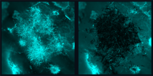
Candida albicans is a species of yeast that is a normal part of the human microbiota but can also cause severe infections that pose a significant global health risk due to their resistance to existing treatments, so much so that the World Health Organization has highlighted this as a priority issue. The picture above shows a before (left) and after (right) fluorescence image of fungal biofilms being precisely targeted by nanozyme microrobots without bonding to or disturbing the tissue sample. (Image: Min Jun Oh and Seokyoung Yoon)
Infections caused by fungi, such as Candida albicans, pose a significant global health risk due to their resistance to existing treatments, so much so that the World Health Organization has highlighted this as a priority issue.
Although nanomaterials show promise as antifungal agents, current iterations lack the potency and specificity needed for quick and targeted treatment, leading to prolonged treatment times and potential off-target effects and drug resistance.
“Candida forms tenacious biofilm infections that are particularly hard to treat,” Koo says. “Current antifungal therapies lack the potency and specificity required to quickly and effectively eliminate these pathogens, so this collaboration draws from our clinical knowledge and combines Ed’s team and their robotic expertise to offer a new approach.”
The team of researchers is a part of Penn Dental’s Center for Innovation & Precision Dentistry, an initiative that leverages engineering and computational approaches to uncover new knowledge for disease mitigation and advance oral and craniofacial health care innovation.
For this paper, published in Advanced Materials, the researchers capitalized on recent advancements in catalytic nanoparticles, known as nanozymes, and they built miniature robotic systems that could accurately target and quickly destroy fungal cells. They achieved this by using electromagnetic fields to control the shape and movements of these nanozyme microrobots with great precision.
“The methods we use to control the nanoparticles in this study are magnetic, which allows us to direct them to the exact infection location,” Steager says. “We use iron oxide nanoparticles, which have another important property, namely that they’re catalytic.”
Other authors include Min Jun Oh, Alaa Babeer, Yuan Liu, Zhi Ren, Zhenting Xiang, Yilan Miao, and Chider Chen of Penn Dental; and David P. Cormode and Seokyoung Yoon of the Perelman School of Medicine. Cormode also holds a secondary appointment in Bioengineering.
This research was supported in part by the National Institute for Dental and Craniofacial Research (R01 DE025848, R56 DE029985, R90DE031532 and; the Basic Science Research Program through the National Research Foundation of Korea of the Ministry of Education (NRF-2021R1A6A3A03044553).
Sharon Kuo, a graduating senior in Mechanical Engineering and Applied Mechanics (MEAM), is the inaugural recipient of the Madison “Maddie” Magee Award for Undergraduate Excellence.
Kuo, who is also minoring in Mathematics, comes to Penn from Taipei, Taiwan. Her interests within her major include mechanical design and product design, and she is passionate about space exploration and advancing human spaceflight.
This award will continue to be presented each year to a Penn Engineering senior who best exemplifies the energy, enthusiasm and excellence that was Maddie.
The award for Undergraduate Excellence was established in honor of Madison “Maddie” N. Magee, who graduated with both a bachelor’s degree in Mechanical Engineering and Applied Mechanics (MEAM) and a master’s degree in Bioengineering (BE) in 2021. Maddie passed away while hiking the Pacific Crest Trail on May 28, 2022. Read more about this award here.
Penn Engineering is proud to announce the establishment of the Madison “Maddie” Magee Award for Undergraduate Excellence, named in honor of the memory of Madison “Maddie” N. Magee, who graduated with both a bachelor’s degree in Mechanical Engineering and Applied Mechanics (MEAM) and a master’s degree in Bioengineering (BE) in 2021. Following her time at Penn, Maddie joined the Integrative Baseball Performance department of the Philadelphia Phillies, where she collaborated with a group in developing the next generation of baseball players by analyzing biomechanics data.
To establish this award, 130 donors, including the Philadelphia Phillies, came together in 2022 to raise over $50,000, meaning that undergraduate students will be able to receive this award in perpetuity. Recipients will be Penn Engineering seniors who “exemplify the energy, enthusiasm, and excellence that was Maddie.”
“Maddie was full of life and promise and brought unmatched passion and spirit to everything she did,” says Kevin T. Turner, Professor and Chair of MEAM. “It was impossible to not see the impact that she was having on our Department and the School.” Magee excelled as a student at Penn, working as a Teaching Assistant at both Penn and Drexel and providing countless hours of tutoring to fellow students.
It is with deep gratitude for Maddie’s profound and lasting impact on many students, faculty and staff that this award is established.
Maddie passed away while hiking the Pacific Crest Trail on May 28, 2022.
Each year, the the Department of Bioengineering seeks exceptional candidates to conduct summer research in bioengineering with the support of two scholarships: the Abraham Noordergraaf Student Summer Bioengineering Research Fund and the Blair Undergraduate Research Fund in the Department of Bioengineering. These scholarships provide a living stipend for students to conduct research on campus in a Penn research lab under the mentorship of a faculty member. The Abraham Noordergraaf Student Summer Bioengineering Research Fund provides financial support for undergraduate or graduate summer research opportunities in bioengineering with a preference for study in the area of cardiovascular systems. Dr. Noordergraaf, who died in 2014, was a founding member and first chair of Penn Bioengineering. The Blair Undergraduate Research Fund in the Department of Bioengineering supports three to five undergraduate research scholars each year with the support of Dr. James C. Blair II. After a competitive round of proposals, the following six scholars were chosen for the Summer 2022 semester. Keep reading below for the research abstracts and bios of the awardees.
The Blair Undergraduate Research Fund in the Department of Bioengineering (Blair Scholars)
Ella Atsavapranee
Student: Ella Atsavapranee (BE Class of 2023)
PI: Michael J. Mitchell, J. Peter and Geri Skirkanich Assistant Professor of Innovation, Bioengineering
“Lipid nanoparticle-mediated delivery of RAS protease to inhibit cancer cell growth”
Mutations in RAS, a family of proteins found in all human cells, drive a third of cancers, including many pancreatic, colorectal, and lung cancers. However, there are still no therapies that can effectively prevent RAS from causing tumor growth. Recently, a protease was engineered to specifically degrade active RAS, offering a promising new tool for treating these cancers. However, many protein-based therapies still cannot be effectively delivered to patients. Lipid nanoparticles (LNPs), which were used in the Pfizer-BioNTech and Moderna COVID-19 vaccines, have emerged as a promising platform for safe and effective delivery of both nucleic acids and proteins. We formulated a library of LNPs using different cationic lipids. We characterized the LNPs by size, charge, and pKa, and tested their ability to deliver fluorescently labeled protease. The LNPs were able to encapsulate and deliver a RAS protease, successfully reducing proliferation of colon cancer cells.
Ella is a senior from Maryland studying bioengineering and chemistry. She works in Dr. Michael Mitchell’s lab, developing lipid nanoparticles to deliver proteins that reduce cancer cell proliferation. She has also conducted research on early-stage cancer detection and therapy monitoring (at Stanford University) and drug delivery across the blood-brain barrier for neurodegenerative diseases (at University of Maryland). She is passionate about translational research, science communication, and promoting diversity in STEM.
Chiadika Eleh
Student: Chiadika Eleh (BE and CIS Class of 2024)
PI: Eric J. Brown, Associate Professor of Cancer Biology, Perelman School of Medicine
“Investigating Viability in ATR and WEE1 Inhibitor Treated Ovarian Cancer Cells”
High-grade serous ovarian cancers (HGSOCs) are an aggressive subtype of ovarian cancer, accounting for up to 80% of all ovarian cancer-related deaths. More than half of HGSOCs are homologous recombination deficient; thus, they lack a favorable response when treated with common chemotherapeutic trials. Therefore, new treatment strategies must be developed to increase the life expectancy and quality of life of HGSOC patients. To address the lack of effective treatment options, the Brown Lab is interested in combining ATR and WEE1 inhibition (ATRi/WEE1i) to target HGSOC cells. It has previously been shown that low-dose ATRi/WEE1i is an effective treatment strategy for CCNE1-amplified ovarian cancer-derived PDX tumors (Xu et al., 2021, Cell Reports Medicine). Therefore, the next step is to characterize the HGSOC-specific response to ATRi/WEE1i treatment. This project aims to characterize the viability phenotype of ovarian cancer (OVCAR3) cells in the presence of ATRi/WEE1i in both single and combination treatments. With further research, Eleh hopes to prove the hypothesis low-dose combination ATRi/WEE1i treatment will result in the synergistic loss of viability in OVCAR3 cells. This goal will be achieved through the treatment of OVCAR3 cells with ranging doses of ATRi and Wee1i over 24 and 48 hour time intervals. We hope that this data will help set a treatment baseline that can be used for all OVCAR30-based viability experiments in the future.
Chiadika Eleh is a Bioengineering and Computer Science junior and a member of Penn Engineering’s Rachleff Scholar program. As a Blair Scholar, she worked in Dr. Eric Brown’s cancer biology lab, where she studied cell cycle checkpoint inhibitors as a form of cancer treatment.
“Tbc1d2b regulates vascular formation during development and tissue repair after ischemia”
The mechanisms behind endothelial cells forming blood vessels remains unknown. We have identified Tbc1d2b as a protein that is integral to the regulation of vascular formation. In order to investigate the role of Tbc1d2b in tubule formation, fibrin gel bead assays will be conducted to evaluate how the presence of Tbc1d2b is required for angiogenesis. Fibrin gel bead assays simulate the extracellular matrix environment to support the in vitro development of vessels from human umbilical vein endothelial cells (HUVEC) coated on cytodex beads. In order to confirm the success of angiogenesis, immunostaining for Phalloidin and CD31 will be conducted. After confirmation that fibrin gel bead assays can produce in vitro tubules, sgRNA CRISPR knockout of Tbc1d2b will be performed on HUVEC cells which will then be used to conduct more fibrin gel bead assays. We hypothesize that HUVEC with the Tbc1d2b knockout phenotype will be unable to form tubules while wild type HUVEC will be able to.
Gloria Lee is a rising senior studying Bioengineering and Physics in the VIPER program from Denver, Colorado. Her research in Dr. Yi Fan’s lab focuses on the role that proteins play in cardiovascular tubule formation.
Abraham Noordergraaf Student Summer Bioengineering Research Fund (Noordergraaf Fellows)
Gary Lin
Student: Gary Lin (Master’s in MEAM Class of 2023)
PI: Michelle J. Johnson, Associate Professor in Physical Medicine and Rehabilitation, Perelman School of Medicine, and in Bioengineering
“Development and Integration of Dynamically Modulating Control Systems in the Rehabilitation Using Community-Based Affordable Robotic Exercise System (Rehab CARES)”
As the number of stroke patients requiring rehabilitative care continues to increase, strain is being put onto the US health infrastructure which already has a shortage of rehabilitation practitioners. To help alleviate this pressure, a cost-effective robotic rehabilitative platform was developed to increase access to rehabilitative care. The haptic TheraDrive, a one-degree of freedom actuated hand crank that can apply assistive and resistive forces, was modified to train pronation and supination at the elbow and pinching of the fingers in addition to flexion and extension of the elbow and shoulder. Two controllers were created including an open-loop force controller and a closed-loop proportional-integral (PI) with adaptive control gains based on subject performance in therapy-game tasks as well as galvanic skin response. Stroke subjects (n=11) with a range of cognitive and motor impairment completed 4 therapy games in both adaptive and non-adaptive versions of the controllers (n=8) while measuring force applied on the TheraDrive handle. Resulting normalized average power versus Upper Extremity Fugl-Meyer (UE-FM) and Montreal Cognitive Assessment (MoCA) correlation analyses showed that power was strongly correlated with UE-FM in 2 of the conditions and moderately correlated with the other 6 while MoCA was moderate correlated to 2 of the conditions and weakly correlated to the rest. Mann-Whitney U-tests between adaptive and non-adaptive versions of each therapy game showed no significant differences with regards to power between controller types (p<0.05).
Gary is a master’s student in the School of Engineering studying Mechanical Engineering and Applied Mechanics with a concentration in Robotic and Mechatronic systems. His research primarily focuses on developing affordable rehabilitation robotics for use in assessment and game-based therapies post neural injury. Many of his interests revolve around the design of mechatronic systems and the algorithms used to control them for use in healthcare spaces.
Priya Shah
Student: Priya Shah (BE Class of 2024)
PI: Alex J. Hughes, Assistant Professor in Bioengineering
“Optogenetic Control of Developing Kidney Cells for Future Treatment of End-Stage Renal Disease”
This project sought to build from prior research in the Hughes Lab on the geometric and mechanical consequences of kidney form on cell and tissue-scale function. While the developmental trajectory of the kidney is well understood, little is currently known about many factors affecting nephron progenitor differentiation rate. Insufficient differentiation of nephron progenitor cells during kidney formation can result in lower nephron number and glomerular density, which is a risk factor for progression to end-stage renal disease later in life. Prior studies indicated that the amount of nephron differentiation – and thus function of the adult kidney – is correlated to the packing of ureteric tubule tips present at the surface of the kidney. Building off of research conducted in the Bugaj Lab, we found that inserting an optogenetic construct into the genome of human embryonic kidney (HEK) cells allowed us to manipulate the contraction of those cells through exposing them to blue light. Manipulating the contraction of the cells allows for the manipulation of the packing of ureteric tubule tips at the kidney surface. We used a lentiviral vector to transduce HEK293 cells with the optogenetic construct and witnessed visible contraction of the cells when they were exposed to blue light. Future work will include using CRISPR-Cas9 to introduce the optogenetic construct into IPS cells.
Priya is a junior studying bioengineering and had the opportunity to work on manipulating developing kidney cells using an optogenetic construct in the Hughes Lab this summer. She is thrilled to continue this research throughout the coming school year. Outside of the lab, Priya is involved with the PENNaach dance team and the Society of Women Engineers, as well as other mentorship roles.
Cosette Tomita
Student: Cosette Tomita (Master’s in MEAM Class of 2023)
“Expression and Characterization of an Anti-Aβ42 scFv”
Background: Amyloid Beta (Aβ42) fibrils contribute to the pathology of Alzheimer’s Disease. Numerous monoclonal antibodies have been developed against Aβ42. In this study we have designed and expressed a short chain variable fragment specific to Aβ42 (Anti-Aβ42 scFv). To characterize our anti-Aβ42 scFv we have performed structural analysis using transmission electron microscopy (TEM) and binding kinetics using microscale thermophoresis (MST) compared to commercially available antibodies 6E10, Aducanumab, and an IgG isotype control. The goal of this study is to determine if labeling densities and binding constants for Aducanumab and anti-Aβ42 scFv are not significantly different.
Method: To characterize Aβ42 fibril associated antibodies we used negative stain TEM. Aβ42 fibrils were stained on a glow discharged copper grid, and incubated with gold conjugated anti-Aβ42 scFv, 6E10—which binds all Aβ species, aducanumab, or IgG isotype control. Labeling densities were calculated as the number of fibril-associated gold particles per 1 μm2 for each image. Next, we used microscale thermophoresis determine the binding kinetics. Antibodies or anti-Aβ42 scFv were labeled with Alexa Fluor-647 and unlabeled Aβ42 was titrated in a serial dilution over 16 capillaries. The average fluorescence intensity was plotted against the antibody or scFv concentration and the curves were analyzed using the GraphPad Prism software to calculate the dissociation constant (KD) values.
Results: We found a significant difference, tested with a one-way ANOVA (P <0.0001), in gold particle associated Aβ fibrils per 1 μm2 between anti-Aβ42 scFv, 6E10, aducanumab, and IgG isotype control. Further analysis of aducanumab and 6CO3 with unpaired student t-test indicates significant differences in fibril associated gold particles between aducanumab vs. 6E10 (P=0.0003), Aducanumab vs. Isotype control (P <0.0001), anti-Aβ42 scFv vs 6E10 (p=0.0072), and anti-Aβ42 scFv vs Isotype Control (P=0.0029) with no significant difference in labeling densities between Aducanumab and anti-Aβ42 scFv. The expected KD values from MST were 1.8μM for Aducanumab and anti-Aβ42 scFv, 10.3nM for 6E10 and no expected binding for the isotype control. The experimental KD values for anti-Aβ42 scFv and 6E10 are 0.1132μM and 1.467μM respectively. The KD value for Isotype control was undetermined, as expected, however, the KD for Aducanumab was undetermined due to suboptimal assay conditions. Due to confounding variables in the experimental set up such as the use of Aβ1-16 compared to Aβ42 and the use of different fluorophores—5-TAMRA, Alexa Fluor 647 or FITC— the experimental KD values were off by several orders of magnitude.
Conclusion: We have illustrated similar labeling densities between Aducanumab and our anti-Aβ42 scFv. In the future, we will further optimize the MST assay conditions and compare the KD values obtained by MST with other techniques such as surface plasma resonance.
Cosette was born and raised in Chicago land area. Go Sox! She attended University of Missouri where she majored in Chemistry and Biology. She synthesized sigma-2 radiotracers and developed advanced skills in biochemical techniques in Dr. Susan Lever’s lab. After graduation, she moved to NJ to work at Lantheus, a radiopharmaceutical company. She missed academia and the independence of program and project development, so she came to work at the Penn Cyclotron facility before entering the Bioengineering master’s program.


 Penn Engineering is proud to announce the establishment of the Madison “Maddie” Magee Award for Undergraduate Excellence, named in honor of the memory of Madison “Maddie” N. Magee, who graduated with both a bachelor’s degree in Mechanical Engineering and Applied Mechanics (MEAM) and a master’s degree in Bioengineering (BE) in 2021. Following her time at Penn, Maddie joined the Integrative Baseball Performance department of the Philadelphia Phillies, where she collaborated with a group in developing the next generation of baseball players by analyzing biomechanics data.