Leveraging the Body’s Postal System to Understand and Treat Disease

by Nathi Magubane

Microwell device with a solution in the reservoir (Image: Courtesy of David E. Reynolds)

Akin to the packages sent from one person to another via an elaborate postal system, cells send tiny parcels that bear contents and packaging material that serve key purposes: To protect the contents from the outside world and to make sure it gets to the right place via a label with an address. 

These packages are known as extracellular vesicles (EVs)—lipid-bound molecules that serve a variety of regulatory and maintenance functions throughout the body. They assist in the removal of unwanted materials within the cell, and they transport proteins, aid in DNA and RNA transfer, and promote tumorigeneses in cancerous cells. 

Given their myriad roles, EVs have taken center stage for many researchers in the biomedical space as they have the potential to improve current methods of disease detection and treatment. The main challenge, however, is accurately identifying the molecular contents of EVs while also characterizing the EVs, which, unlike other cellular components that are more homogenous, have more heterogeneity.

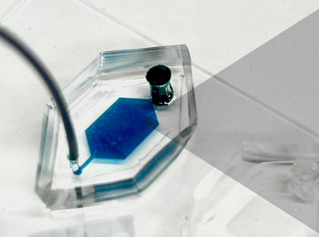
Now, a team of researchers at the University of Pennsylvania has developed a novel platform, droplet-free double digital assay, for not only profiling individual EVs but also accurately discerning their molecular contents. The researchers took the digital assay, which quantifies the contents of a molecule via binary metric—a 1 corresponds to the presence of a molecule and a zero to the lack thereof—and applies it to the EV. The work is published in Advanced Science.

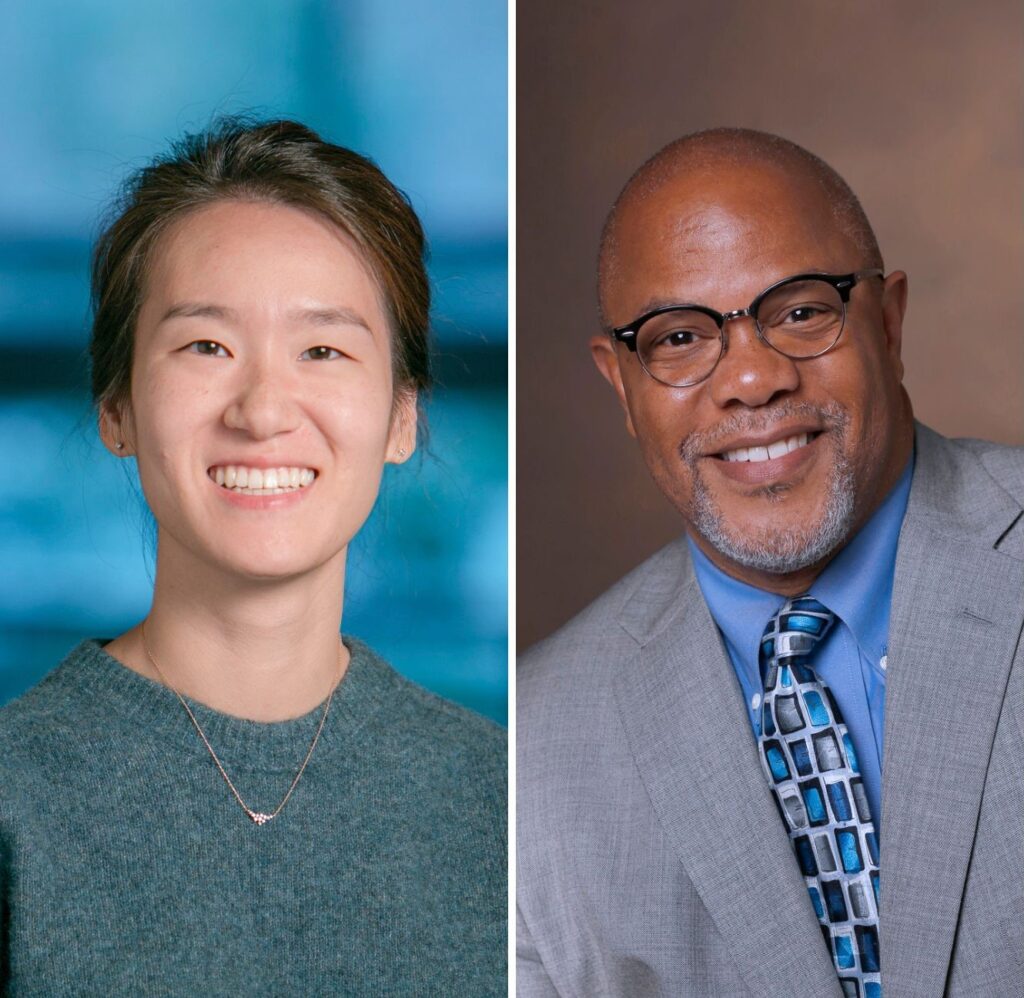
The team was led by Jina Ko, an assistant professor with appointments in the School of Engineering and Applied Science and Perelman School of Medicine. “Our method allows for highly accurate quantification of the individual molecules inside an EV,” Ko says . “This opens up many doors in the realm of early disease detection and treatment.”

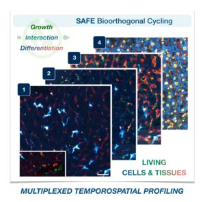
The researchers first compartmentalized individual EVs utilizing a microwell approach to isolate the EVs. Next, they captured individual molecules within the EVs and amplified the signal for clarity. The team then was able to determine the expression levels of pivotal EV biomarkers with remarkable precision via fluorescence.

Read the full story in Penn Today.

Jina Ko is an assistant professor in the Department of Pathology and Laboratory Medicine in the Perelman School of Medicine and an assistant professor in the Department of Bioengineering in the School of Engineering and Applied Science at the University of Pennsylvania.

David Reynolds is a Ph.D. candidate in the Department of Bioengineering in Penn Engineering.

Other authors include, Menghan Pan, George Galanis, Yoon Ho Roh, Renee-Tyler T. Morales, Shailesh Senthil Kumar, and Su-Jin Heo of the Department of Bioengineering at Penn Engineering; Jingbo Yang and Xiaowei Xu of the Department of Pathology and Laboratory Medicine at Penn Medicine; and Wei Guo of the Department of Biology in the School of Arts & Sciences at Penn.

The research was supported by the National Institutes of Health: grants R00CA256353, R35 GM141832, and CA174523 (SPORE).

Two Penn Bioengineers Receive NIH Director Award

by Nathi Magubane

Jina Ko (left) and Kevin Johnson (right), both from the School of Engineering and the Perelman School of Medicine with appointments in Bioengineering, have received the National Institute of Health Director’s Award to support their “highly innovative and broadly impactful” research projects through the High-Risk, High-Reward program.

The National Institutes of Health (NIH) has awarded grants to three researchers from the University of Pennsylvania through the NIH Common Fund’s High-Risk, High-Reward Research program. The research of Kevin B. Johnson, Jina Ko, and Sheila Shanmugan will be supported through the program, which funds “highly innovative and broadly impactful” biomedical or behavioral research by exceptionally creative scientists.

The High-Risk, High-Reward Research program catalyzes scientific discovery by supporting highly innovative research proposals that, due to their inherent risk, may struggle in the traditional peer-review process despite their transformative potential. Program applicants are encouraged to think “outside the box” and pursue trail-blazing ideas in any area of research relevant to the NIH’s mission to advance knowledge and enhance health.

Two Penn Bioengineering faculty, Johnson and Ko, are among 85 recipients for 2023.

Johnson, the David L. Cohen University Professor of Pediatrics, is a Penn Integrates Knowledge University Professor who holds appointments in the Department of Computer and Information Science in the School of Engineering and Applied Science and the Department of Biostatistics, Epidemiology, and Informatics in the Perelman School of Medicine. He also holds secondary appointments in Bioengineering, Pediatrics, and in the Annenberg School for Communication. He is widely known for his work with e-prescribing and computer-based documentation and, more recently, work communicating science to lay audiences, which includes a documentary about health-information exchange. Johnson has authored more than 150 publications and was elected to the American College of Medical Informatics, Academic Pediatric Society, National Academy of Medicine, International Association of Health Science Informatics, and American Institute for Medical and Biological Engineering.

Ko is an assistant professor in the Department of Pathology and Laboratory Medicine in the Perelman School of Medicine and Department of Bioengineering in the School of Engineering and Applied Science. She focuses on developing single molecule detection from single extracellular vesicles and multiplexed molecular profiling to better diagnose diseases and monitor treatment efficacy. Ko earned her Ph.D. in bioengineering at Penn in 2018, during which time she developed machine learning-based microchip diagnostics that can detect blood-based biomarkers to diagnose pancreatic cancer and traumatic brain injury. For her postdoctoral training, she worked at the Massachusetts General Hospital and the Wyss Institute at Harvard University as a Schmidt Science Fellow and a NIH K99/R00 award recipient. Ko developed new methods to profile single cells and single extracellular vesicles with high throughput and multiplexing.

Read the full announcement in Penn Today.

2023 Graduate Research Fellowships for Bioengineering Students

Congratulations to the fourteen Bioengineering students to receive 2023  National Science Foundation Graduate Research Fellowship Program (NSF GRFP) fellowships. The prestigious NSF GRFP program recognizes and supports outstanding graduate students in NSF-supported fields. The recipients honorees were selected from a highly-competitive, nationwide pool. Further information about the program can be found on the NSF website.

Carlos Armando Aguila, Ph.D. student in Bioengineering, is a member of the Center of Neuroengineering and Therapeutics, advised by Erin Conrad, Assistant Professor in Neurology, and Brian Litt, Professor in Bioengineering and Neurology. His research focuses on analyzing electroencephalogram (EEG) signals to better understand epilepsy.

Joseph Lance Victoria Casila is a Ph.D. student in Bioengineering in the lab of Riccardo Gottardi, Assistant Professor in Pediatrics and Bioengineering. His research focuses on probing environmental factors that influence stem cell differentiation towards chondrogenesis for cartilage engineering and regeneration.

Trevor Chan is a Ph.D. student in Bioengineering in the lab of Felix Wehrli, Professor of Radiologic Science. His research is in developing computational methods for medical image refinement and analysis. Two ongoing projects are: self-supervised methods for CT super-resolution and assessment of osteoporosis, and semi-supervised segmentation of 3D and 4D echocardiograms for surgical correction of congenital heart-valve defects.

Rakan El-Mayta is an incoming Ph.D. student in the lab of Drew Weissman, Roberts Family Professor in Vaccine Research. Rakan studies messenger RNA-lipid nanoparticle vaccines for the treatment and prevention of infectious diseases. Prior to starting in the Bioengineering graduate program, he worked as a Research Assistant in Weissman lab and in the lab of Michael Mitchell, Associate Professor in Bioengineering.

Austin Jenk is a Ph.D. student in the lab of Robert Mauck, Mary Black Ralston Professor in Orthopaedic Surgery and Bioengineering. Austin aims to develop early intervention, intra-articular therapeutics to combat the onset of post-traumatic osteoarthritis following acute joint injuries. His work focuses on developing a therapeutic that can be employed not only in conventional healthcare settings, but also emergency and battlefield medicine.

Jiageng Liu is a Ph.D. student in the lab of Alex Hughes, Assistant Professor in Bioengineering. His work aims to precisely control the bio-physical/chemical properties of iPSC-derived organoids with advanced synthetic biology approaches to create functional replacement renal tissues.

Alexandra Neeser is a Ph.D. student in the lab of Leyuan Ma, Assistant Professor of Pathology and Laboratory Medicine. Her research focuses on solid tumor microenvironment delivery of therapeutics.


William Karl Selboe Ojemann, a Ph.D. Student in Bioengineering, is a member of the Center for Neuroengineering and Therapeutics directed by Brian Litt, Professor in Bioengineering and Neurology. His research is focused on developing improved neurostimulation therapies for epilepsy and other neurological disorders.

Savan Patel (BSE Class of 2023) conducted research in the lab of Michael Mitchell, Associate Professor in Bioengineering, where he worked to develop lipid nanoparticle formulations for immunotherapy and extrahepatic delivery of mRNA. He will be joining the Harvard-MIT HST MEMP Ph.D. program in the fall of 2023.

David E. Reynolds, a Ph.D. student in Bioengineering, is a member of the lab of Jina Ko, Assistant Professor in Bioengineering and Pathology and Laboratory Medicine. His research focuses on developing novel and translatable technologies to address currently intractable diagnostic challenges for precision medicine.

Andre Roots is a Ph.D. student in the lab of Christopher Madl, Assistant Professor in Materials Science and Engineering. His research focuses on the use of protein engineering techniques and an optimized 3D human skeletal muscle microtissue platform to study the effects of biophysical material properties on cells.

Emily Sharp, a second year Ph.D. student in Bioengineering, is a member of the lab of Robert Mauck, Mary Black Ralston Professor in Orthopaedic Surgery and Bioengineering, part of the McKay Orthopaedic Research Laboratories. Her research focuses on designing multi-functional biomaterials to enhance tissue repair, specifically intervertebral disc repair following herniation and discectomy.

Nat Thurlow is a Ph.D. student in the lab of Louis J. Soslowsky, Fairhill Professor in Orthopedic Surgery and Bioengineering. Their current work focuses on delineating the roles of collagens V and XI in tendon mechanics, fibril structure, and gene expression during tendon development and healing.

Maggie Wagner, Ph.D. student in Bioengineering, is a member in the labs of Josh Baxter, Assistant Professor of Orthopaedic Surgery, and Flavia Vitale, Assistant Professor in Neurology and Bioengineering. Her research focuses on the development of novel sensors to record and monitor muscle neuromechanics.

Researchers Develop Technology to Keep Track of Living Cells and Tissues

SAFE Bioorthogonal Cycling

Cells in complex organisms undergo frequent changes, and researchers have struggled to monitor these changes and create a comprehensive profile for living cells and tissues. Historically researchers have been limited to only 3-5 markers due to spectral overlaps in fluorescence microscopy, an essential tool required for imaging cells. With only this small handful of markers, it is difficult to monitor protein expressions of live cells and a comprehensive profile of cellular dynamics cannot be created. However, a new study in Nature Biotechnology addresses these limitations by demonstrating a new method for comprehensive profiling of living cells.

Jina Ko, PhD

Jina Ko, Assistant Professor in Bioengineering in the School of Engineering and Applied Science and in Pathology and Laboratory Medicine in the Perelman School of Medicine, conducted postdoctoral research at Massachusetts General Hospital (MGH) and the Wyss Institute at Harvard University, and the work for this study was done under the supervision of Jonathan Carlson M.D., Ph.D. and Ralph Weissleder M.D., Ph.D. of MGH. Ko’s lab at Penn develops novel technologies using bioengineering, molecular biology, and chemistry to address diagnostic challenges for precision medicine.

To address these limitations in microscopy, the team developed a new chemistry tool which was highly gentle to cells. This “scission-accelerated fluorophore exchange (or SAFE)” method utilizes “click” chemistry, a type of chemistry that follows examples found in nature to create fast and simple reactions. This new SAFE method functions with non-toxic conditions to living cells and tissues, whereas previous methods have used harsh chemicals that would strip off fluorophores and consequently would not work with living cells and tissues.

With the development of SAFE, the authors demonstrated that researchers can now effectively perform multiple cycles of cell profiling and can monitor cellular changes over the course of their observations. Instead of the previous limitation of 3-5 markers total, SAFE allows for many more cycles and can keep track of almost as many markers as the researcher wants. One can now stain cells and quench/release fluorophores and repeat the cycle multiple times for multiplexing on living cells. Each cycle can profile 3 markers, and so someone interested in profiling 15 markers could easily perform 5 cycles to achieve this much more comprehensive cell profile. With this breakthrough in more detailed imaging of cells, SAFE demonstrates broad applicability for allowing researchers to better investigate the physiologic dynamics in living systems.

Read the paper, “Spatiotemporal multiplexed immunofluorescence imaging of living cells and tissues with bioorthogonal cycling of fluorescent probes,” in Nature Biotechnology.

This study was supported by the Schmidt Science Fellows in Partnership with the Rhodes Trust and National Institutes of Health, National Cancer Institute (K99CA256353).

Week in BioE (May 1, 2018)

To Bee or Not To Bee

beesYou might have heard reporting over the last few years that honeybees are dying at faster-than-usual rates. Over the last decade, colony collapse rates increased significantly, causing precipitous losses in the overall bee population. The consequences could be grave: in addition to providing honey, bee pollination is an important factor in agriculture, affecting major crops such as melons. squashes, and several kinds of nuts. Loss of this factor could substantially increase prices or even result in shortages.


To address this crisis, scientists at Washington State University focused on the role played by pesticides in colony collapse disorder. These poisons are particularly toxic to bees in tiny amounts, with the problem compounded by the ability of these toxins to build up in the bees’ bodies. A group of students led by Waled Suliman, PhD, a postdoctoral research associate in WSU’s Department of Biological Systems Engineering, developed a powder that acts like a magnet to draw pesticide out of the insects’ bodies. The bees then excrete the pesticide-laden particles like any other kind of waste.


The initiative, called Gaminus, has already tested its material in bees and found that the design works as planned. In coming months, they intend to continue their research by measuring toxin levels in the excreted particles.

Advances in Visualization

An important field within bioengineering is visualization, or the ability to use technology to enable scientists to see biological processes not normally visible to the naked eye. If you’ve seen a fetal ultrasound, for instance, then you’ve seen how one part of this area has advanced enormously in recent years. However, integrating visualization technologies with surgery remains a major challenge, particularly for minimally invasive surgeries. One key obstacle is that surgeons must rely on video screens during surgery, rather than being able to look down and feel the tissue with their hands.


A startup at the Cleveland Clinic is attempting to integrate perioperative visualization with HoloLens, a brand of smart glasses developed by Microsoft, to produce “mixed reality,” i.e., a combination of actual vision and virtual reality. With a grant from the National Heart, Lung, and Blood Institute awarded to Centerline Biomedical, the Cleveland Clinic startup, and to Karl West, Director of Medical Device Solutions at Cleveland Clinic and a staff member in the Lerner Research Institute’s Department of Biomedical Engineering, the integrated visualization device will be tested in a preclinical model of cardiac stent placement.


Elsewhere in the Midwest, Nathan Gianneschi, PhD, Professor of Chemistry, Biomedical Engineering and Materials Science and Engineering at Northwestern University, has been leading an effort to augment transmission electron microscopy (TEM). In its common form, TEM provides highly detailed images of submicroscopic organisms and structures and can provide visualization of nanomaterials as they grow. Gianneschi’s new approach, called liquid cell TEM (LCTEM), uses an irradiated region of a liquid cell to facilitate real-time visualization. The work is detailed in a recent article in ACS Central Science. You can see video posted online at the journal website.

Turning Red

Ultraviolet and infrared light appear beyond either end of the visible light spectrum. Past work using either ultraviolet or infrared light to activate fluorescent proteins can help visualize biochemistry in vivo, but it can also damage cells because of the activating light or the chemicals produced by illuminating the proteins. Recently, Young L. Kim, PhD,  Associate Professor of Biomedical Engineering at Purdue, led a team of scientists who produced red fluorescent silk to kill harmful bacteria when the protein is activated by external green light. Dr. Kim and his colleagues report their findings in Advanced Science. The silk requires further testing, but if ultimately proved successful, it could overcome a current limitation of the use light-activated fluorescent biomaterials in controlling pathogens, which is that the light itself, often in the ultraviolet part of the spectrum, comes with its own potentially negative effects on health.

Absorbable Stents for Cardiac Care

Vascular stents to reopen blocked coronary arteries are usually the treatments used for patients with mild coronary artery disease. These simple devices are a small tube, sometimes coated with a drug to prevent clotting, inserted into the artery to restore flow. Stents can fail over time, requiring reimplantation, and the stents may also narrow over time and reduce blood flow to the surrounding tissue. To overcome this problem, Donghui Zhu, PhD, Associate Professor in the Department of Biomedical Engineering at the University of North Texas, developed a stent that is fully biodegradable and disappears over time as the damaged tissue heals. Dr. Zhu recently won a $2 million grant from the National Institutes of Health to test the stent in a series of trials.


People and Places

Penn State University has won a research grant from the American Heart Association, which will be used to support its 10-week Penn State Summer Translational Cardiovascular Science Institute (STCSI). Led by Keefe Manning, PhD, Professor of Biomedical Engineering at Penn State, the STCSI will provide $4,000 stipends for undergraduate students to conduct summer research on cardiovascular disease.


Finally, here at Penn Bioengineering, we are immensely proud to announce that our PhD student Jina Ko was named one of 14 PhD candidates in the inaugural class of Schmidt Science Fellows. Schmidt Fellows are each awarded a $100,000 stipend to cover the cost of living while conducting postdoctoral research. Congratulations, Jina!

Jina Ko Named Schmidt Scholar

Jina Ko
Jina Ko

Jina Ko, a PhD student in the Department of Bioengineering at the University of Pennsylvania, was among the 14 PhD candidates from the U.S., Canada, and Germany to be named to the inaugural class of Schmidt Science Fellows. The announcement was made on April 23 at the Apella Alexandria Center for Life Science in New York. Jina will receive a $100,000 to cover living expenses while working as a postdoctoral fellow under the auspices of the Rhodes Trust, which also administers the Rhodes Scholarships for student-athletes. The placement is preceded by a five-week orientation at the University of Oxford beginning this July.

An alumna of Rice University in Houston, Jina came to Penn in 2013 and has worked in the lab of Professor David Issadore, working on microfluidics and lab-on-a-chip technologies. “Jina is the ideal person for the Schmidt Fellowship,” Dr. Issadore said, “and I am very pleased that Eric and Wendy Schmidt agreed! Her work in my lab has brought together microfluidics with machine learning to develop diagnostics for diseases that do not have conventional biomarkers. By working with collaborators at Abramson Cancer Center and Presbyterian hospital, Jina demonstrated an ability to accurately diagnose pancreatic cancer at its earliest stages and prognose specific states of traumatic brain injury, both of which were not possible with previous technology. This fellowship will allow Jina to take a much deeper dive into machine learning and its application to next generation medical diagnostics during her post-doc, and I can’t wait to see what she develops.”

Pancreatic Cancer Detection With Micropore Chip

Pancreatic cancer remains one of the deadliest types of cancer, with one- and five-year survival rates of only 20% and 7%, respectively, according to the American Cancer Society. The mortality is so high because the disease does not typically cause symptoms until it is too late. Therefore, earlier detection could be the key to better survival rates.

In a new paper published by Lab on a Chip, a research team from the lab of David Issadore, assistant professor of Bioengineering, reports on its development of a micropore chip, callled the circulating tumor cell fluorescence in situ hybridization (CaTCh FISH) chip, that could detect circulating tumor cells (CTCs) from mice and patients with pancreatic cancer, even at very low, previously undetectable levels.

pancreatic cancer
Jin A Ko

Jin A (Jina) Ko, who is a Ph.D. student in Bioengineering and first author on the paper, says that CTCs are a key mechanism underlying metastasis, which is another reason why pancreatic cancer has such a low survival rate. Not only can the chip that she helped design detect these cells, which circulate in the bloodstream, but more importantly, pancreatic tumors shed these cells even in their very early stages before any spread has occurred. Therefore, provided the test is performed early enough, the tumor can be detected and treated. Patients with family histories of pancreatic cancer or who have tested positive for certain gene mutations would likely benefit from this sort of test.

The study authors also tested the CaTCh FISH chip using blood samples from 14 patients with advanced pancreatic cancer and from healthy controls. They found that their micropore chip could detect several RNA markers of cancer in 10-mL samples — around 2 tsp. In addition, there were no false-negative results among the healthy controls, demonstrating a high level of reliability in that regard.

“We have developed a microchip platform that combines fast, magnetic micropore-based negative immunomagnetic selection with rapid on-chip in situ RNA profiling,” Jina said. “This integrated chip can isolate both rare circulating cells and cell clusters directly from whole blood and allow individual cells to be profiled for multiple RNA cancer biomarkers.”