Penn Engineers’ ‘LADL’ Uses Light to Serve Up On-demand Genome Folding

Every cell in your body has a copy of your genome, tightly coiled and packed into its nucleus. Since every copy is effectively identical, the difference between cell types and their biological functions comes down to which, how and when the individual genes in the genome are expressed, or translated into proteins.

Scientists are increasingly understanding the role that genome folding plays in this process. The way in which that linear sequence of genes are packed into the nucleus determines which genes come into physical contact with each other, which in turn influences gene expression.
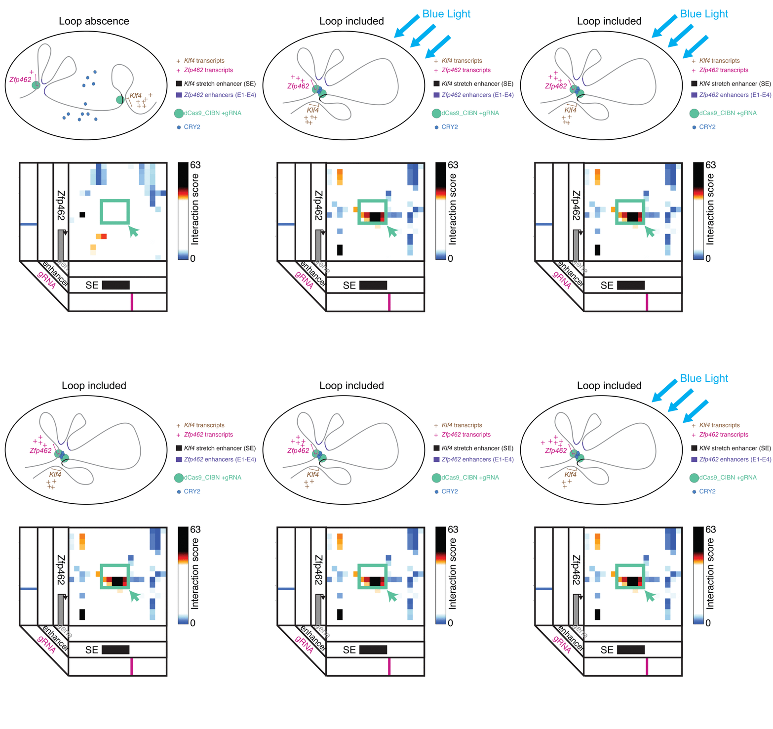
LADL combines CRISPR/Cas9 and optogenetics to bring two distant points in a linear gene sequence into physical contact, forming a folding pattern known as a “loop.” Looping interactions influence gene expression, so the researchers envision LADL as being a powerful tool for studying these dynamics.

Jennifer Phillips-Cremins, assistant professor in Penn Engineering’s Department of Bioengineering, is a pioneer in this field, known as “3-D Epigenetics.” She and her colleagues have now demonstrated a new technique for quickly creating specific folding patterns on demand, using light as a trigger.

The technique, known as LADL or light-activated dynamic looping, combines aspects of two other powerful biotechnological tools: CRISPR/Cas9 and optogenetics. By using the former to target the ends of a specific genome fold, or loop, and then using the latter to snap the ends together like a magnet, the researchers can temporarily create loops between exact genomic segments in a matter of hours.

The ability to make these genome folds, and undo them, on such a short timeframe makes LADL a promising tool for studying 3D-epigenetic mechanisms in more detail. With previous research from the Phillips-Cremins lab implicating these mechanisms in a variety of neurodevelopmental diseases, they hope LADL will eventually play a role in future studies, or even treatments.

Jennifer Phillips-Cremins, Ji Hun Kim and Mayuri Rege

Alongside Phillips-Cremins, lab members Ji Hun Kim and Mayuri Rege led the study, and Jacqueline Valeri, Aryeh Metzger, Katelyn R. Titus, Thomas G. Gilgenast, Wanfeng Gong and Jonathan A. Beagan contributed to it. They collaborated with associate professor of Bioengineering Arjun Raj and Margaret C. Dunagin, a member of his lab.

The study was published in the journal Nature Methods.

“In recent years,” Phillips-Cremins says, “scientists in our fields have overcome technical and experimental challenges in order to create ultra-high resolution maps of how the DNA folds into intricate 3D patterns within the nucleus. Although we are now capable of visualizing the topological structures, such as loops, there is a critical gap in knowledge in how genome structure configurations contribute to genome function.”

In order to conduct experiments on these relationships, researchers studying these 3D patterns were in need of tools that could manipulate specific loops on command. Beyond the intrinsic physical challenges — putting two distant parts of the linear genome in physical contact is quite literally like threading a needle with a thread that is only a few atoms thick — such a technique would need to be rapid, reversible and work on the target regions with a minimum of disturbance to neighboring sequences.

The advent of CRISPR/Cas9 solved the targeting problem. A modification of the gene editing tool allowed researchers to home in on the desired sequences of DNA on either end of the loop they wanted to form. If those sequences could be engineered to seek one another out and snap together under the other necessary conditions, the loop could be formed on demand.

Cremins Lab members then sought out biological mechanisms that could bind the ends of the loops together, and found an ideal one in the toolkit of optogenetics. The proteins CIB1 and CRY2, found in Arabidopsis, a flowering plant that’s a common model organism for geneticists, are known to bind together when exposed to blue light.

“Once we turn the light on, these mechanisms begin working in a matter of milliseconds and make loops within four hours,” says Rege. “And when we turn the light off, the proteins disassociate, meaning that we expect the loop to fall apart.”

“There are tens of thousands of DNA loops formed in a cell,” Kim says. “Some are formed slowly, but many are fast, occurring within the span of a second. If we want to study those faster looping mechanisms, we need tools that can act on a comparable time scales.”

As shown in a 2013 Nature Methods paper by fellow Penn bioengineer Lukasz Bugaj, the optical response of the CRY2 protein is a key component of LADL. When the blue light is turned on, CRY2 proteins in cell immediately find one another and bind together into clumps large enough to be seen under magnification. When the light is turned off, the clumps begin to dissolve away.”

Fast acting folding mechanisms also have an advantage in that they lead to fewer perturbations of the surrounding genome, reducing the potential for unintended effects that would add noise to an experiment’s results.

The researchers tested LADL’s ability to create the desired loops using their high-definition 3D genome mapping techniques. With the help of Arjun Raj, an expert in measuring the activity of transcriptional RNA sequences, they also were able to demonstrate that the newly created loops were impacting gene expression.

The promise of the field of 3D-epigenetics is in investigating the relationships between these long-range loops and mechanisms that determine the timing and quantity of the proteins they code for. Being able to engineer those loops means researchers will be able to mimic those mechanisms in experimental conditions, making LADL a critical tool for studying the role of genome folding on a variety of diseases and disorders.

“It is critical to understand the genome structure-function relationship on short timescales because the spatiotemporal regulation of gene expression is essential to faithful human development and because the mis-expression of genes often goes wrong in human disease,” Phillips-Cremins says. “The engineering of genome topology with light opens up new possibilities to understanding the cause-and-effect of this relationship. Moreover we anticipate that, over the long term, the use of light will allow us to target specific human tissues and even to control looping in specific neuron subtypes in the brain.”

The research was supported by the New York Stem Cell Foundation; Alfred P. Sloan Foundation; the National Institutes of Health through its Director’s New Innovator Award from the National Institute of Mental Health, grant no. 1DP2MH11024701, and a 4D Nucleome Common Fund, grant no. 1U01HL1299980; and the National Science Foundation through a joint NSF-National Institute of General Medical Sciences grant to support research at the interface of the biological and mathematical sciences, grant no. 1562665, and a Graduate Research Fellowship, grant no. DGE-1321851.

Originally published on the Penn Engineering Medium blog.

Week in BioE (August 9, 2018)

Converting Fat to Fight Obesity

White fat stories calories and provides the body with insulation.

There are two types of fat in the human body: brown and white. Brown fat, the “good” fat, is rich in mitochondria, which gives it its brown appearance. Whereas white fat stores calories and acts as an insulator, mitochondria-rich brown fat burns energy to produce heat throughout the body and maintains body temperature. White fat, conversely, uses its stored energy to insulate the body and keep its temperature level. While all fat serves a purpose in the body, an excess of white fat cells causes obesity, a condition affecting one in three adults in the U.S. and the root cause of many potential health problems. Finding ways to convert white fat to brown opens a possibility of treating this problem naturally.

A new study in Scientific Reports proposes a clever way to convert fat types. Professor of Biomedical Engineering Samuel Sia, PhD, of the Columbia University School of Engineering and Applied Science, led a team which developed a method of converting white fat into brown using a tissue-grafting technique. After extracting and converting the fat, it can then be transplanted back into the patient. White fat is hard-wired to convert to brown under certain conditions, such as exposure to cold temperatures, so the trick for Dr. Sia’s team was finding a way to make the conversion last for long periods. The studies conducted with mice suggested that using these methods, newly-converted fat stayed brown for a period of two months.

Dr. Sia’s team will proceed to conduct further tests, especially on the subjects’ metabolism and overall weight after undergoing the procedure, and they hope that eventual clinical trials will result in new methods to treat or even prevent obesity in humans.

Cremins Lab Student Appointed Blavatnik Fellow

Linda Zhou is currently pursuing her MD/PhD in Genomics and Computational Biology under the supervision of Dr. Jennifer Phillips-Cremins.

The Perelman School of Medicine named Linda Zhou, a student in BE’s Cremins Laboratory, a Blavatnik Fellow for the 2018-2019 academic year. The selection process for this award is highly competitive, and Linda’s selection speaks to the excellent quality of her scholarship and academic performance. The fellows will be honored in a special ceremony at the Museum of Natural History in New York City.

Linda received her B.S. in Biophysics and Biochemistry from Yale University and is currently pursuing her M.D./Ph.D. in the Genomics and Computational Biology Program at Penn. “I am honored to be named a Blavatnik Fellow and am extremely excited to continue my graduate studies investigating neurological disorders and the 3D genome,” she said. “This support will be integral to achieving my long term goal of driving scientific discovery that will help treat human disease.”

Linda’s research is overseen by Penn Bioengineering Assistant Professor Jennifer Phillips-Cremins, PhD. “Linda is an outstanding graduate student,” said Dr. Cremins. “It is a true delight to work with her. She is hard working, intelligent, kind, and has extraordinary leadership ability. Her unrelenting search for ground-state truth makes her a shining star.”

The Blavatnik Family Fellowship in Biomedical Research is a new award announced by the Perelman School of Medicine in May of this year. This generous gift from the Blavatnik Family Foundation awards $2 million to six recipients in the Biomedical Graduate Studies Program at Penn for each of the next four years.

Growing Lungs in a Lab

As the demand for lung transplants continues to rise, so does the need for safe and effective transplanted lungs. Bioengineered lungs grown or created in labs are one way of meeting this demand. The problem – as is ever the case with transplants – is the high rate of rejection. The results of success are always better when cells from the patient herself (or autologous cells) are used in the transplanted organ.

Recently Joan Nichols, PhD, Professor of Internal Medicine, and Microbiology and Immunology, at the University of Texas Medical Branch at Galveston, successfully bioengineered the first human lung. Her latest study published in Science Translational Medicine describes the next milestone for Dr. Nichols’ lab: successfully transplanting a bioengineered lung into a pig.

These advances are possible due to Dr. Nichols’ work with autologous cells, continuing the trend of “on demand” medicine (i.e. medicine tailor for a specific patient) which we track on this blog. Dr. Nichols’ particular method is to build the structure of a lung (using the harvested organs of dead pigs in this case), de-cellularize the tissue, and then repopulate it with autologous cells from the intended recipient. This way, the host body recognizes the cells as friendly and the likelihood of acceptance increases. While further study is needed before clinical trials can begin, Dr. Nichols and her team see the results as extremely promising and believe that we are on the way to bioengineered human lungs.

Nanoparticles Combat Dental Plaque

Combine a diet high in sugar with poor oral hygiene habits and dental cavities likely result. The sugar triggers the formation of an acidic biofilm (plaque) on the teeth, eroding the surface. Early childhood dental cavities affect one in every four children in the United States and hundreds of millions more globally. It’s a particularly severe problem in underprivileged populations.

In a study published in Nature Communications this week, researchers led by Hyun (Michel) Koo of the University of Pennsylvania School of Dental Medicine in collaboration with David Cormode of Penn’s Perelman School of Medicine and School of Engineering and Applied Science used FDA-approved nanoparticles to effectively disrupt biofilms and prevent tooth decay in both an experimental human-plaque-like biofilm and in an animal model that mimics early-childhood caries.

Dr. David Cormode is Assistant Professor of Radiology and Secondary Faculty in Bioengineering at Penn. His research includes Bioengineering Therapeutics, Devices and Drug Delivery and Biomaterials.

Read the full story at Penn Today. Media contact Katherine Unger Baillie.

Stopping the Flu from Catching On

The flu virus is notoriously contagious, but there may be a way to stop it before it starts. In order for the influenza virus to successfully transport itself into the cells of a human host, it needs a certain protein called hemagglutinin which mediates its entry. By interfering with this vital ingredient, researchers can effectively kill the virus.

A new study in the Proceedings of the National Academy of Sciences discusses a method of disrupting the process by which this protein causes the virus to infect its host cells. This discovery could lead to more effective flu vaccines that target the flu virus at its root, rather than current ones which have to keep up with the ongoing changes and mutations of the virus itself. Indeed, the need for different vaccines to address various “strains” of the flu is moot if a vaccine can stop the virus from infecting people in the first place.

This breakthrough results from grants provided by the NSF, the Welch Foundation, and the NIH to Rice University and Baylor College of Medicine. Lead researchers José Onuchic, PhD, Harry C. and Olga K. Wiess Chair of Physics and Professor of Chemistry and BioSciences at Rice University; Jianpeng Ma, PhD, Professor of Bioengineering at Rice University and Lodwick T. Bolin Professor of Biochemistry at Baylor College of Medicine; and Qinghua Wang, PhD, Assistant Professor of Biochemistry at Baylor College of Medicine. Their team will continue to study the important role proteins play in how the flu virus operates.

People and Places

This week, we congratulate a few new leadership appointments in bioengineering. First, the Georgia Institute of Technology appointed Penn BE alumnus Andréas García, PhD, the new Executive Director of the Parker H. Petit Institute for Bioengineering and Bioscience. In addition to his new role, Dr. García is also the George W. Woodruff School of Mechanical Engineering Regents Professor. He conducts research in biomolecular, cellular, and tissue engineering and collaborates with a number of research centers across Georgia Tech. Dr. García graduated with both his M.S.E. and Ph.D. from the University of Pennsylvania’s Department of Bioengineering.

Secondly, the University of Minnesota Institute for Engineering in Medicine (IEM) named the Distinguished McKnight University Professor John Bischof, PhD, their new director. This follows Dr. Bischof’s recent position as interim director for the IEM. Dr. Bischof earned his Ph.D. in Mechanical Engineering at the University of California at Berkeley, and is currently a faculty member in both the Mechanical Engineering and Biomedical Engineering Departments at the University of Minnesota. Dr. Bischof holds the Carl and Janet Kuhrmeyer Chair in Mechanical Engineering.

At an earlier, but no less impressive, point in his academic career, Tanishq Abraham became the youngest person to graduate with a degree in biomedical engineering. The fifteen year old recently graduated summa cum laude from the University of California, Davis. As part of his graduating research, Abraham – a first-generation Indian-American – designed a device to measure the heart rates of burn victims. Abraham has already been accepted by U.C. Davis for his Ph.D. and plans to continue on to his M.D.

Finally, the work continues to create affordable and well-fitted prosthetics, especially for remote, rural, and underfunded areas both in the U.S. and abroad. Unfortunately, recent studies published by the Centre for Biomedical Engineering at the India Institute of Technology Delhi (IIT) demonstrate the uphill nature of this battle; stating that India alone contains over half a million upper limb amputees. To address this explosive population, researchers and entrepreneurs are using new bioengineering technologies such as digital manufacturing, 3D scanning and printing, and more. The best innovations are those that save time, resources, and money, without sacrificing quality in the prosthetic or patient comfort. Penn Engineering’s Global Biomedical Service (GBS) program similarly responds to this need, as each year students follow an academically rigorous course with a two-week immersive trip to China, where they learn how to create and fit prosthetic limbs for local children in conjunction with Hong Kong Polytechnic University.

Collaboration in Research by Bioengineering Faculty

Jennifer Phillips-Cremins
Danielle Bassett

In faculty matters, specialization is the name of game. The areas in which individual professors conduct their research and teach are highly specific, with often no overlap between the areas of expertise of people in the same departments. Given the broad range of topics covered by the term, bioengineering is particularly complex in the array of subjects researched by faculty.

Now and then, however, these paths converge. Most recently, Jennifer Phillips-Cremins, Ph.D., Assistant Professor of Bioengineering, and Danielle Bassett, Ph.D., Eduardo D. Glandt Faculty Fellow and Associate Professor of Bioengineering, collaborated on a paper published in Nature Methods. Dr. Cremins’s research has focused on genome folding, an intricate process by which DNA in the nuclei of cells creates loops that result in  specific forms of gene regulation. Dr. Bassett’s area is network science and systems theory. Both professors apply their research in the area of central nervous development.

In the new paper, Drs. Cremins and Bassett, along with members of both their labs and colleagues from the Department of Genetics, developed a a graph theory-based method for detecting genome folding, called 3DNetMod, which outperformed earlier models used for the same purpose. In addition, Dr. Cremins is profiled in the same issue of Nature Methods, where she discusses how her past education and experience have resulted in her career achievements thus far.

Phillips-Cremins Research Identifies Protein Involved in Brain Development

Phillips-Cremins
Jennifer Phillips-Cremins, Ph.D.

The vast majority of genetic mutations that are associated with disease occur at sites in the genome that aren’t genes. These sequences of DNA don’t code for proteins themselves, but provide an additional layer of instructions that determine if and when particular genes are expressed. Researchers are only beginning to understand how the non-coding regions of the genome influence gene expression and might be disrupted in disease.

​​​​​​​​​​​​Jennifer Phillips-Cremins, assistant professor in the Department of Bioengineering in the University of Pennsylvania’s School of Engineering and Applied Science, studies the three-dimensional folding of the genome and the role it plays in brain development. When a stretch of DNA folds, it creates a higher-order structure called a looping interaction, or “loop.” In doing so, it brings non-coding sites into physical contact with their target genes, precisely regulating gene expression in space and time during development.

Phillips-Cremins and lab member Jonathan Beagan have led a new study identifying a new protein that connects loops in embryonic stem cells as they begin to differentiate into types of neurons. Though the study was conducted in mice, these findings inform aspects of human brain development, including how the genetic material folds in the 3-D nucleus and is reconfigured as stem cells become specialized. Better understanding of these mechanisms may be relevant to a wide range of neurodevelopmental disorders.

Cremins lab members Michael Duong, Katelyn Titus, Linda Zhou, Zhendong Cao, Jingjing Ma, Caroline Lachanski and Daniel Gillis also contributed to the study, which was published in the journal Genome Research.​​​​​​

Continue reading at the SEAS blog.