New Chip Opens Door to AI Computing at Light Speed

by Ian Scheffler

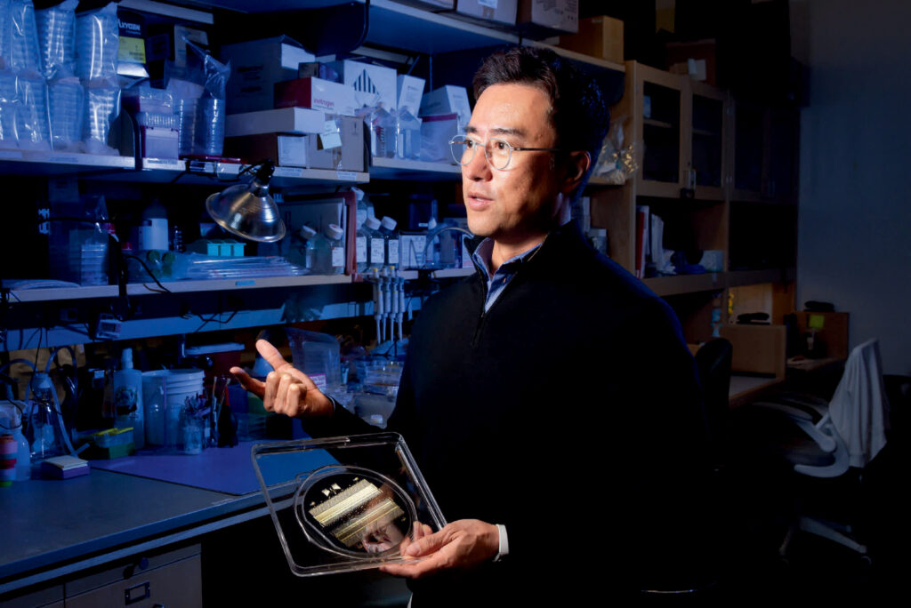
Computing at the speed of light may reduce the energy cost of training AI. (Narongrit Doungmanee via Getty Images)

Penn Engineers have developed a new chip that uses light waves, rather than electricity, to perform the complex math essential to training AI. The chip has the potential to radically accelerate the processing speed of computers while also reducing their energy consumption.

The silicon-photonic (SiPh) chip’s design is the first to bring together Benjamin Franklin Medal Laureate and H. Nedwill Ramsey Professor Nader Engheta’s pioneering research in manipulating materials at the nanoscale to perform mathematical computations using light — the fastest possible means of communication — with the SiPh platform, which uses silicon, the cheap, abundant element used to mass-produce computer chips.

The interaction of light waves with matter represents one possible avenue for developing computers that supersede the limitations of today’s chips, which are essentially based on the same principles as chips from the earliest days of the computing revolution in the 1960s.

In a paper in Nature Photonics, Engheta’s group, together with that of Firooz Aflatouni, Associate Professor in Electrical and Systems Engineering, describes the development of the new chip. “We decided to join forces,” says Engheta, leveraging the fact that Aflatouni’s research group has pioneered nanoscale silicon devices.

Their goal was to develop a platform for performing what is known as vector-matrix multiplication, a core mathematical operation in the development and function of neural networks, the computer architecture that powers today’s AI tools.

Read the full story in Penn Engineering Today.

Nader Engheta is the H. Nedwill Ramsey Professor in Electrical and Systems Engineering, Bioengineering, Materials Science and Engineering, and in Physics and Astronomy.

What Makes a Breakthrough? “Eight Steps Back” Before Making it to the Finish Lit

by Meagan Raeke

(From left to right) Breakthrough Prize recipients Drew Weissman, Virginia M-Y Lee, Katalin Karikó, and Carl June at a reception on Feb. 13. (Image: Courtesy of Penn Medicine News)

In popular culture, scientific discovery is often portrayed in “Eureka!” moments of sudden realization: a lightbulb moment, coming sometimes by accident. But in real life—and in Penn Medicine’s rich history as a scientific innovator for more than 250 years—scientific breakthroughs can never truly be distilled down to a single, “ah-ha” moment. They’re the result of years of hard work, perseverance, and determination to keep going, despite repeated, often discouraging, barriers and setbacks. 

“Research is [like taking], four, or six, or eight steps back, and then a little stumble forward,” said Drew Weissman, MD, PhD, the Roberts Family Professor of Vaccine Research. “You keep doing that over and over and somehow, rarely, you can get to the top of the step.” 

For Weissman and his research partner, Katalin Karikó, PhD, an adjunct professor of Neurosurgery, that persistence—documented in thousands of news stories across the globe—led to the mRNA technology that enabled two lifesaving COVID-19 vaccines, earning the duo numerous accolades, including the highest scientific honor, the 2023 Nobel Prize in Medicine

Weissman and Karikó were also the 2022 recipients of the Breakthrough Prize in Life Sciences, the world’s largest science awards, popularly known as the “Oscars of Science.” Founded in 2012 by a group of web and tech luminaries including Google co-founder Sergey Brin and Meta CEO Mark Zuckerberg, the Breakthrough Prizes recognize “the world’s top scientists working in the fundamental sciences—the disciplines that ask the biggest questions and find the deepest explanations.” With six total winners, including four from the Perelman School of Medicine (PSOM), Penn stands alongside Harvard and MIT as the institutions whose researchers have been honored with the most Breakthrough Prizes. 

Virginia M.Y. Lee, PhD, the John H. Ware 3rd Professor in Alzheimer’s Research, was awarded the Prize in 2020 for discovering how different forms of misfolded proteins can move from cell to cell and lead to neurodegenerative disease progression. Carl June, MD, the Richard W. Vague Professor in Immunotherapy, is the most recent recipient and will be recognized at a star-studded red-carpet event in April for pioneering the development of CAR T cell therapy, which programs patients’ own immune cells to fight their cancer.

The four PSOM Breakthrough Prize recipients were honored on Tuesday, Feb. 13, 2024, when a new large-scale installation was unveiled in the lobby of the Biomedical Research Building to celebrate each laurate and their life-changing discoveries. During a light-hearted panel discussion, the honorees shared how a clear purpose, dogged determination, and a good sense of humor enabled their momentum forward. 

Read the full story in Penn Medicine News.

Carl June and Jon Epstein are members of the Penn Bioengineering Graduate Group. Read more stories featuring them in the BE Blog here and here, respectively.

Weissman presented the Department of Bioengineering’s 2022 Herman P. Schwan Distinguished Lecture: “Nucleoside-modified mRNA-LNP therapeutics.” Read more stories featuring Weissman in the BE Blog here.

Illuminating the Invisible: Bringing the Smallest Protein Clusters into Focus

by Ian Scheffler

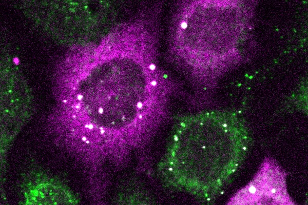
The bright white spots represent tiny clusters of proteins detected by CluMPS. (Photo by: Thomas Mumford)

Penn Engineers have pioneered a new way to visualize the smallest protein clusters, skirting the physical limitations of light-powered microscopes and opening new avenues for detecting the proteins implicated in diseases like Alzheimer’s and testing new treatments.

In a paper in Cell Systems, Lukasz Bugaj, Assistant Professor in Bioengineering, describes the creation of CluMPS, or Clusters Magnified by Phase Separation, a molecular tool that activates by forming conspicuous blobs in the presence of target protein clusters as small as just a few nanometers. In essence, CluMPS functions like an on/off switch that responds to the presence of clusters of the protein it is programmed to detect.

Normally, says Bugaj, detecting such clusters requires laborious techniques. “With CluMPS, you don’t need anything beyond the standard lab microscope.” The tool fuses with the target protein to form condensates orders of magnitude larger than the protein clusters themselves that resemble the colorful blobs in a lava lamp. “We think the simplicity of the approach is one of its main benefits,” says Bugaj. “You don’t need specialized skills or equipment to quickly see whether there are small clusters in your cells.”

Read the full story in Penn Engineering Today.

Penn Bioengineering Cockroach Lab Featured in Popular Mechanics

Every Penn Bioengineering semester culminates in a series of “demo days” — dedicated time in which undergraduate Bioengineering students demonstrate projects made in their Bioengineering lab courses or in Senior Design for their classmates and faculty. These are held in the George H. Stephenson Foundation Educational Laboratory & Bio-MakerSpace (or the Penn BE Labs), the dedicated teaching lab for the Bioengineering Department which also functions as an interdisciplinary bio-makerspace open to the entire Penn community.

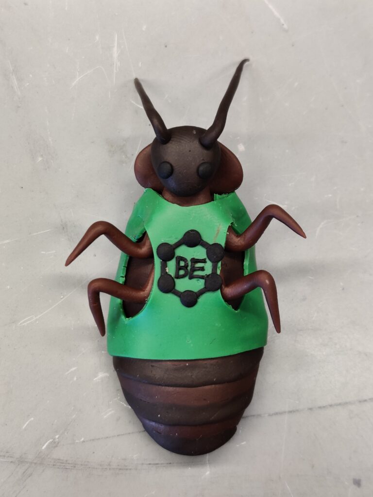
For the Fall 2023 demos, Popular Mechanics paid a visit to the BE Labs to witness the (in)famous “cockroach lab,” a staple of the third year course “Bioengineering, Modeling, Analysis, and Design Laboratory” (affectionately known as BE MAD). This year’s cockroach demos featured a miniature Taylor Swift — flaunting a cockroach limb — and several projects featuring the faces of course faculty, David Meaney, Solomon R. Pollack Professor in Bioengineering and Senior Associate Dean in Penn Engineering, and Michael Patterson, Director of Educational Laboratories in Bioengineering.

Read “How Severed Cockroach Legs Could Help Us ‘Fully Rebuild’ Human Bodies” in Popular Mechanics.

Read more stories featuring the Penn BE Labs in the BE Blog here.

Researchers Breathe New Life into Lung Repair

by Nathi Magubane

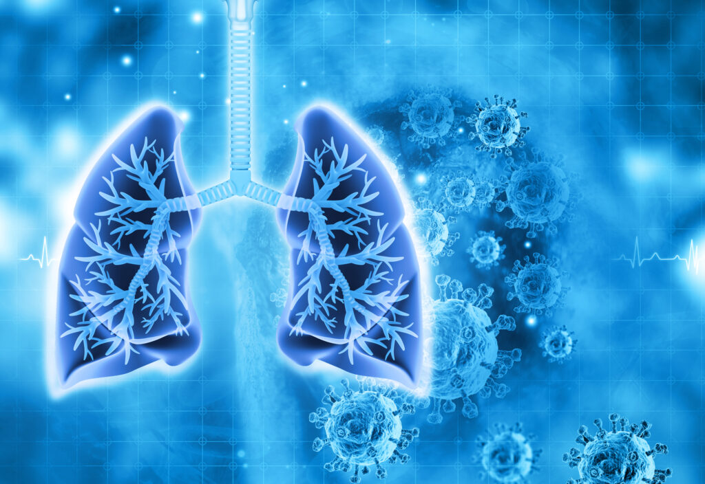
Image: iStock/Mohammed Haneefa Nizamudeen

In the human body, the lungs and their vasculature can be likened to a building with an intricate plumbing system. The lungs’ blood vessels are the pipes essential for transporting blood and nutrients for oxygen delivery and carbon dioxide removal. Much like how pipes can get rusty or clogged, disrupting normal water flow, damage from respiratory viruses, like SARS-CoV-2 or influenza, can interfere with this “plumbing system.”

In a recent study, researchers looked at the critical role of vascular endothelial cells in lung repair. Their work, published in Science Translational Medicine, was led by Andrew Vaughan of the University of Pennsylvania’s School of Veterinary Medicine and shows that, by using techniques that deliver vascular endothelial growth factor alpha (VEGFA) via lipid nanoparticles (LNPs), that they were able to greatly enhance modes of repair for these damaged blood vessels, much like how plumbers patch sections of broken pipes and add new ones.

“While our lab and others have previously shown that endothelial cells are among the unsung heroes in repairing the lungs after viral infections like the flu, this tells us more about the story and sheds light on the molecular mechanisms at play,” says Vaughan, assistant professor of biomedical sciences at Penn Vet. “Here we’ve identified and isolated pathways involved in repairing this tissue, delivered mRNA to endothelial cells, and consequently observed enhanced recovery of the damaged tissue. These findings hint at a more efficient way to promote lung recovery after diseases like COVID-19.”

They found VEGFA’s involvement in this recovery, while building on work in which they used single cell RNA sequencing to identify transforming growth factor beta receptor 2 (TGFBR2) as a major signaling pathway. The researchers saw that when TGFBR2 was missing it stopped the activation of VEGFA. This lack of signal made the blood vessel cells less able to multiply and renew themselves, which is vital for the exchange of oxygen and carbon dioxide in the tiny air sacs of the lungs.

“We’d known there was a link between these two pathways, but this motivated us to see if delivering VEGFA mRNA into endothelial cells could improve lung recovery after disease-related injury,” says first author Gan Zhao, a postdoctoral researcher in the Vaughan Lab.

The Vaughan Lab then reached out to Michael Mitchell of the School of Engineering and Applied Science, whose lab specializes in LNPs, to see if delivery of this mRNA cargo would be feasible.

“LNPs have been great for vaccine delivery and have proven incredibly effective delivery vehicles for genetic information. But the challenge here was to get the LNPs into the bloodstream without them heading to the liver, which is where they tend to congregate as its porous structure lends favor to substances passing from the blood into hepatic cells for filtration,” says Mitchell, an associate professor of bioengineering at Penn Engineering and a coauthor of the paper. “So, we had to devise a way to specifically target the endothelial cells in the lungs.”

Lulu Xue, a postdoctoral researcher in the Mitchell Lab and a co-first author of the paper, explains that they engineered the LNP to have an affinity for lung endothelial cells, this is known as extra hepatic delivery, going beyond the liver.

Read the full story in Penn Today.

Secondary Cancers Following CAR T Cell Therapy Are Rare, Penn Medicine Analysis Shows

by Meagan Raeke

3d illustration of a damaged and disintegrating cancer cell. (Image: iStock/vitanovski)

The development of any type of second cancer following CAR T cell therapy is a rare occurrence, as found in an analysis of more than 400 patients treated at Penn Medicine, researchers from the Perelman School of Medicine at the University of Pennsylvania reported today in Nature Medicine. The team also described a single case of an incidental T cell lymphoma that did not express the CAR gene and was found in the lymph node of a patient who developed a secondary lung tumor following CAR T cell therapy.

CAR T cell therapy, a personalized form of immunotherapy in which each patient’s T cells are modified to target and kill their cancer cells, was pioneered at Penn. More than 30,000 patients with blood cancers in the United States—many of whom had few, if any, remaining treatment options available—have been treated with CAR T cell therapy since the first such therapy was approved in 2017. Some of the earliest patients treated in clinical trials have gone on to experience long-lasting remissions of a decade or more.

Secondary cancers, including T cell lymphomas, are a known, rare risk of several types of cancer treatment, including chemotherapy, radiation, and stem cell transplant. CAR T cell therapy is currently only approved to treat blood cancers that have relapsed or stopped responding to treatment, so patients who receive CAR T cell therapies have already received multiple other types of treatment and are facing dire prognoses.

In November 2023, the FDA announced an investigation into several reported cases of secondary T cell malignancies, including CAR-positive lymphoma, in patients who previously received CAR T cell therapy products. In January 2024, the FDA began requiring drugmakers to add a safety label warning to CAR T cell products. While the FDA review is still ongoing, it remains unclear whether the secondary T cell malignancies were caused by CAR T cell therapy.

As a leader in CAR T cell therapy, Penn has longstanding, clearly established protocols to monitor each patient both during and after treatment – including follow-up for 15 years after infusion – and participates in national reporting requirements and databases that track outcomes data from all cell therapy and bone marrow transplants.

Marco Ruella, M.D.

“When this case was identified, we did a detailed analysis and concluded the T cell lymphoma was not related to the CAR T cell therapy. As the news of other cases came to light, we knew we should go deeper, to comb through our own data to better understand and help define the risk of any type of secondary cancer in patients who have received CAR T cell products,” said senior author Marco Ruella, MD, an assistant professor of Hematology-Oncology and Scientific Director of the Lymphoma Program. “What we found was very encouraging and reinforces the overall safety profile for this type of personalized cell therapy.”

Read the full story in Penn Medicine News.

Marco Ruella is Assistant Professor of Medicine in the Perelman School of Medicine. He is a member of the Penn Bioengineering Graduate Group.

Building Tiny Organs

by David Levin

Dan Huh, Ph.D. (Photo credit: Leslie Barbaro)

More than 34 million Americans suffer from pulmonary diseases like asthma, emphysema and chronic bronchitis. While medical treatments can keep these ailments in check, there are currently no cures. Part of the reason, notes Dan Huh, is that it’s incredibly hard to study how these diseases actually work. While researchers can grow cells taken from human lungs in a dish, they cannot expect them to act like they would in the body. In order to mimic the real deal, it’s necessary to recreate the complex, 3D environment of the lung — right down to its tiny air sacs and blood vessels — and to gently stretch and release the tissue to simulate breathing.

Huh, Associate Professor in Bioengineering, is the cofounder of Vivodyne, a Penn Engineering biotech spinoff that is creating tissues like these in the lab. Vivodyne uses a bioengineering technology that Huh has been developing for more than a decade. While a postdoctoral fellow at Harvard’s Wyss Institute, he played a central role in creating a novel device called an “organ on a chip,” which, as the name implies, assembles multiple cell types on a tiny piece of engineered plastic to create an approximation of an organ.

“While those chips represented a major innovation,” says Huh, “they still weren’t truly lifelike. They lacked many of the essential features of their counterparts in the human body, such as the network of blood vessels running between different kinds of tissue, which are essential for transporting oxygen, nutrients, waste products and various biochemical signals.”

Read the full article in the Fall 2023 issue of the Penn Engineering Magazine.

The Heart and Soul of Innovation: Noor Momin Harnesses the Immune System to Treat Heart Disease

by Ian Scheffler

Noor Momin, Stephenson Foundation Term Assistant Professor of Innovation

While growing up, Noor Momin, who joined the Department of Bioengineering in January as the Stephenson Foundation Term Assistant Professor of Innovation, imagined becoming a physician. Becoming a doctor seemed like a tangible way for someone interested in science to make a difference. Not until college did she realize the impact she could have as a bioengineer instead.

“I was taping microscope slides together,” Momin recalls of her initial experience as an undergraduate researcher at the University of Texas at Austin. “I didn’t even know what a Ph.D. was.”

It wasn’t until co-authoring her first paper, which explores how lipids, the water-repelling molecules that make up cell membranes (and also fats and oils), can switch between more fluid and less fluid arrangements, that Momin understood the degree to which bioengineering can influence medicine. “Someone could potentially use that paper for drug design,” Momin says.

Today, Momin’s research applies her molecular expertise to heart disease, which despite numerous advances in treatment — from coronary artery bypass surgery to cholesterol-lowering statins — remains the primary cause of mortality worldwide.

As Momin sees it, the conventional wisdom of treating the heart like a mechanical pump, whose pipes can be replaced or whose throughput can be treated to prevent clogging in the first place, overshadows the immune system’s critical role in the development of heart disease.

Read the full story in Penn Engineering Today.

Bioengineers on the Brink of Breaching Blood-brain Barrier

by Nathi Magubane

From left: Emily Han, Rohan Palanki, Jacqueline Li, Michael Mitchell, Dongyoon Kim, and Marshall Padilla of Penn Engineering.

Imagine the brain as an air traffic control tower, overseeing the crucial and complex operations of the body’s ‘airport.’ This tower, essential for coordinating the ceaseless flow of neurological signals, is guarded by a formidable layer that functions like the airport’s security team, diligently screening everything and everyone, ensuring no unwanted intruders disrupt the vital workings inside.

However, this security, while vital, comes with a significant drawback: sometimes, a ‘mechanic’—in the form of critical medication needed for treating neurological disorders—is needed inside the control tower to fix arising issues. But if the security is too stringent, denying even these essential agents entry, the very operations they’re meant to protect could be jeopardized.

Now, researchers led by Michael Mitchell of the University of Pennsylvania are broaching this long-standing boundary in biology, known as the blood-brain barrier, by developing a method akin to providing this mechanic with a special keycard to bypass security. Their findings, published in the journal Nano Letters, present a model that uses lipid nanoparticles (LNPs) to deliver mRNA, offering new hope for treating conditions like Alzheimer’s disease and seizures—not unlike fixing the control tower’s glitches without compromising its security.

“Our model performed better at crossing the blood-brain barrier than others and helped us identify organ-specific particles that we later validated in future models,” says Mitchell, associate professor of bioengineering at Penn’s School of Engineering and Applied Science, and senior author on the study. “It’s an exciting proof of concept that will no doubt inform novel approaches to treating conditions like traumatic brain injury, stroke, and Alzheimer’s.”

Read the full story in Penn Today.

Protein Partners Identified as Potential Key for Fetal Bone Development

Image: iStock/Christoph Burgstedt

A pair of proteins, YAP and TAZ, has been identified as conductors of bone development in the womb and could provide insight into genetic diseases such as osteogenesis imperfecta, known commonly as “brittle bone disease.” This research, published in Developmental Cell and led by members of the McKay Orthopaedic Research Laboratory of the Perelman School of Medicine, adds understanding to the field of mechanobiology, which studies how mechanical forces influence biology.

“Despite more than a century of study on the mechanobiology of bone development, the cellular and molecular basis largely has remained a mystery,” says the study’s senior author, Joel Boerckel, an associate professor of orthopaedic surgery. “Here, we identify a new population of cells that are key to turning the body’s early cartilage template into bone, guided by the force-activated gene regulating proteins, YAP and TAZ.”

Read the full story in Penn Medicine News.

Joel D. Boerckel is Associate Professor in Orthopaedic Surgery and in Bioengineering.