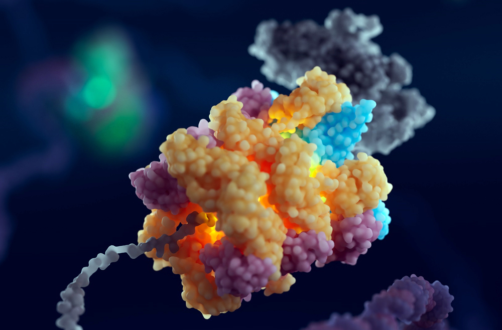
Illustration of the 55LCC complex. (Image: Courtesy of Cameron Baines/Phospho Biomedical Animation)
When cells in the human body divide, they must first make accurate copies of their DNA. The DNA replication exercise is one of the most important processes in all living organisms and is fraught with risks of mutation, which can lead to cell death or cancer. Now, findings from biologists from the Perelman School of Medicine and from the University of Leeds have identified a multiprotein “machine” in cells that helps govern the pausing or stopping of DNA replication to ensure its smooth progress. Illustration of the 55LCC complex. (Image: Courtesy of Cameron Baines/Phospho Biomedical Animation)
The discovery, published in Cell, advances the understanding of DNA replication, helps explain a puzzling set of genetic diseases, and could inform the development of future treatments for neurologic and developmental disorders.
“We’ve found what appears to be a critical quality-control mechanism in cells,” says senior co-corresponding author Roger Greenberg, the J. Samuel Staub, M.D. Professor in the department of Cancer Biology, director of the Penn Center for Genome Integrity, and director of basic science at the Basser Center for BRCA at Penn Medicine. “Trillions of cells in our body divide every single day, and this requires accurate replication of our genomes. Our work describes a new mechanism that regulates protein stability in replicating DNA. We now know a bit more about an important step in this complex biological process.”
Fat is a normal and necessary part of the body. Fat cells store and release energy, as well as play significant roles in hormonal regulation and immunity.
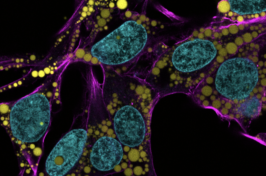
Engineers and scientists at the University of Pennsylvania are the first to discover fat-filled lipid droplets’ (pictured above in green) surprising capability to indent and puncture the nucleus, the organelle which contains and regulates a cell’s DNA.
In recent decades, a concerning rise in metabolic illnesses – such as cardiovascular disease, high blood pressure and diabetes – has focused scientific attention on the biology and chemistry of fat, resulting in a wealth of information about how fat cells work.
But fat cells and their metabolic activities are only part of the story.
Fat-filled lipid droplets, tiny spheres of fat many times smaller than fat cells, are a growing subject of scientific interest. Found inside many different cell types, these lipid particles have long been little understood. Studies have begun to illuminate these droplets’ participation in metabolic functions and cellular protection, but we still know next to nothing about the physical nature of fat.
Now, researchers at the University of Pennsylvania School of Engineering and Applied Science have looked beyond biochemistry to publish groundbreaking work on the physics of these droplets, revealing them to be a potential threat to a cell’s nucleus. In the August issue of the Journal of Cell Biology, they are the first to discover fat-filled lipid droplets’ surprising capability to indent and puncture the nucleus, the organelle which contains and regulates a cell’s DNA.
The stakes of their findings are high: a ruptured nucleus can lead to elevated DNA damage that is characteristic of many diseases, including cancer.
“Intuitively, people think of fat as soft,” says Discher. “And on a cellular level it is. But at this small size of droplet— measuring just a few microns rather than the hundreds of microns of a mature fat cell—it stops being soft. Its shape has a much higher curvature, bending other objects very sharply. This changes its physics in the cell. It can deform. It can damage. It can rupture.”
Every spring, the Graduate Association of Bioengineers (GABE) at Penn partners up with iPraxis, an educational non-profit organization based in Philadelphia, to organize BETA Day, an event that brings together Bioengineering graduate students and local Philadelphia grade school students to introduce them to the field of bioengineering, the life of graduate students, and hands-on scientific demonstrations. Due to COVID-19 restrictions, we adapted the traditional in-person BETA Day into a virtual event on Zoom. This year, we assembled kits containing the necessary materials for our chosen demonstrations and worked with iPraxis to coordinate their delivery to partner schools and their students. This enabled students to perform their demonstrations in a hands-on manner from their own homes; over 40 students were able to participate in extracting their own DNA and making biomaterials with safe household materials.
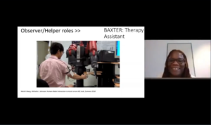
Michelle Johnson presents on her work in robotics
The day began with a fantastic lecture by Michelle Johnson, Associate Professor in Bioengineering and Physical Medicine and Rehabilitation, who introduced students to the field of rehabilitation robotics and shared her experience as a scientist. Students then learned about DNA and biomaterials through lectures mediated by the graduate students Dayo Adetu and Puneeth Guruprasad. After each lecture, students broke into breakout rooms with graduate student facilitators where they were able to get some hands-on scientific experience as they extracted DNA from their cheek cells and fabricated alginate hydrogels. Michael Sobrepera, a graduate student in Dr. Johnson’s lab, concluded the event by giving a lecture on the process of robotics development and discussed where the field is heading and some important considerations for the field.
Dayo Adetu, Bioengineering Master’s student and GABE President, teaches the students about Genetic Engineering
While yet another online event may seem unexciting, throughout the lectures students remained exceptionally engaged and raised fantastic questions ranging from the accessibility of low income communities to novel robotic therapeutic technologies to the bioethical questions robotic engineers will face as technologies advance. The impact of BETA day was evident as the high school students began to discuss the possible majors they would like to pursue for their bachelor’s degrees. Events like BETA Day give a glimpse into possible STEM fields and careers students can pursue.
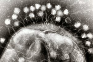
Transmission electron micrograph of multiple bacteriophages, viruses that infect bacteria, attached to a cell wall. New research describes how bacteria can optimize their “memory” of past viral infections in order to launch an effective immune response against a new invader. (Image: Graham Beards)
Before CRISPR became a household name as a tool for gene editing, researchers had been studying this unique family of DNA sequences and its role in the bacterial immune response to viruses. The region of the bacterial genome known as the CRISPR cassette contains pieces of viral genomes, a genomic “memory” of previous infections. But what was surprising to researchers is that rather than storing remnants of every single virus encountered, bacteria only keep a small portion of what they could hold within their relatively large genomes.
In recent years, CRISPR has become the go-to biotechnology platform, with the potential to transform medicine and bioengineering. In bacteria, CRISPR is a heritable and adaptive immune system that allows cells to fight viral infections: As bacteria come into contact with viruses, they acquire chunks of viral DNA called spacers that are incorporated into the bacteria’s genome. When the bacteria are attacked by a new virus, spacers are copied from the genome and linked onto molecular machines known as Cas proteins. If the attached sequence matches that of the viral invader, the Cas proteins will destroy the virus.
Bacteria have a different type of immune system than vertebrates, explains senior author Vijay Balasubramanian, but studying bacteria is an opportunity for researchers to learn more about the fundamentals of adaptive immunity. “Bacteria are simpler, so if you want to understand the logic of immune systems, the way to do that would be in bacteria,” he says. “We may be able to understand the statistical principles of effective immunity within the broader question of how to organize an immune system.”
This research was supported by the Simons Foundation (Grant 400425) and National Science Foundation Center for the Physics of Biological Function (Grant PHY-1734030).
Hammer will offer a course on COVID-19 and the coronavirus pandemic during Penn’s Summer II session, which will be held online this year. The course will be co-taught with Miriam Wattenbarger, senior lecturer in CBE.
The course, “Biotechnology, Immunology, and COVID-19,” will culminate with a case study of the coronavirus pandemic including the types of drugs proposed and their mechanism of action, as well as the process of vaccine development.
“Obviously, the pandemic has been a life-altering event, causing an immense dislocation for everyone in our community, especially the students. Between me and Miriam, who has been trumpeting the importance of vaccines for some time in her graduate-level CBE courses, we have the expertise to inform students about this disease and how we might combat it,” says Hammer.
For more than ten years, Wattenbarger has run courses and labs focused on drug delivery and biotechnology, key elements of the vaccine development process.
“I invite both researchers and industry speakers to meet with my students,” Wattenbarger says, “so that they learn the crucial role engineers play in both vaccine development and manufacturing.”
Beyond studying the interactions between the immune system and viruses — including HIV, influenza, adenovirus and coronavirus — students will cover a variety of biotechnological techniques relevant to tracking and defending against them, including recombinant DNA technology, polymerase chain reaction, DNA sequencing, gene therapy, CRISPR-Cas9 editing, drug discovery, small molecule inhibitors, vaccines and the clinical trial process.
Students will also learn the mathematical principles used to quantify biomolecular interactions, as well as those found behind simple epidemiological models and methods for making and purifying drugs and vaccines.
“We all have to contribute in the ways that we can. Having taught biotechnology to freshmen for the past decade, this is something that I can do that can both inform and build community,” says Hammer. “Never has it been more important to have an informed and scientifically literate community that can fight this or any future pandemic.”
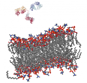
Getting a complex protein like an antibody through the membrane of a cell without damaging either is a long-standing challenge in the life sciences. Penn Engineers have found a plug-and-play solution that makes antibodies compatible with the delivery vehicles commonly used to ferry nucleic acids across that barrier.
For almost any conceivable protein, corresponding antibodies can be developed to block it from binding or changing shape, which ultimately prevents it from carrying out its normal function. As such, scientists have looked to antibodies as a way of shutting down proteins inside cells for decades, but there is still no consistent way to get them past the cell membrane in meaningful numbers.
Now, Penn Engineering researchers have figured out a way for antibodies to hitch a ride with transfection agents, positively charged bubbles of fat that biologists routinely use to transport DNA and RNA into cells. These delivery vehicles only accept cargo with a highly negative charge, a quality that nucleic acids have but antibodies lack. By designing a negatively charged amino acid chain that can be attached to any antibody without disrupting its function, they have made antibodies broadly compatible with common transfection agents.
Beyond the technique’s usefulness towards studying intracellular dynamics, the researchers conducted functional experiments with antibodies that highlight the technique’s potential for therapeutic applications. One antibody blocked a protein that decreases the efficacy of certain drugs by prematurely ejecting them from cells. Another blocked a protein involved in the transcription process, which could be an even more fundamental way of knocking out proteins with unwanted effects.
DNA Microscopy Gives a Better Look at Cell and Tissue Organization
A new technique that researchers from the Broad Institute of MIT and Harvard University are calling DNA microscopy could help map cells for better understanding of genetic and molecular complexities. Joshua Weinstein, Ph.D., a postdoctoral associate at the Broad Institute, who is also an alumnus of Penn’s Physics and Biophysics department and former student in Penn Bioengineering Professor Ravi Radhakrishnan’s lab, is the first author of this paper on optics-free imaging published in Cell.
The primary goal of the study was to find a way of improving analysis of the spatial organization of cells and tissues in terms of their molecules like DNA and RNA. The DNA microscopy method that Weinstein and his team designed involves first tagging DNA, and allowing the DNA to replicate with those tags, which eventually creates a cloud of sorts that diffuses throughout the cell. The DNA tags subsequent interactions with molecules throughout the cell allowed Weinstein and his team to calculate the locations of those molecules within the cell using basic lab equipment. While the researchers on this project focused their application of DNA microscopy on tracking human cancer cells through RNA tags, this new method opens the door to future study of any condition in which the organization of cells is important.
If you’ve ever pressed a picture-hanging strip onto the wall only to realize it’s slightly off-center, you know the disappointment behind adhesion as we typically experience it: it may be strong, but it’s mostly irreversible. While you can un-stick the used strip from the wall, you can’t turn its stickiness back on to adjust its placement; you have to start over with a new strip or tolerate your mistake. Beyond its relevance to interior decorating, durable, reversible adhesion could allow for reusable envelopes, gravity-defying boots, and more heavy-duty industrial applications like car assembly.
Such adhesion has eluded scientists for years but is naturally found in snail slime. A snail’s epiphragm — a slimy layer of moisture that can harden to protect its body from dryness — allows the snail to cement itself in place for long periods of time, making it the ultimate model in adhesion that can be switched on and off as needed. In a new study, Penn Engineers demonstrate a strong, reversible adhesive that uses the same mechanisms that snails do.
Low-Dose Radiation CT Scans Could Be Improved by Machine Learning
Machine learning is a type of artificial intelligence growing more and more popular for applications in bioengineering and therapeutics. Based on learning from patterns in a way similar to the way we do as humans, machine learning is the study of statistical models that can perform specific tasks without explicit instructions. Now, researchers at Rensselaer Polytechnic Institute (RPI) want to use these kinds of models in computerized tomography (CT) scanning by lowering radiation dosage and improving imaging techniques.
A recent paper published in Nature Machine Intelligence details the use of modularized neural networks in low-dose CT scans by RPI bioengineering faculty member Ge Wang, Ph.D., and his lab. Since decreasing the amount of radiation used in a scan will also decrease the quality of the final image, Wang and his team focused on a more optimized approach of image reconstruction with machine learning, so that as little data as possible would be altered or lost in the reconstruction. When tested on CT scans from Massachusetts General Hospital and compared to current image reconstruction methods for the scans, Wang and his team’s method performed just as well if not better than scans performed without the use of machine learning, giving promise to future improvements in low-dose CT scans.
A Mind-Controlled Robotic Arm That Requires No Implants
A new mind-controlled robotic arm designed by researchers at Carnegie Mellon University is the first successful noninvasive brain-computer interface (BCI) of its kind. While BCIs have been around for a while now, this new design from the lab of Bin He, Ph.D., a Trustee Professor and the Department Head of Biomedical Engineering at CMU, hopes to eliminate the brain implant that most interfaces currently use. The key to doing this isn’t in trying to replace the implants with noninvasive sensors, but in improving noisy EEG signals through machine learning, neural decoding, and neural imaging. Paired with increased user engagement and training for the new device, He and his team demonstrated that their design enhanced continuous tracking of a target on a computer screen by 500% when compared to typical noninvasive BCIs. He and his team hope that their innovation will help make BCIs more accessible to the patients that need them by reducing the cost and risk of a surgical implant while also improving interface performance.
KIChE is an organization that aims “to promote constructive and mutually beneficial interactions among Korean Chemical Engineers in the U.S. and facilitate international collaboration between engineers in U.S. and Korea.”
We would also like to congratulate Natalia Trayanova, Ph.D., of the Department of Biomedical Engineering at Johns Hopkins University on being inducted into the Women in Tech International (WITI) Hall of Fame. Beginning in 1996, the Hall of Fame recognizes significant contributions to science and technology from women. Trayanova’s research specializes in computational cardiology with a focus on virtual heart models for the study of individualized heart irregularities in patients. Her research helps to improve treatment plans for patients with cardiac problems by creating virtual simulations that help reduce uncertainty in either diagnosis or courses of therapy.
Finally, we would like to congratulate Andre Churchwell, M.D., on being named Vanderbilt University’s Chief Diversity Officer and Interim Vice Chancellor for Equity, Diversity, and Inclusion. Churchwell is also a professor of medicine, biomedical engineering, and radiology and radiological sciences at Vanderbilt, with a long career focused in cardiology.
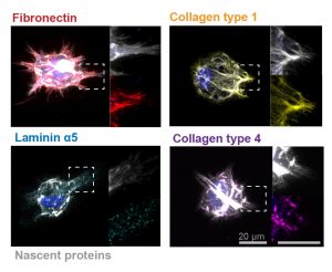
In these images, the researchers labeled new proteins white, and antibodies against other proteins in different colors. The co-localization of new proteins and antibodies show how cells can impact their local environments.
As different as muscle, blood, brain and skin cells are from one another, they all share the same DNA. Stem cells’ transformation into these specialized cells — a process called cell fate determination — is controlled through various signals from their surroundings.
A recent Penn Engineering study suggests that cells may have more control over their fate than previously thought.
Jason Burdick, Robert D. Bent Professor of Bioengineering, and Claudia Loebel, a postdoctoral researcher in his lab, led the study. Robert Mauck, Mary Black Ralston Professor for Education and Research in Orthopaedic Surgery at Penn’s Perelman School of Medicine, also contributed to the research.
Since Watson and Crick published their initial studies detailing the double helix structure of DNA in the early 1960s, what we know about genetics and the nucleic acids underlying them has grown enormously. Consequently, what bioengineering can do with DNA and genes continually expands.
One fascinating bioengineering field that emerged in the past decade was DNA origami, which uses the well-established binding across DNA elements to create three-dimensional structures out of linear DNA sequences. Recent work has utilized this feature of DNA construction to make machines, rather than just parts, out of DNA.
Yonggang Ke, Ph.D., of Georgia Tech/Emory’s Department of Biomedical Engineering, constructed machines made of DNA that consist of arrays of units that can “switch” between “settings” by changing shape. A change in shape of one unit of an array can cause the other units in the array to shift; these changes are stimulated by inserting a previously deleted strand of DNA into the array. Although it has been known for some time that DNA could be used to store and transmit information, Dr. Ke’s research team proved for the first time that these arrays could be shaped physically into machines in the shapes of rectangles and tubes.
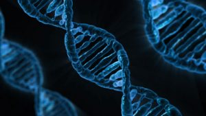
DNA under a microscope
While we learn more about how to make DNA-based devices, we are also creating new technologies to manipulate DNA more rapidly. Scientists at Rutgers and Harvard developed a process whereby thousands of genes could be cloned at one time to create enormous libraries of proteins. To achieve this goal, the authors used a technology called LASSO (long-adapter single-strand oligonucleotide) probes, which they have already used to clone a library using a human microbiome sample.
Instead of the traditional process of cloning one gene at a time, the team led by Professor Biju Parekkadan, Ph.D. at Rutgers, invented a technology to clone hundred of genes simultaneously. These cloned DNA segments are much longer than the length of DNA cloned with standard techniques, allowing us to test the functional significance of these much longer DNA segments. The technology could impact a number of scientific fields because we will finally learn how long stretches of protein function — some parts may degrade other proteins, while other parts will interact and modify other proteins (e.g., phosphorylation, a key process in epigenetics). These new discoveries can be key for discovering new ways to engineer proteins and to manufacture new drugs that mimic the function of nature’s DNA products.
Using Sweat as a Biosensor
While the field learns more about the molecular-level control of DNA, we are also taking advantage of new micro- and nanoscale manufacturing processes to capture diagnostic information from easily accessible body fluids. Many clinical diagnostics use chemical measurements from blood to diagnose a disease or to take corrective action. This is not an ideal procedure because it requires either the collection of blood at a laboratory or the repeated collection of small blood volumes through a pinprick. Either one hurts.
Bioengineers at the University of Texas at Dallas developed a wearable diagnostic device to detect cortisol, glucose, and IL-6 in body sweat, eliminating any painful needle sticks. Its transmissions vary, but if optimized, the device could replace the painful and inconvenient practice of sticking one’s finger to obtain a drop of blood for glucose testing, which many patients with diabetes must do several times per day. Although insulin pumps have been available for some time, these are invasive devices that must be worn at all times.


 A new technique that researchers from the Broad Institute of MIT and Harvard University are calling DNA microscopy could help map cells for better understanding of genetic and molecular complexities.
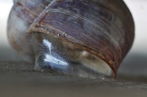
