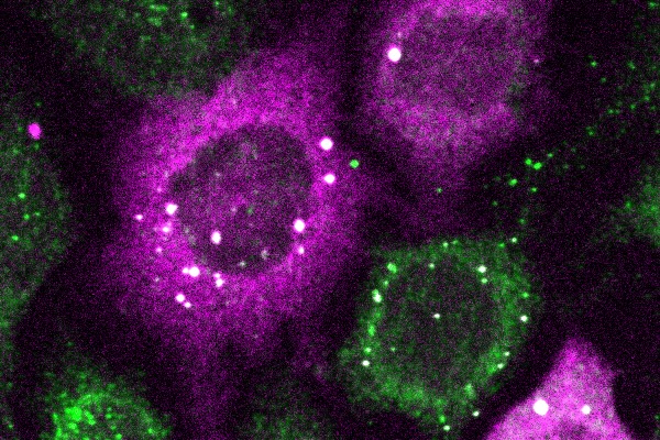
The bright white spots represent tiny clusters of proteins detected by CluMPS. (Photo by: Thomas Mumford)
Penn Engineers have pioneered a new way to visualize the smallest protein clusters, skirting the physical limitations of light-powered microscopes and opening new avenues for detecting the proteins implicated in diseases like Alzheimer’s and testing new treatments.
In a paper in Cell Systems, Lukasz Bugaj, Assistant Professor in Bioengineering, describes the creation of CluMPS, or Clusters Magnified by Phase Separation, a molecular tool that activates by forming conspicuous blobs in the presence of target protein clusters as small as just a few nanometers. In essence, CluMPS functions like an on/off switch that responds to the presence of clusters of the protein it is programmed to detect.
Normally, says Bugaj, detecting such clusters requires laborious techniques. “With CluMPS, you don’t need anything beyond the standard lab microscope.” The tool fuses with the target protein to form condensates orders of magnitude larger than the protein clusters themselves that resemble the colorful blobs in a lava lamp. “We think the simplicity of the approach is one of its main benefits,” says Bugaj. “You don’t need specialized skills or equipment to quickly see whether there are small clusters in your cells.”
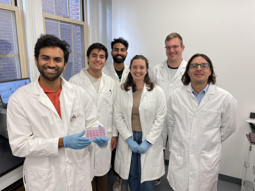
Ajay Thatte, Benjamin Nachod, Rohan Palanki, Kelsey Swingle, Alex Hamilton, and Michael Mitchell (Left to Right – Courtesy of the Mitchell Lab)
Autoimmune disorders are among the most prevalent chronic diseases across the globe, affecting approximately 5-7% of the world’s population. Emerging treatments for autoimmune disorders focus on “adoptive cell therapies,” or those using cells from a patient’s own body to achieve immunosuppression. These therapeutic cells are recognized by the patient’s body as ‘self,’ therefore limiting side effects, and are specifically engineered to localize the intended therapeutic effect.
In treating autoimmune diseases, current adoptive cell therapies have largely centered around the regulatory T cell (Treg), which is defined by the expression of the Forkhead box protein 3, orFoxp3. Although Tregs offer great potential, using them for therapeutic purposes remains a major challenge. In particular, current delivery methods result in inefficient engineering of T cells.
Tregs only compose approximately 5-10% of circulating peripheral blood mononuclear cells. Furthermore, Tregs lack more specific surface markers that differentiate them from other T cell populations. These hurdles make it difficult to harvest, purify and grow Tregs to therapeutically relevant numbers. Although there are additional tissue-resident Tregs in non-lymphoid organs such as in skeletal muscle and visceral adipose tissue, these Tregs are severely inaccessible and low in number.
“The major challenges associated with ex vivo (outside the body) cell engineering are efficiency, toxicity, and scale-up: our mRNA lipid nanoparticles (mRNA LNPs) allow us to overcome all of these issues,” says Mitchell. “Our work’s novelty comes from three major components: first, the use of mRNA, which allows for the generation of transient immunosuppressive cells; second, the use of LNPs, which allow for effective delivery of mRNA and efficient cell engineering; and last, the ex vivo engineering of primary human T cells for autoimmune diseases, offering the most direct pipeline for clinical translation of this therapy from bench to bedside.”
“To our knowledge, this is one of the first mRNA LNP platforms that has been used to engineer T cells for autoimmune therapies,” he continues. “Broadly, this platform can be used to engineer adoptive cell therapies for specific autoimmune diseases and can potentially be used to create therapeutic avenues for allergies, organ transplantation and beyond.”
Delivering the Foxp3 protein to T cells has been difficult because proteins do not readily cross the cell membrane. “The mRNA encodes for Foxp3 protein, which is a transcription factor that makes the T cells immunosuppressive rather than active,” explains first author Ajay Thatte, a doctoral student in Bioengineering and NSF Fellow in the Mitchell Lab. “These engineered T cells can suppress effector T cell function, which is important as T cell hyperactivity is a common phenotype in autoimmune diseases.”
Penn Bioengineering is proud to congratulate Sunghee Estelle Park, Ph.D. on her appointment as Assistant Professor in the Weldon School of Biomedical Engineering at Purdue University. Park earned her Ph.D. at Penn Bioengineering, graduating in July 2023. She conducted doctoral research in the BIOLines Lab of Dan Huh, Associate Professor in Bioengineering. Her appointment at Purdue will begin January 2024.
During her Ph.D. research, Park forged a unique path that combined principles in developmental biology, stem cell biology, organoids, and organ-on-a-chip technology to develop innovative in vitro models that can faithfully replicate the pathophysiology of various human diseases. Using a microengineered model of the human retina, she discovered previously unknown roles of the MAPK, IL-17, PI3K-AKT, and TGF-β signaling pathways in the pathogenesis of age-related macular degeneration (AMD), presenting novel therapeutic targets that could be further investigated for the development of AMD treatments. More recently, she tackled a significant challenge in the organoid field, the limited tissue growth and maturity in conventional organoid cultures, by designing microengineered systems that enabled organoids to grow with unprecedented levels of maturity and human-relevance. By integrating these platforms with bioinformatics and computational analyses, she identified novel disease-specific biomarkers of inflammatory bowel disease (IBD) and intestinal fibrosis, including previously unknown link between the presence of lncRNA and the development of IBD.
“The unique interdisciplinary expertise I gained from these projects has shaped me into a scholar with a strong collaborative ethos, a quality I hold in high esteem as we work towards advancing our knowledge and management of health and disease,” says Park.
Her vision as an independent researcher is to become a leading faculty who makes impactful contributions to our fundamental understanding of the factors influencing the structural and functional changes of human organs in health and disease. To achieve this, she plans to lead a stem cell bioengineering laboratory with a primary focus on tissue engineering and regenerative medicine. This will involve developing human organoids-on-a-chip systems and establishing next-generation biomedical devices and therapies tailored for regenerative and personalized medicine.
“I am grateful to all my Ph.D. mentors and lab mates at the BIOLines lab and especially my advisor Dr. Dan Huh, for his exceptional guidance, unwavering support, and invaluable mentorship throughout my Ph.D. journey,” says Park. “Dan’s expertise, dedication, and commitment to excellence have been instrumental in shaping both my research and professional development, while also training me to become an independent scientist and mentor.”
Congratulations to Dr. Park from everyone at Penn Bioengineering!
Cosette Tomita, a master’s student in Bioengineering, spoke with Penn Engineering Graduate Admissions about her research in cellular therapy and her path to Penn Engineering.
“What were you doing before you came to Penn Engineering?
After college I wanted to get some industry experience before going to graduate school, so I spent a year working for a pharmaceutical company in New Jersey. I learned a lot—but mostly I learned that I wanted to go back into academia. So I was looking for a more research-oriented position to boost my graduate school applications, and I found a position at Penn’s cyclotron facility. Shortly after that, I applied to the master’s program. I’m still working at the cyclotron, so I’m doing the program part time.
How has your experience in the program been so far?
I love the research I’m doing here. I love the collaboration we have and the fact that I’m able to work with whoever I want to. And I can only say good things about my PI, Robert Mach. He’s a very busy man, but he makes time for his people. And he recognizes when somebody has a lot on their plate and he will go to bat for that person.
What’s your research all about?
The focus of my PI’s lab is on neurodegenerative diseases and opiate use, so we’re looking to make imaging agents and antagonists that can help with the opioid crisis.
For my project, I wanted to look at treating neurodegenerative disease from the perspective of cellular therapy. My PI doesn’t have that expertise, so when I came to him with this idea, he said I should talk to Mark Sellmyer in the bioengineering department. He does a lot of cellular therapies, cell engineering, protein engineering and things of that nature. So his lab is more biological.
I don’t have a grant for my research, so my advisors are supporting it out of their own pockets. They could have said, no, you need to work on this project that’s already going on in the lab. But they gave me the intellectual freedom to do what I wanted to do.”
Folding@home is led by Gregory Bowman, a Penn Integrates Knowledge Professor who has appointments in the Departments of Biochemistry and Biophysics in the Perelman School of Medicine and the Department of Bioengineering in the School of Engineering and Applied Science. (Image: Courtesy of Penn Medicine News)
Two heads are better than one. The ethos behind the scientific research project Folding@home is that same idea, multiplied: 50,000 computers are better than one.
Folding@home is a distributed computing project which is used to simulate protein folding, or how protein molecules assemble themselves into 3-D shapes. Research into protein folding allows scientists to better understand how these molecules function or malfunction inside the human body. Often, mutations in proteins influence the progression of many diseases like Alzheimer’s disease, cancer, and even COVID-19.
Penn is home to both the computer brains and human minds behind the Folding@home project which, with its network, forms the largest supercomputer in the world. All of that computing power continually works together to answer scientific questions such as what areas of specific protein implicated in Parkinson’s disease may be susceptible to medication or other treatment.
Using the network hub at Penn, Bowman and his team assign experiments to each individual computer which communicates with other computers and feeds info back to Philly. To date, the network is comprised of more than 50,000 computers spread across the world.
“What we do is like drawing a map,” said Bowman, explaining how the networked computers work together in a type of system that experts call Markov state models. “Each computer is like a driver visiting different places and reporting back info on those locations so we can get a sense of the landscape.”
Individuals can participate by signing up and then installing software to their standard personal desktop or laptop. Participants can direct the software to run in the background and limit it to a certain percentage of processing power or have the software run only when the computer is idle.
When the software is at work, it’s conducting unique experiments designed and assigned by Bowman and his team back at Penn. Users can play scientist and watch the results of simulations and monitor the data in real time, or they can simply let their computer do the work while they go about their lives.


 Cosette Tomita, a master’s student in Bioengineering, spoke with Penn Engineering Graduate Admissions about her research in cellular therapy and her path to Penn Engineering.