Enamored by the chemical processes of life, Yihui Shen, J. Peter and Geri Skirkanich Assistant Professor of Innovation in Bioengineering, started her research career as a chemist studying the way that proteins fold and the intricate dynamics underlying life processes.
“As an undergraduate, I studied physical chemistry, thinking that one day I’d be addressing challenges in hardcore STEM fields,” she says. “It wasn’t until I observed the dynamics of a single protein molecule that I fell in love with microscopy. I realized that this imaging tool could not only help us observe biological processes on a small scale, but it could also provide new insight at the interface of engineering, chemistry and physics and solve problems on a large scale.”
When Shen turned her attention to microscopy, the field itself was advancing quickly, with improvements being made and new techniques being released every month. Without missing a beat, Shen dove deeper into the most current tools available when she joined Dr. Wei Min’s lab at Columbia University as a doctoral student.
“Professor Wei Min is a pioneer in a new imaging technique called coherent Raman imaging,” says Shen. “In this type of microscopy, we focus light on a very specific point in the cell and measure the amount of scattered light that comes back after exchanging energy with the molecular vibration. This approach allows us to visualize the spatial distribution of different molecules, the very chemistry of life I had studied as an undergraduate, at a high enough resolution to gain insights into biological processes, such as tissue organization, drug distribution and cellular metabolism.”
With this new tool under her belt, Shen was able to ask the kinds of questions that could connect the use of this observation tool to practical applications for real-world challenges.
“I started thinking outside the box,” says Shen. “What if we could observe the chemical exchanges involved in metabolism as they are happening on the scale of a single cell, and then use that insight to pinpoint the exact metabolic pathways and molecules that facilitate tumor growth and disease?”
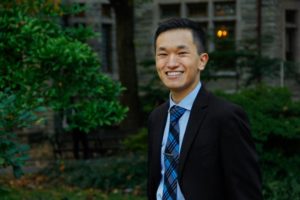
Cesar de la Fuente (left), Fangping Wan (center), and Marcelo der Torossian Torres (right). Fangping holds a 3D model of a unique ATP synthase fragment, identified by their lab’s deep learning model, APEX, as having potent antibiotic properties.
“Make sure you finish your antibiotics course, even if you start feeling better’ is a medical mantra many hear but ignore,” says Cesar de la Fuente of the University of Pennsylvania.
He explains that this phrase is, however, crucial as noncompliance could hamper the efficacy of a key 20th century discovery, antibiotics. “And in recent decades, this has led to the rise of drug-resistant bacteria, a growing global health crisis causing approximately 4.95 million deaths per year and threatens to make even common infections deadly,” he says.
De la Fuente, a Presidential Assistant Professor, and a team of interdisciplinary researchers have been working on biomedical innovations tackling this looming threat. In a new study, published in Nature Biomedical Engineering, they developed an artificial intelligence tool to mine the vast and largely unexplored biological data—more than 10 million molecules of both modern and extinct organisms— to discover new candidates for antibiotics.
“With traditional methods, it takes around six years to develop new preclinical drug candidates to treat infections and the process is incredibly painstaking and expensive,” de la Fuente says. “Our deep learning approach can dramatically reduce that time, driving down costs as we identified thousands of candidates in just a few hours, and many of them have preclinical potential, as tested in our animal models, signaling a new era in antibiotic discovery.” César de la Fuente holds a 3D model of a unique ATP synthase fragment, identified by his lab’s deep learning model, APEX, as having potent antibiotic properties. This molecular structure, resurrected from ancient genetic data, represents a promising lead in the fight against antibiotic-resistant bacteria.
These latest findings build on methods de la Fuente has been working on since his arrival at Penn in 2019. The team asked a fundamental question: Can machines be used to accelerate antibiotic discovery by mining the world’s biological information? He explains that this idea is based on the notion that biology, at its most basic level, is an information source, which could theoretically be explored with AI to find new useful molecules.
Almost a century ago, the discovery of antibiotics like penicillin revolutionized medicine by harnessing the natural bacteria-killing abilities of microbes. Today, a new study co-led by researchers at the Perelman School of Medicine at the University of Pennsylvania suggests that natural-product antibiotic discovery is about to accelerate into a new era, powered by artificial intelligence (AI).
The study, published in Cell, the researchers used a form of AI called machine learning to search for antibiotics in a vast dataset containing the recorded genomes of tens of thousands of bacteria and other primitive organisms. This unprecedented effort yielded nearly one million potential antibiotic compounds, with dozens showing promising activity in initial tests against disease-causing bacteria.
“AI in antibiotic discovery is now a reality and has significantly accelerated our ability to discover new candidate drugs. What once took years can now be achieved in hours using computers” said study co-senior author César de la Fuente, PhD, a Presidential Assistant Professor in Psychiatry, Microbiology, Chemistry, Chemical and Biomolecular Engineering, and Bioengineering.
Nature has always been a good place to look for new medicines, especially antibiotics. Bacteria, ubiquitous on our planet, have evolved numerous antibacterial defenses, often in the form of short proteins (“peptides”) that can disrupt bacterial cell membranes and other critical structures. While the discovery of penicillin and other natural-product-derived antibiotics revolutionized medicine, the growing threat of antibiotic resistance has underscored the urgent need for new antimicrobial compounds.
In recent years, de la Fuente and colleagues have pioneered AI-powered searches for antimicrobials. They have identified preclinical candidates in the genomes of contemporary humans, extinct Neanderthals and Denisovans, woolly mammoths, and hundreds of other organisms. One of the lab’s primary goals is to mine the world’s biological information for useful molecules, including antibiotics.
The growing threat of antimicrobial resistance demands innovative solutions in drug discovery. Scientists are turning to artificial intelligence (AI) and machine learning (ML) to accelerate the discovery and development of antimicrobial peptides (AMPs). These short strings of amino acids are promising for combating bacterial infections, yet transitioning them into clinical use has been challenging. Leveraging novel AI-driven models, researchers aim to overcome these obstacles, heralding a new era in antimicrobial therapy.
A new article in Nature Reviews Bioengineering illuminates the promises and challenges of using AI for antibiotic discovery. Cesar de la Fuente, Presidential Assistant Professor in Microbiology and Psychiatry in the Perelman School of Medicine, in Bioengineering and Chemical and Biomolecular Engineering in the School of Engineering and Applied Science, and Adjunct Assistant Professor in Chemistry in the School of Arts and Sciences, collaborated with James J. Collins, Termeer Professor of Medical Engineering and Science at MIT, to provide an introduction to this emerging field, outlining both its current limitations and its massive potential.
In the past five years, groundbreaking work in the de la Fuente Lab has dramatically accelerated the discovery of new antibiotics, reducing the timeline from years to mere hours. AI-driven approaches employed in his laboratory have already yielded numerous preclinical candidates, showcasing the transformative potential of AI in antimicrobial research and offering new potential solutions against currently untreatable infections.
Recent advancements in AI and ML are revolutionizing drug discovery by enabling the precise prediction of biomolecular properties and structures. By training ML models on high-quality datasets, researchers can accurately forecast the efficacy, toxicity and other crucial attributes of novel peptides. This predictive power expedites the screening process, identifying promising candidates for further evaluation in a fraction of the time required by conventional methods.
Traditional approaches to AMP development have encountered hurdles such as toxicity and poor stability. AI models help overcome these challenges by designing peptides with enhanced properties, improving stability, efficacy and safety profiles, and fast-tracking the peptides’ clinical application.
While AI-driven drug discovery has made significant strides, challenges remain. The availability of high-quality data is a critical bottleneck, necessitating collaborative efforts to curate comprehensive datasets to train ML models. Furthermore, ensuring the interpretability and transparency of AI-generated results is essential for fostering trust and wider adoption in clinical settings. However, the future is promising, with AI set to revolutionize antimicrobial therapy development and address drug resistance.
Integrating AI and ML into antimicrobial peptide development marks a paradigm shift in drug discovery. By harnessing these cutting-edge technologies, researchers can address longstanding challenges and accelerate the discovery of novel antimicrobial therapies. Continuous innovation in AI-driven approaches is likely to spearhead a new era of precision medicine, augmenting our arsenal against infectious diseases.
The de la Fuente Lab uses use the power of machines to accelerate discoveries in biology and medicine. The lab’s current projects include using AI for antibiotic discovery, molecular de-extinction, reprogramming venom-derived peptides to discover new antibiotics, and developing low-cost diagnostics for bacterial and viral infections. Read more posts featuring de la Fuente’s work in the BE Blog.
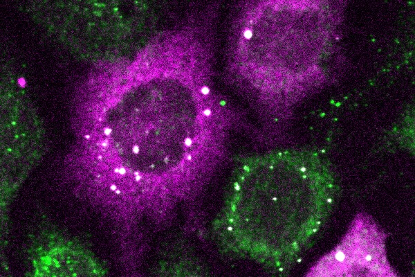
The bright white spots represent tiny clusters of proteins detected by CluMPS. (Photo by: Thomas Mumford)
Penn Engineers have pioneered a new way to visualize the smallest protein clusters, skirting the physical limitations of light-powered microscopes and opening new avenues for detecting the proteins implicated in diseases like Alzheimer’s and testing new treatments.
In a paper in Cell Systems, Lukasz Bugaj, Assistant Professor in Bioengineering, describes the creation of CluMPS, or Clusters Magnified by Phase Separation, a molecular tool that activates by forming conspicuous blobs in the presence of target protein clusters as small as just a few nanometers. In essence, CluMPS functions like an on/off switch that responds to the presence of clusters of the protein it is programmed to detect.
Normally, says Bugaj, detecting such clusters requires laborious techniques. “With CluMPS, you don’t need anything beyond the standard lab microscope.” The tool fuses with the target protein to form condensates orders of magnitude larger than the protein clusters themselves that resemble the colorful blobs in a lava lamp. “We think the simplicity of the approach is one of its main benefits,” says Bugaj. “You don’t need specialized skills or equipment to quickly see whether there are small clusters in your cells.”
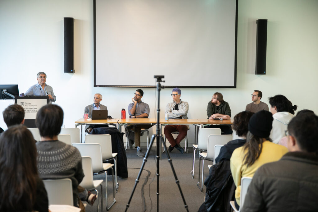
René Vidal, at the podium, introduces the event “ChatGPT turns one: How is generative AI reshaping science?” Bhuvnesh Jain, left at the table, moderated the discussion with Sudeep Bhatia, Konrad Kording, Andrew Zahrt, and Nick Pangakis.
As a neuroscientist surveying the landscape of generative AI—artificial intelligence capable of generating text, images, or other media—Konrad Kording cites two potential directions forward: One is the “weird future” of political use and manipulation, and the other is the “power tool direction,” where people use ChatGPT to get information as they would use a drill to build furniture.
“I’m not sure which of those two directions we’re going but I think a lot of the AI people are working to move us into the power tool direction,” says Kording, a Penn Integrates Knowledge (PIK) University professor with appointments in the Perelman School of Medicine and School of Engineering and Applied Science. Reflecting on how generative AI is shifting the paradigm of science as a discipline, Kording said he thinks “it will push science as a whole into a much more collaborative direction,” though he has concerns about ChatGPT’s blind spots.
Kording joined three University of Pennsylvania researchers from the chemistry, political science, and psychology departments sharing their perspectives in the recent panel “ChatGPT turns one: How is generative AI reshaping science?” PIK Professor René Vidal opened the event, which was hosted by the School of Arts & Sciences’ Data Driven Discovery Initiative (DDDI), and Bhuvnesh Jain, physics and astronomy professor and co-faculty director of DDDI, moderated the discussion.
“Generative AI is moving so rapidly that even if it’s a snapshot, it will be very interesting for all of us to get that snapshot from these wonderful experts,” Jain said. OpenAI launched ChatGPT, a large language model (LLM)-based chatbot, on Nov. 30, 2022, and it rapidly ascended to ubiquity in news reports, faculty discussions, and research papers. Colin Twomey, interim executive director of DDDI, told Penn Today that it’s an open question as to how it will change the landscape of scientific research, and the` idea of the event was to solicit colleagues’ opinions on interesting directions in their fields.
Konrad Paul Kording is Nathan Francis Mossell University Professor in Bioengineering and Computer and Information Science in Penn Engineering and in Neuroscience in the Perelman School of Medicine.
Daeyeon Lee (left), Oren Friedman (center) and Sergei Vinogradov (right)
Each year, the Nemirovsky Engineering and Medicine Opportunity (NEMO) Prize, funded by Penn Health-Tech, awards $80,000 to a collaborative team of researchers from the University of Pennsylvania’s Perelman School of Medicine and the School of Engineering and Applied Science for early-stage, interdisciplinary ideas.
This year, the NEMO Prize has been awarded to Penn Engineering’s Daeyeon Lee, Russel Pearce and Elizabeth Crimian Heuer Professor in Chemical and Biomolecular Engineering, Oren Friedman, Associate Professor of Clinical Otorhinolaryngology in the Perelman School of Medicine, and Sergei Vinogradov, Professor in the Department of Biochemistry and Biophysics in the Perelman School of Medicine and the Department of Chemistry in the School of Arts & Sciences. Together, they are developing a new therapy that improves the survival and success of soft-tissue grafts used in reconstructive surgery.
More than one million people receive soft-tissue reconstructive surgery for reasons such as tissue trauma, cancer or birth defects. Autologous tissue transplants are those where cells and tissue such as fat, skin or cartilage are moved from one part of a patient’s body to another. As the tissue comes from the patient, there is little risk of transplant rejection. However, nearly one in four autologous transplants fail due to tissue hypoxia, or lack of oxygen. When transplants fail the only corrective option is more surgery. Many techniques have been proposed and even carried out to help oxygenate soft tissue before it is transplanted to avoid failures, but current solutions are time consuming and expensive. Some even have negative side effects. A new therapy to help oxygenate tissue quickly, safely and cost-effectively would not only increase successful outcomes of reconstructive surgery, but could be widely applied to other medical challenges.
The therapy proposed by this year’s NEMO Prize recipients is a conglomerate or polymer of microparticles that can encapsulate oxygen and disperse it in sustainable and controlled doses to specific locations over periods of time up to 72 hours. This gradual release of oxygen into the tissue from the time it is transplanted to the time it functionally reconnects to the body’s vascular system is essential to keeping the tissue alive.
“The microparticle design consists of an oxygenated core encapsulated in a polymer shell that enables the sustained release of oxygen from the particle,” says Lee. “The polymer composition and thickness can be controlled to optimize the release rate, making it adaptable to the needs of the hypoxic tissue.”
These life-saving particles are designed to be integrated into the tissue before transplantation. However, because they exist on the microscale, they can also be applied as a topical cream or injected into tissue after transplantation.
“Because the microparticles are applied directly into tissues topically or by interstitial injection (rather than being administered intravenously), they surpass the need for vascular channels to reach the hypoxic tissue,” says Friedman. “Their micron-scale size combined with their interstitial administration, minimizes the probability of diffusion away from the injury site or uptake into the circulatory system. The polymers we plan to use are FDA approved for sustained-release drug delivery, biocompatible and biodegrade within weeks in the body, presenting minimal risk of side effects.”
The research team is currently testing their technology in fat cells. Fat is an ideal first application because it is minimally invasive as an injectable filler, making it versatile in remodeling scars and healing injury sites. It is also the soft tissue type most prone to hypoxia during transplant surgeries, increasing the urgency for oxygenation therapy in this particular tissue type.
Bella Mirro, a fourth year student in Bioengineering who also minors in Chemistry, spoke with 34th Street Magazine about her many roles at Penn, including being Co–President of Shelter Health Outreach Program (SHOP), a Research Assistant in lab of Michal A. Elovitz, the Hilarie L. Morgan and Mitchell L. Morgan President’s Distinguished Professor in Women’s Health at Penn Medicine, and a Penn Engineering Council Marketing Team Member. In this Q&A, she discusses her research in women’s health and her passions for accessible healthcare, serving Philadelphia’s homeless community, and good food.
Each year, the the Department of Bioengineering seeks exceptional candidates to conduct summer research in bioengineering with the support of two scholarships: the Abraham Noordergraaf Student Summer Bioengineering Research Fund and the Blair Undergraduate Research Fund in the Department of Bioengineering. These scholarships provide a living stipend for students to conduct research on campus in a Penn research lab under the mentorship of a faculty member. The Abraham Noordergraaf Student Summer Bioengineering Research Fund provides financial support for undergraduate or graduate summer research opportunities in bioengineering with a preference for study in the area of cardiovascular systems. Dr. Noordergraaf, who died in 2014, was a founding member and first chair of Penn Bioengineering. The Blair Undergraduate Research Fund in the Department of Bioengineering supports three to five undergraduate research scholars each year with the support of Dr. James C. Blair II. After a competitive round of proposals, the following six scholars were chosen for the Summer 2022 semester. Keep reading below for the research abstracts and bios of the awardees.
The Blair Undergraduate Research Fund in the Department of Bioengineering (Blair Scholars)
Ella Atsavapranee
Student: Ella Atsavapranee (BE Class of 2023)
PI: Michael J. Mitchell, J. Peter and Geri Skirkanich Assistant Professor of Innovation, Bioengineering
“Lipid nanoparticle-mediated delivery of RAS protease to inhibit cancer cell growth”
Mutations in RAS, a family of proteins found in all human cells, drive a third of cancers, including many pancreatic, colorectal, and lung cancers. However, there are still no therapies that can effectively prevent RAS from causing tumor growth. Recently, a protease was engineered to specifically degrade active RAS, offering a promising new tool for treating these cancers. However, many protein-based therapies still cannot be effectively delivered to patients. Lipid nanoparticles (LNPs), which were used in the Pfizer-BioNTech and Moderna COVID-19 vaccines, have emerged as a promising platform for safe and effective delivery of both nucleic acids and proteins. We formulated a library of LNPs using different cationic lipids. We characterized the LNPs by size, charge, and pKa, and tested their ability to deliver fluorescently labeled protease. The LNPs were able to encapsulate and deliver a RAS protease, successfully reducing proliferation of colon cancer cells.
Ella is a senior from Maryland studying bioengineering and chemistry. She works in Dr. Michael Mitchell’s lab, developing lipid nanoparticles to deliver proteins that reduce cancer cell proliferation. She has also conducted research on early-stage cancer detection and therapy monitoring (at Stanford University) and drug delivery across the blood-brain barrier for neurodegenerative diseases (at University of Maryland). She is passionate about translational research, science communication, and promoting diversity in STEM.
Chiadika Eleh
Student: Chiadika Eleh (BE and CIS Class of 2024)
PI: Eric J. Brown, Associate Professor of Cancer Biology, Perelman School of Medicine
“Investigating Viability in ATR and WEE1 Inhibitor Treated Ovarian Cancer Cells”
High-grade serous ovarian cancers (HGSOCs) are an aggressive subtype of ovarian cancer, accounting for up to 80% of all ovarian cancer-related deaths. More than half of HGSOCs are homologous recombination deficient; thus, they lack a favorable response when treated with common chemotherapeutic trials. Therefore, new treatment strategies must be developed to increase the life expectancy and quality of life of HGSOC patients. To address the lack of effective treatment options, the Brown Lab is interested in combining ATR and WEE1 inhibition (ATRi/WEE1i) to target HGSOC cells. It has previously been shown that low-dose ATRi/WEE1i is an effective treatment strategy for CCNE1-amplified ovarian cancer-derived PDX tumors (Xu et al., 2021, Cell Reports Medicine). Therefore, the next step is to characterize the HGSOC-specific response to ATRi/WEE1i treatment. This project aims to characterize the viability phenotype of ovarian cancer (OVCAR3) cells in the presence of ATRi/WEE1i in both single and combination treatments. With further research, Eleh hopes to prove the hypothesis low-dose combination ATRi/WEE1i treatment will result in the synergistic loss of viability in OVCAR3 cells. This goal will be achieved through the treatment of OVCAR3 cells with ranging doses of ATRi and Wee1i over 24 and 48 hour time intervals. We hope that this data will help set a treatment baseline that can be used for all OVCAR30-based viability experiments in the future.
Chiadika Eleh is a Bioengineering and Computer Science junior and a member of Penn Engineering’s Rachleff Scholar program. As a Blair Scholar, she worked in Dr. Eric Brown’s cancer biology lab, where she studied cell cycle checkpoint inhibitors as a form of cancer treatment.
“Tbc1d2b regulates vascular formation during development and tissue repair after ischemia”
The mechanisms behind endothelial cells forming blood vessels remains unknown. We have identified Tbc1d2b as a protein that is integral to the regulation of vascular formation. In order to investigate the role of Tbc1d2b in tubule formation, fibrin gel bead assays will be conducted to evaluate how the presence of Tbc1d2b is required for angiogenesis. Fibrin gel bead assays simulate the extracellular matrix environment to support the in vitro development of vessels from human umbilical vein endothelial cells (HUVEC) coated on cytodex beads. In order to confirm the success of angiogenesis, immunostaining for Phalloidin and CD31 will be conducted. After confirmation that fibrin gel bead assays can produce in vitro tubules, sgRNA CRISPR knockout of Tbc1d2b will be performed on HUVEC cells which will then be used to conduct more fibrin gel bead assays. We hypothesize that HUVEC with the Tbc1d2b knockout phenotype will be unable to form tubules while wild type HUVEC will be able to.
Gloria Lee is a rising senior studying Bioengineering and Physics in the VIPER program from Denver, Colorado. Her research in Dr. Yi Fan’s lab focuses on the role that proteins play in cardiovascular tubule formation.
Abraham Noordergraaf Student Summer Bioengineering Research Fund (Noordergraaf Fellows)
Gary Lin
Student: Gary Lin (Master’s in MEAM Class of 2023)
PI: Michelle J. Johnson, Associate Professor in Physical Medicine and Rehabilitation, Perelman School of Medicine, and in Bioengineering
“Development and Integration of Dynamically Modulating Control Systems in the Rehabilitation Using Community-Based Affordable Robotic Exercise System (Rehab CARES)”
As the number of stroke patients requiring rehabilitative care continues to increase, strain is being put onto the US health infrastructure which already has a shortage of rehabilitation practitioners. To help alleviate this pressure, a cost-effective robotic rehabilitative platform was developed to increase access to rehabilitative care. The haptic TheraDrive, a one-degree of freedom actuated hand crank that can apply assistive and resistive forces, was modified to train pronation and supination at the elbow and pinching of the fingers in addition to flexion and extension of the elbow and shoulder. Two controllers were created including an open-loop force controller and a closed-loop proportional-integral (PI) with adaptive control gains based on subject performance in therapy-game tasks as well as galvanic skin response. Stroke subjects (n=11) with a range of cognitive and motor impairment completed 4 therapy games in both adaptive and non-adaptive versions of the controllers (n=8) while measuring force applied on the TheraDrive handle. Resulting normalized average power versus Upper Extremity Fugl-Meyer (UE-FM) and Montreal Cognitive Assessment (MoCA) correlation analyses showed that power was strongly correlated with UE-FM in 2 of the conditions and moderately correlated with the other 6 while MoCA was moderate correlated to 2 of the conditions and weakly correlated to the rest. Mann-Whitney U-tests between adaptive and non-adaptive versions of each therapy game showed no significant differences with regards to power between controller types (p<0.05).
Gary is a master’s student in the School of Engineering studying Mechanical Engineering and Applied Mechanics with a concentration in Robotic and Mechatronic systems. His research primarily focuses on developing affordable rehabilitation robotics for use in assessment and game-based therapies post neural injury. Many of his interests revolve around the design of mechatronic systems and the algorithms used to control them for use in healthcare spaces.
Priya Shah
Student: Priya Shah (BE Class of 2024)
PI: Alex J. Hughes, Assistant Professor in Bioengineering
“Optogenetic Control of Developing Kidney Cells for Future Treatment of End-Stage Renal Disease”
This project sought to build from prior research in the Hughes Lab on the geometric and mechanical consequences of kidney form on cell and tissue-scale function. While the developmental trajectory of the kidney is well understood, little is currently known about many factors affecting nephron progenitor differentiation rate. Insufficient differentiation of nephron progenitor cells during kidney formation can result in lower nephron number and glomerular density, which is a risk factor for progression to end-stage renal disease later in life. Prior studies indicated that the amount of nephron differentiation – and thus function of the adult kidney – is correlated to the packing of ureteric tubule tips present at the surface of the kidney. Building off of research conducted in the Bugaj Lab, we found that inserting an optogenetic construct into the genome of human embryonic kidney (HEK) cells allowed us to manipulate the contraction of those cells through exposing them to blue light. Manipulating the contraction of the cells allows for the manipulation of the packing of ureteric tubule tips at the kidney surface. We used a lentiviral vector to transduce HEK293 cells with the optogenetic construct and witnessed visible contraction of the cells when they were exposed to blue light. Future work will include using CRISPR-Cas9 to introduce the optogenetic construct into IPS cells.
Priya is a junior studying bioengineering and had the opportunity to work on manipulating developing kidney cells using an optogenetic construct in the Hughes Lab this summer. She is thrilled to continue this research throughout the coming school year. Outside of the lab, Priya is involved with the PENNaach dance team and the Society of Women Engineers, as well as other mentorship roles.
Cosette Tomita
Student: Cosette Tomita (Master’s in MEAM Class of 2023)
“Expression and Characterization of an Anti-Aβ42 scFv”
Background: Amyloid Beta (Aβ42) fibrils contribute to the pathology of Alzheimer’s Disease. Numerous monoclonal antibodies have been developed against Aβ42. In this study we have designed and expressed a short chain variable fragment specific to Aβ42 (Anti-Aβ42 scFv). To characterize our anti-Aβ42 scFv we have performed structural analysis using transmission electron microscopy (TEM) and binding kinetics using microscale thermophoresis (MST) compared to commercially available antibodies 6E10, Aducanumab, and an IgG isotype control. The goal of this study is to determine if labeling densities and binding constants for Aducanumab and anti-Aβ42 scFv are not significantly different.
Method: To characterize Aβ42 fibril associated antibodies we used negative stain TEM. Aβ42 fibrils were stained on a glow discharged copper grid, and incubated with gold conjugated anti-Aβ42 scFv, 6E10—which binds all Aβ species, aducanumab, or IgG isotype control. Labeling densities were calculated as the number of fibril-associated gold particles per 1 μm2 for each image. Next, we used microscale thermophoresis determine the binding kinetics. Antibodies or anti-Aβ42 scFv were labeled with Alexa Fluor-647 and unlabeled Aβ42 was titrated in a serial dilution over 16 capillaries. The average fluorescence intensity was plotted against the antibody or scFv concentration and the curves were analyzed using the GraphPad Prism software to calculate the dissociation constant (KD) values.
Results: We found a significant difference, tested with a one-way ANOVA (P <0.0001), in gold particle associated Aβ fibrils per 1 μm2 between anti-Aβ42 scFv, 6E10, aducanumab, and IgG isotype control. Further analysis of aducanumab and 6CO3 with unpaired student t-test indicates significant differences in fibril associated gold particles between aducanumab vs. 6E10 (P=0.0003), Aducanumab vs. Isotype control (P <0.0001), anti-Aβ42 scFv vs 6E10 (p=0.0072), and anti-Aβ42 scFv vs Isotype Control (P=0.0029) with no significant difference in labeling densities between Aducanumab and anti-Aβ42 scFv. The expected KD values from MST were 1.8μM for Aducanumab and anti-Aβ42 scFv, 10.3nM for 6E10 and no expected binding for the isotype control. The experimental KD values for anti-Aβ42 scFv and 6E10 are 0.1132μM and 1.467μM respectively. The KD value for Isotype control was undetermined, as expected, however, the KD for Aducanumab was undetermined due to suboptimal assay conditions. Due to confounding variables in the experimental set up such as the use of Aβ1-16 compared to Aβ42 and the use of different fluorophores—5-TAMRA, Alexa Fluor 647 or FITC— the experimental KD values were off by several orders of magnitude.
Conclusion: We have illustrated similar labeling densities between Aducanumab and our anti-Aβ42 scFv. In the future, we will further optimize the MST assay conditions and compare the KD values obtained by MST with other techniques such as surface plasma resonance.
Cosette was born and raised in Chicago land area. Go Sox! She attended University of Missouri where she majored in Chemistry and Biology. She synthesized sigma-2 radiotracers and developed advanced skills in biochemical techniques in Dr. Susan Lever’s lab. After graduation, she moved to NJ to work at Lantheus, a radiopharmaceutical company. She missed academia and the independence of program and project development, so she came to work at the Penn Cyclotron facility before entering the Bioengineering master’s program.
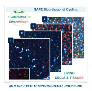
Cells in complex organisms undergo frequent changes, and researchers have struggled to monitor these changes and create a comprehensive profile for living cells and tissues. Historically researchers have been limited to only 3-5 markers due to spectral overlaps in fluorescence microscopy, an essential tool required for imaging cells. With only this small handful of markers, it is difficult to monitor protein expressions of live cells and a comprehensive profile of cellular dynamics cannot be created. However, a new study in Nature Biotechnology addresses these limitations by demonstrating a new method for comprehensive profiling of living cells.
Jina Ko, PhD
Jina Ko, Assistant Professor in Bioengineering in the School of Engineering and Applied Science and in Pathology and Laboratory Medicine in the Perelman School of Medicine, conducted postdoctoral research at Massachusetts General Hospital (MGH) and the Wyss Institute at Harvard University, and the work for this study was done under the supervision of Jonathan Carlson M.D., Ph.D. and Ralph Weissleder M.D., Ph.D. of MGH. Ko’s lab at Penn develops novel technologies using bioengineering, molecular biology, and chemistry to address diagnostic challenges for precision medicine.
To address these limitations in microscopy, the team developed a new chemistry tool which was highly gentle to cells. This “scission-accelerated fluorophore exchange (or SAFE)” method utilizes “click” chemistry, a type of chemistry that follows examples found in nature to create fast and simple reactions. This new SAFE method functions with non-toxic conditions to living cells and tissues, whereas previous methods have used harsh chemicals that would strip off fluorophores and consequently would not work with living cells and tissues.
With the development of SAFE, the authors demonstrated that researchers can now effectively perform multiple cycles of cell profiling and can monitor cellular changes over the course of their observations. Instead of the previous limitation of 3-5 markers total, SAFE allows for many more cycles and can keep track of almost as many markers as the researcher wants. One can now stain cells and quench/release fluorophores and repeat the cycle multiple times for multiplexing on living cells. Each cycle can profile 3 markers, and so someone interested in profiling 15 markers could easily perform 5 cycles to achieve this much more comprehensive cell profile. With this breakthrough in more detailed imaging of cells, SAFE demonstrates broad applicability for allowing researchers to better investigate the physiologic dynamics in living systems.
This study was supported by the Schmidt Science Fellows in Partnership with the Rhodes Trust and National Institutes of Health, National Cancer Institute (K99CA256353).