Researchers Develop Technology to Keep Track of Living Cells and Tissues

SAFE Bioorthogonal Cycling

Cells in complex organisms undergo frequent changes, and researchers have struggled to monitor these changes and create a comprehensive profile for living cells and tissues. Historically researchers have been limited to only 3-5 markers due to spectral overlaps in fluorescence microscopy, an essential tool required for imaging cells. With only this small handful of markers, it is difficult to monitor protein expressions of live cells and a comprehensive profile of cellular dynamics cannot be created. However, a new study in Nature Biotechnology addresses these limitations by demonstrating a new method for comprehensive profiling of living cells.

Jina Ko, PhD

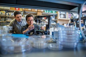
Jina Ko, Assistant Professor in Bioengineering in the School of Engineering and Applied Science and in Pathology and Laboratory Medicine in the Perelman School of Medicine, conducted postdoctoral research at Massachusetts General Hospital (MGH) and the Wyss Institute at Harvard University, and the work for this study was done under the supervision of Jonathan Carlson M.D., Ph.D. and Ralph Weissleder M.D., Ph.D. of MGH. Ko’s lab at Penn develops novel technologies using bioengineering, molecular biology, and chemistry to address diagnostic challenges for precision medicine.

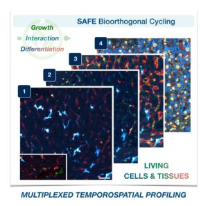
To address these limitations in microscopy, the team developed a new chemistry tool which was highly gentle to cells. This “scission-accelerated fluorophore exchange (or SAFE)” method utilizes “click” chemistry, a type of chemistry that follows examples found in nature to create fast and simple reactions. This new SAFE method functions with non-toxic conditions to living cells and tissues, whereas previous methods have used harsh chemicals that would strip off fluorophores and consequently would not work with living cells and tissues.

With the development of SAFE, the authors demonstrated that researchers can now effectively perform multiple cycles of cell profiling and can monitor cellular changes over the course of their observations. Instead of the previous limitation of 3-5 markers total, SAFE allows for many more cycles and can keep track of almost as many markers as the researcher wants. One can now stain cells and quench/release fluorophores and repeat the cycle multiple times for multiplexing on living cells. Each cycle can profile 3 markers, and so someone interested in profiling 15 markers could easily perform 5 cycles to achieve this much more comprehensive cell profile. With this breakthrough in more detailed imaging of cells, SAFE demonstrates broad applicability for allowing researchers to better investigate the physiologic dynamics in living systems.

Read the paper, “Spatiotemporal multiplexed immunofluorescence imaging of living cells and tissues with bioorthogonal cycling of fluorescent probes,” in Nature Biotechnology.

This study was supported by the Schmidt Science Fellows in Partnership with the Rhodes Trust and National Institutes of Health, National Cancer Institute (K99CA256353).

New data reveals cell size sparks genome awakening in embryos

Awakening of the zygote genome over time as decreasing individual cell size triggers early embryo transcription. (Image: Hui Chen, Penn Medicine; Cell Press)

There is a transition during early development when an embryo undergoes biochemical changes, switching from being controlled by maternal molecules to being governed by its own genome.

For the first time, a team from the Perelman School of Medicine found in an embryo that activation of its genome does not happen all at once, instead it follows a specific pattern controlled primarily by the various sizes of its cells. The researchers published their results as the cover story in Developmental Cell.

In an early embryo undergoing cell division, maternally loaded RNA and proteins regulate the cell cycle. The genomes of the zygote—a term for the fertilized egg—are initially in sleep mode. However, at a point in the early life of the embryo, these zygotic nuclei “wake up” and expression from their genomes takes biochemical control over subsequent embryo development. But how an embryo “recognizes” when to undergo this transition has remained unknown.

“How an embryo ‘hands over’ control of development from mother to zygote is a fundamental question in developmental biology,” says senior author Matthew C. Good, an assistant professor of both cell and developmental biology and bioengineering. “Previously it was not appreciated that different regions of a vertebrate embryo can undergo genome activation at different times, or how directly cell size regulates the awakening of a zygote’s genome.”

Read more at Penn Medicine News.

Organs-on-a-Chip Hurtle Toward the Final Frontier

Graduate student Andrei Georgescu and Assistant Professor Dan Huh in Huh’s lab. Adapting the organ-on-a-chip technology for a trip to the International Space Station presented Huh’s team with a number of engineering challenges. (Photo: Kevin Monko)

Throughout the 60-year history of the U.S. space program—from the Mercury capsules of the 1960s to today’s International Space Station—astronauts have been getting sick. Researchers know being in orbit seems to suppress their immune systems, creating a more fertile ground for infections to grow. But nobody really understands why.

Early on the morning of April 26, a SpaceX Falcon 9 rocket will launch a cargo mission to the ISS from Cape Canaveral Air Force Station. Along with fresh water, food, and other necessities for the crew, the craft will be carrying two experiments designed by Penn scientists that could help shed light on why bugs have bedeviled space travelers.

For more than a decade, Dan Huh, the Wilf Family Term Assistant Professor of Bioengineering in the School of Engineering and Applied Science, has been developing super-small devices that use living cells to stand in for larger organs. These organs-on-a-chip hold great promise for all kinds of research, from diagnosing disease to curing them. They’re also a way to test things, including drugs and cosmetics, in a way that mimics real life without relying on animal subjects.

Read the full story at Penn TodayMedia contact Gwyneth K. Shaw

Penn Bioengineers: Cells Control Their Own Fate by Manipulating Their Environment

by Lauren Salig

In these images, the researchers labeled new proteins white, and antibodies against other proteins in different colors. The co-localization of new proteins and antibodies show how cells can impact their local environments.

As different as muscle, blood, brain and skin cells are from one another, they all share the same DNA. Stem cells’ transformation into these specialized cells — a process called cell fate determination — is controlled through various signals from their surroundings.

A recent Penn Engineering study suggests that cells may have more control over their fate than previously thought.

Jason Burdick, Robert D. Bent Professor of Bioengineering, and Claudia Loebel, a postdoctoral researcher in his lab, led the study. Robert Mauck, Mary Black Ralston Professor for Education and Research in Orthopaedic Surgery at Penn’s Perelman School of Medicine, also contributed to the research.

Their study was published in Nature Materials.

Read the full story in the Penn Engineering Medium Blog. Media contact Evan Lerner.