BE Seminar: “Designing Biology for Detection and Control” (Pamela A. Silver)

Speaker: Pamela A. Silver, Ph.D.
Elliot T. and Onie H. Adams Professor of Biochemistry and Systems Biology
Harvard Medical School

Date: Thursday, January 28, 2021
Time: 3:00-4:00 PM EST
Zoom – check email for link or contact ksas@seas.upenn.edu

Title: “Designing Biology for Detection and Control”

Abstract:

The engineering of Biology presents infinite opportunities for therapeutic design, diagnosis, and prevention of disease. We use what we know from Nature to engineer systems with predictable behaviors. We also seek to discover new natural strategies to then re-engineer. I will present concepts and experiments that address how we approach these problems in a systematic way. Conceptually, we seek to both design cells and proteins to control disease states and to detect and predict the severity of emerging pathogens. For example, we have engineered components of the gut microbiome to act therapeutics for infectious disease, proteins to prolong cell states, living pathogen sensors and high throughput analysis to predict immune response of emerging viruses.

Bio:

Pamela Silver is the Adams Professor of Biochemistry and Systems Biology at Harvard Medical School and the Wyss Institute for Biologically Inspired Engineering. She received her BS in Chemistry and PhD in Biochemistry from the University of California. Her work has been recognized by an Established Investigator of the American Heart Association, a Research Scholar of the March of Dimes, an NSF Presidential Young Investigator Award, Claudia Adams Barr Investigator, an NIH MERIT award, the Philosophical Society Lecture, a Fellow of the Radcliffe Institute, and election to the American Academy of Arts and Sciences. She is among the top global influencers in Synthetic Biology and her work was named one of the top 10 breakthroughs by the World Economic Forum. She serves on the board of the Internationally Genetics Engineering Machines (iGEM) Competition and is member of the National Science Advisory Board for Biosecurity. She has led numerous projects for ARPA-E, iARPA and DARPA. She is the co-founder of several Biotech companies including most recently KulaBio and serves on numerous public and private advisory boards.

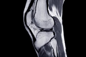
Magnetic Field and Hydrogels Could Be Used to Grow New Cartilage

by Frank Otto

MRI Knee joint or Magnetic resonance imaging sagittal view for detect tear or sprain of the anterior cruciate ligament (ACL).

Using a magnetic field and hydrogels, a team of researchers in the Perelman School of Medicine have demonstrated a new possible way to rebuild complex body tissues, which could result in more lasting fixes to common injuries, such as cartilage degeneration. This research was published in Advanced Materials.

“We found that we were able to arrange objects, such as cells, in ways that could generate new, complex tissues without having to alter the cells themselves,” says the study’s first author, Hannah Zlotnick, a graduate student in bioengineering who works in the McKay Orthopaedic Research Laboratory at Penn Medicine. “Others have had to add magnetic particles to the cells so that they respond to a magnetic field, but that approach can have unwanted long-term effects on cell health. Instead, we manipulated the magnetic character of the environment surrounding the cells, allowing us to arrange the objects with magnets.”

In humans, tissues like cartilage can often break down, causing joint instability or pain. Often, the breakdown isn’t in total, but covers an area, forming a hole. Current fixes are to fill those holes in with synthetic or biologic materials, which can work but often wear away because they are not the same exact material as what was there before. It’s similar to fixing a pothole in a road by filling it with gravel and making a tar patch: The hole will be smoothed out but eventually wear away with use because it’s not the same material and can’t bond the same way.

What complicates fixing cartilage or other similar tissues is that their makeup is complex.

“There is a natural gradient from the top of cartilage to the bottom, where it contacts the bone,” Zlotnick explains. “Superficially, or at the surface, cartilage has a high cellularity, meaning there is a higher number of cells. But where cartilage attaches to the bone, deeper inside, its cellularity is low.”

So the researchers, which included senior author Robert Mauck, PhD, director of the McKay Lab and a professor of Orthopaedic Surgery and Bioengineering, sought to find a way to fix the potholes by repaving them instead of filling them in. With that in mind, the research team found that if they added a magnetic liquid to a three-dimensional hydrogel solution, cells, and other non-magnetic objects including drug delivery microcapsules, could be arranged into specific patterns that mimicked natural tissue through the use of an external magnetic field.

Read more at Penn Medicine News.