
To Bee or Not To Bee
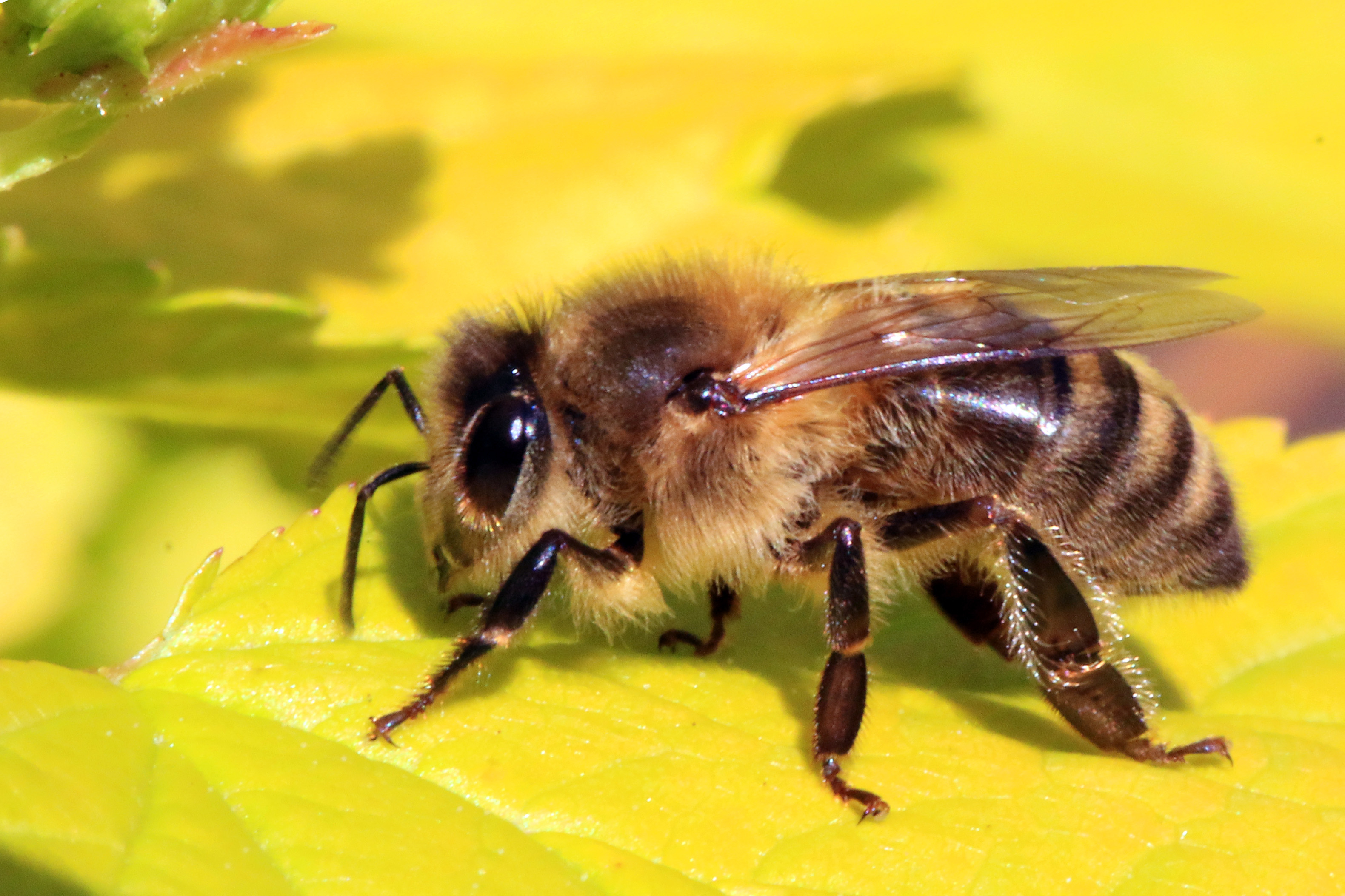 You might have heard reporting over the last few years that honeybees are dying at faster-than-usual rates. Over the last decade, colony collapse rates increased significantly, causing precipitous losses in the overall bee population. The consequences could be grave: in addition to providing honey, bee pollination is an important factor in agriculture, affecting major crops such as melons. squashes, and several kinds of nuts. Loss of this factor could substantially increase prices or even result in shortages.
To address this crisis, scientists at Washington State University focused on the role played by pesticides in colony collapse disorder. These poisons are particularly toxic to bees in tiny amounts, with the problem compounded by the ability of these toxins to build up in the bees’ bodies. A group of students led by Waled Suliman, PhD, a postdoctoral research associate in WSU’s Department of Biological Systems Engineering, developed a powder that acts like a magnet to draw pesticide out of the insects’ bodies. The bees then excrete the pesticide-laden particles like any other kind of waste.
The initiative, called Gaminus, has already tested its material in bees and found that the design works as planned. In coming months, they intend to continue their research by measuring toxin levels in the excreted particles.
Advances in Visualization
An important field within bioengineering is visualization, or the ability to use technology to enable scientists to see biological processes not normally visible to the naked eye. If you’ve seen a fetal ultrasound, for instance, then you’ve seen how one part of this area has advanced enormously in recent years. However, integrating visualization technologies with surgery remains a major challenge, particularly for minimally invasive surgeries. One key obstacle is that surgeons must rely on video screens during surgery, rather than being able to look down and feel the tissue with their hands.
A startup at the Cleveland Clinic is attempting to integrate perioperative visualization with HoloLens, a brand of smart glasses developed by Microsoft, to produce “mixed reality,” i.e., a combination of actual vision and virtual reality. With a grant from the National Heart, Lung, and Blood Institute awarded to Centerline Biomedical, the Cleveland Clinic startup, and to Karl West, Director of Medical Device Solutions at Cleveland Clinic and a staff member in the Lerner Research Institute’s Department of Biomedical Engineering, the integrated visualization device will be tested in a preclinical model of cardiac stent placement.
Elsewhere in the Midwest, Nathan Gianneschi, PhD, Professor of Chemistry, Biomedical Engineering and Materials Science and Engineering at Northwestern University, has been leading an effort to augment transmission electron microscopy (TEM). In its common form, TEM provides highly detailed images of submicroscopic organisms and structures and can provide visualization of nanomaterials as they grow. Gianneschi’s new approach, called liquid cell TEM (LCTEM), uses an irradiated region of a liquid cell to facilitate real-time visualization. The work is detailed in a recent article in ACS Central Science. You can see video posted online at the journal website.
Turning Red
Ultraviolet and infrared light appear beyond either end of the visible light spectrum. Past work using either ultraviolet or infrared light to activate fluorescent proteins can help visualize biochemistry in vivo, but it can also damage cells because of the activating light or the chemicals produced by illuminating the proteins. Recently, Young L. Kim, PhD, Associate Professor of Biomedical Engineering at Purdue, led a team of scientists who produced red fluorescent silk to kill harmful bacteria when the protein is activated by external green light. Dr. Kim and his colleagues report their findings in Advanced Science. The silk requires further testing, but if ultimately proved successful, it could overcome a current limitation of the use light-activated fluorescent biomaterials in controlling pathogens, which is that the light itself, often in the ultraviolet part of the spectrum, comes with its own potentially negative effects on health.
Absorbable Stents for Cardiac Care
Vascular stents to reopen blocked coronary arteries are usually the treatments used for patients with mild coronary artery disease. These simple devices are a small tube, sometimes coated with a drug to prevent clotting, inserted into the artery to restore flow. Stents can fail over time, requiring reimplantation, and the stents may also narrow over time and reduce blood flow to the surrounding tissue. To overcome this problem, Donghui Zhu, PhD, Associate Professor in the Department of Biomedical Engineering at the University of North Texas, developed a stent that is fully biodegradable and disappears over time as the damaged tissue heals. Dr. Zhu recently won a $2 million grant from the National Institutes of Health to test the stent in a series of trials.
People and Places
Penn State University has won a research grant from the American Heart Association, which will be used to support its 10-week Penn State Summer Translational Cardiovascular Science Institute (STCSI). Led by Keefe Manning, PhD, Professor of Biomedical Engineering at Penn State, the STCSI will provide $4,000 stipends for undergraduate students to conduct summer research on cardiovascular disease.
Finally, here at Penn Bioengineering, we are immensely proud to announce that our PhD student Jina Ko was named one of 14 PhD candidates in the inaugural class of Schmidt Science Fellows. Schmidt Fellows are each awarded a $100,000 stipend to cover the cost of living while conducting postdoctoral research. Congratulations, Jina!