
by Sophie Burkholder
A New Sprayable Gel Can Help Prevent Surgical Adhesions
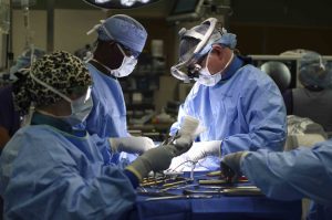
Adhesions are a common kind of scar tissue that can occur after surgery, and though usually not painful, they have the potential to result in complications like chronic pain or decreased heart efficiency, depending on where the scar tissue forms. Now, a sprayable gel developed by researchers at Stanford University will help to prevent adhesions from forming during surgical procedures. The gel, called PNP 1:10 in reference to its polymer-nanoparticle structure, has a similar stiffness to mayonnaise and achieves an ideal balance of slipperiness and stickiness that allows it to adhere easily to tissue of irregular shapes and surfaces. The flexible gel will actually dissolve in the body after two weeks, which is about how long most adhesions take to heal. Though lead author Lyndsay Stapleton, M.S., and senior authors Joseph Woo, M.D., and Eric Appel, Ph.D., have only tested the gel in rats and sheep so far, they hope that human applications are not too far in the future.
Learning to Understand Blood Clots in a New Model
Blood clots are the source of some of the deadliest human conditions and diseases. When a clot forms, blood flow can be interrupted, cutting off supply to the brain, heart, or other vital organs, resulting in potentially serious damage to the mind and body. For patients with certain bleeding disorders, clotting or the lack thereof can hold tremendous importance in surgery, and affect some of the typical procedures of a given operation. To help plan for such situations, researchers at the University of Buffalo created an in vitro model to help better illustrate the complex fluid mechanics of blood flow and clotting. Most importantly, this new model better demonstrates the role of shear stress in blood flow, and the way that it can affect the formation or destruction of blood clots – an aspect that current clinical devices often overlook. Led by Ruogang Zhao, Ph.D., the model can allow surgeons and hematologists to consider the way that certain chemical or physical treatments can affect clot formation on a patient-to-patient basis. The two key factors of the model are its incorporation of blood flow, and its relationship to shear stress, with clot stiffness through microfabrication technology using micropillars as force sensors for the stiffness. Going forward, Zhao and his research team hope to test the model on more patients, to help diversify the different bleeding disorders it can exhibit.
Training the Next Generation of Researchers
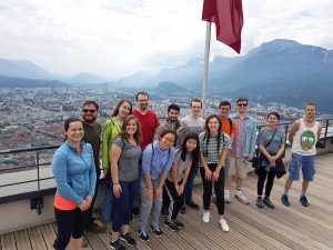
Rebecca Zappala, a junior from Miami, Florida who is majoring in bioengineering, worked in Grenoble this summer on new ways to harvest water from fog. She describes her research project as a “futuristic” way to collect water and says that she’s thankful for the opportunity to work on her first independent research project through the Research and Education in Active Coatings Technology (REACT) program.
After learning the technical skills she needed for her project, Zappala spent her summer independently working on new ways to modify her material’s properties while working closely with her French PI and a post-doc in the lab. She was surprised to see how diverse the lab was, with researchers working on everything from biomolecular research to energy in the same space.
“I learned a lot,” she says about being in such an interdisciplinary setting. “I hadn’t been part of a research team before, and I got a lot of exposure to things that I wouldn’t have been exposed to otherwise.”
Read the rest of the story on Penn Today.
Virginia Tech Course Addresses the Needs of Wounded Veterans
A new course at Virginia Tech encourages students to apply engineering skills to real-life problems in the biomedical world by designing medical devices or other applications to assist veterans suffering from serious injuries or illnesses. Funded by the National Institute of Health, faculty from the Department of Biomedical Engineering and Mechanics hope that the course will allow students to see how theoretical knowledge from the classroom actually works in a clinical setting, and to understand how different stakeholder interests factor into designing a real device. What makes this new class unique from other traditional design-focused courses at other universities is its level of patient interaction. Students at Virginia Tech who choose to take this class will have the chance to gain input from field professionals like clinicians and engineers from the Salem Veterans Affairs Medical Center, while also being able to get direct feedback from the patients that the devices will actually help. Beginning in the spring of 2020, students can take the new course, and volunteer in the veterans clinics to gain even more experience with patients.
People and Places
Sevile Mannickarottu, the Director of the Educational Laboratories in Penn’s Department of Bioengineering and recent recipient of the Staff Recognition Award from the School of Engineering and Applied Sciences, presented a paper to highlight the Stephenson Foundation Bioengineering Educational Lab and Bio-Makerspace at the 126th annual conference of the American Society for Engineering Education. Thanks to the dedication of Mannickarottu and the lab staff to creating a space for simultaneous education and innovation, the Bioengineering Lab continues to be a hub for student community and projects of all kinds.
A week-long program for high school girls interested in STEM allows students to explore ideas and opportunities in the field through lab tours, guest speakers, and hands-on challenges. A collaboration across the University of Virginia Department of Biomedical Engineering, Charlottesville Women in Tech, and St. Anne’s Belfield School, the program gave this year’s students a chance to design therapies for children with disorders like hemiplegia and cerebral palsy, in the hopes that these interactive design challenges will inspire the girls to pursue future endeavors in engineering.
We would like to congratulate Nancy Albritton, Ph.D., on her appointment as the next Frank & Julie Jungers Dean of the College of Engineering at the University of Washington. Albritton brings both a deep knowledge of the research-to-marketplace pipeline and experience in the development of biomedical microdevices and pharmacoengineering to the new position.
We would also like to congratulate Jeffrey Brock, Ph.D., on his appointment as the dean of the Yale School of Engineering and Applied Science. Already both a professor of mathematics and a dean of science in the Faculty of Arts and Sciences at Yale, Brock’s new position will help him to foster collaborations across different departments of academia and research in science and engineering.
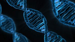 A new technique that researchers from the Broad Institute of MIT and Harvard University are calling DNA microscopy could help map cells for better understanding of genetic and molecular complexities.