Week in BioE (July 26, 2019)

by Sophie Burkholder

New 3D Tumor Models Could Improve Cancer Treatment

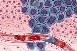
New ways of testing cancer treatments may now be possible thanks to researchers at the University of Akron who developed three-dimensional tumor models of triple-negative breast cancer. Led by Dr. Hossein Tavana, Ph. D., an associate professor of biomedical engineering at the university, the Tissue Engineering Microtechnologies Lab recently received a $1.13 million grant from the prestigious National Cancer Institute (NCI) of the National Institute of Health (NIH) to continue improving these tumor models. Tumors are difficult to fully replicate in vitro, as they are comprised of cancerous cells, connective tissue, and matrix proteins, among several other components. With this new grant, Tavana sees creating a high-throughput system that uses many identical copies of the tumor model for drug testing and better understanding of the way tumors operate. This high-throughput method would allow Tavana and his lab to isolate and test several different approaches at once, which they hope will help change the way tumors are studied and treated everywhere.

Noise-Induced Hearing Loss Poses Greater Threat to Neural Processing

Even though we all know we probably shouldn’t listen to music at high volumes, most of us typically do it anyway. But researchers at Purdue University recently found that noise-induced hearing loss could cause significant changes in neural processing of more complex sound inputs. Led by Kenneth Henry, Ph.D., an assistant professor of otolaryngology at the University of Rochester Medical Center, and Michael Heinz, Ph.D., a professor of biomedical engineering at Purdue University, the study shows that when compared with age-related hearing loss, noise-induced hearing loss will result in a greater decrease in hearing perception even when the two kinds of hearing loss appear to be of the same degree on an audiogram. This is because noise-induced hearing loss occurs because of physical trauma to the ear, rather than the long-term electrochemical degradation of some components that come happen with age. The evidence of this research is yet another reason why we should be more careful about exposing our ears to louder volumes, as they pose a greater risk of serious damage.

Increasing the Patient Populations for Research in Cartilage Therapy and Regenration

Despite the great progress in research of knee cartilage therapy and regeneration, there are still issues with the patient populations that most studies consider. Researchers often want to test new methods on patients that have the greatest chance of injury recovery without complications – often referred to as “green knees” – but this leaves out those patient populations who suffer from conditions or defects that have the potential to cause complications – often referred to as “red knees.” In a new paper published in Regenerative Medicine, the Mary Black Ralston Professor for Education and Research in Orthopaedic Surgery and secondary faculty in the Department of Bioengineering at Penn, Robert Mauck, Ph.D., discusses some cartilage therapies that may be suitable for red knee populations.

Working with James Carey, M.D., the Director of the Penn Center for Cartilage Repair and Osteochondritis, Mauck and his research team realized that even those with common knee cartilage conditions such as the presence of lesions or osteoarthritis were liable to be excluded from most regeneration studies. In discussing alternatives methods and structures of studying cartilage repair and regeneration, Mauck and Carey hope that future therapies will be applicable to a wider range of patient populations, and that there will soon be more options beyond full joint replacement for those with red knee conditions.

Plant-Like Superhydrophobicity Has Applications in Biomedical Engineering

Researchers in the Department of Biomedical Engineering at Texas A&M University recently found ways of incorporating the superhydrophobic properties of some plant leaves into biomedical applications through what they’re calling a “lotus effect.” The Gaharwar Lab, led by principal investigator and assistant professor of biomedical engineering Akhilesh Gaharwar, Ph.D., developed an assembly of two-dimensional atomic layers that they describe as a “nanoflower” to help control surface wetting in a biomedical setting. A recent paper published in Chemical Communications describes Gaharwar and his team’s work as expanding the use of superhydrophobic surface properties in biomedical devices by demonstrating the important role that atomic vacancies play in the wetting characteristic. While Gaharwar hopes to research the impact that controlling superhydrophobicity could have in stem cell applications, his work already allows for innovations in self-cleaning and surface properties of devices involving labs-on-a-chip and biosensing.

People and Places

Nader Engheta, H. Nedwill Ramsey Professor in Electrical and Systems Engineering, Bioengineering and Materials Science and Engineering, has been inducted into the Canadian Academy of Engineering (CAE) as an International Fellow. The CAE comprises many of Canada’s most accomplished engineers and Engheta was among the five international fellows that were inducted this year.

The Academy’s President Eddy Isaacs remarked: “Over our past 32 years, Fellows of Academy have provided insights in the fields of education, infrastructure, and innovation, and we are expecting the new Fellows to expand upon these contributions to public policy considerably.”

Read the full story on Penn Engineering’s Medium Blog. 

We would like to congratulate Anthony Lowman, Ph.D., on his appointment as the Provost and Senior Vice President for Academic Affairs at Rowan University. Formerly the Dean of Rowan’s College of Engineering, Lowman helped the college double in size, and helped foster a stronger research community. Lowman also helped to launch a Ph.D. program for the school, and added two new departments of Biomedical Engineering and Experiential Engineering Education in his tenure as the dean. Widely recognized for his research on hydrogels and drug delivery, Lowman was also formerly a professor of bioengineering at Temple University and Drexel University.

Lastly, we would like to congratulate Daniel Lemons, Ph.D., on his appointment as the Interim President of Lehman College of the City University of New York. Lemons, a professor in the Department of Biology at City College, specializes in cardiovascular and comparative physiology, and was also one of the original faculty members of the New York Center for Biomedical Engineering. With prior research funded by both the National Institute of Health (NIH) and the National Science Foundation (NSF), Lemons also holds patents in biomechanics teaching models and mechanical heart simulators.


Week in BioE (February 13, 2019)

by Sophie Burkholder

Bioengineers Tackle Heart Disease

Heart disease is currently the leading cause of death in the United States, resulting in about 630,000 deaths every year according to the Center for Disease Control. One of the most common side effects of heart disease is damage to blood vessels and cardiac tissue, which can ultimately lead to conditions like high blood pressure, arrhythmia, and even cardiac arrest. In serious cases of irreversible heart damage, often the only option for patients is a full heart transplant, and efforts to engineer vascularized cardiac tissue grafts have proved challenging in research so far.

But researchers Ying Zheng, Ph.D., and Charles Murry, M.D., Ph.D., both of whom have joint appointments in Bioengineering at the University of Washington, have found success in using human microvascular grafts to create working blood vessels in vitro to treat infarcted rat hearts. The new heart muscle, developed from human embryonic stem cell-derived endothelial cells in petri dishes, was grown with a focus on not only being able to easily integrate it in vivo, but also in creating a patch of vasculature that closely mirrored that of the heart. In concentrating more on the mechanical aspects of the blood vessel network, Zheng and Murry were able to better restore normal blood flow to the damaged rat hearts after integration of the grafts. The study appears in a recent edition of Nature Communications.

Another team of bioengineers, led by Michael Sacks, Ph.D. at the University of Texas at Austin, recently invented a software-based method for repairing mitral valves in the heart. Their work, published in the International Journal for Numerical Methods in Biomedical Engineering, uses computational modeling techniques to create a noninvasive way of simulating repairs to the mitral valve, which will allow for a better prediction of surgical procedures and postoperative side effects on a more patient-specific basis. This ability to know which treatment plan may be best-suited for a given patient is important especially for valve repair, as heart valves are notoriously difficult to model or image due to the complexity of their functions. But through the use of advanced technology in 3D echocardiography, Sacks and his team say that their new model is accurate enough to rely on in clinical settings.

Virtual Reality Assists in the Evaluation of Surgery

Any form of surgery is always a high risk procedure, as it is subject to a wide variety of sources of human error and irregularity, even with the best surgeons. Certainly, there should be a system in place to not only continually assess the knowledge of surgeons throughout their careers, but also to evaluate their practices and techniques during operation. Such an evaluation, however, would put patients at risk during the assessment of the surgeon.

But now a team of researchers from Rensselaer Polytechnic Institute has developed a way of simulating colorectal surgical procedures using virtual reality technology. Suvranu De, Sc.D. — the J. Erik Jonsson ‘22 Distinguished Professor of Engineering and Head of the Department of Mechanical, Aerospace and Nuclear Engineering with joint appointments in Biomedical Engineering and Information Technology and Web Science —leads the project which incorporates both visual and tactile feedback for users to employ as a tool for both training and evaluating colorectal surgeons. While virtual reality simulators have been used for similar applications related to procedures like the colonoscopy, they have yet to be fully developed for open surgical procedures, because of the difficulties in creating a fully engaged and immersive environment. Nonetheless, De and his team hope that their work will lead to the creation of the first “Virtual Intelligent Preceptor,” which will allow for more advanced technological innovations in aspects of surgical education that have so far been difficult to standardize. Support for the project comes from the National Institute of Biomedical Imaging and Bioengineering (NBIB).

Penn BE’s Dr. Bassett on Understanding Knowledge Networks in the Brain

Dr. Danielle Bassett, Ph.D., Eduardo D. Glandt Faculty Fellow and Associate Professor of Bioengineering

As a network neuroscientist, Danielle Bassett, Ph.D., Eduardo D. Glandt Faculty Fellow and Associate Professor in the Department Bioengineering, brings together insights from a variety of fields to understand how the brain’s connections form and change: mathematics, physics, electrical engineering and developmental biology, to name a few. Bassett’s recent work on the learning process also draws from linguistics, educational theory and other domains even further afield.

The intersection and interaction of knowledge from multiple sources doesn’t just describe Bassett’s methodology; it’s at the heart of her research itself. At the Society for Industrial and Applied MathematicsAnnual Meeting last year, Bassett provided an address on how the structure of knowledge networks can influence what our brains can do when it comes to learning new things.

Read the full story on Penn Engineering’s Medium blog.

People & Places:

Tammy Dorsey, a graduate student at Wichita State University, created a non-invasive in utero tool to help read the oxygen levels of unborn babies as part of her senior design project. Dorsey says the inspiration for the project came from complications during the birth of her middle child, who despite having a normal heart rate throughout the entire pregnancy, was born blue. The device Dorsey created uses measurements of the baby’s pH to read fetal oxygen levels. She hopes that the design will help doctors better detect when a fetus is in distress during pregnancy and childbirth.

The field of bioengineering is constantly growing, and new programs are always in development. Boise State University has announced the launch of a new doctoral program in bioengineering that will begin in the fall of 2019. Developed through the collaboration of the university’s College of Health Sciences, College of Engineering, Graduate College, and College of Arts and Sciences, this new opportunity to do research in the field of bioengineering will have three study tracks available in biomechanics, mechanobiology, and human performance.

The new biomedical engineering department at the University of Massachusetts Amherst has announced the department’s first faculty appointments. The founding department head will be Professor Tammy L. Haut Donahue, Ph.D., whose research focus is on the biomechanics of the musculoskeletal system. Another professor joining the department’s new faculty is Seth W. Donahue, Ph.D., who has also done research in the field of biomechanics, and specifically how it pertains to tissue regeneration.

Since we last posted, there have also been several significant academic appointments in the field of Bioengineering. This week, we would like to congratulate Bruce Tromberg, Ph.D., on his appointment as the director of the National Institute of Biomedical Imaging and Bioengineering (NIBIB). Dr. Tromberg is currently a Professor with appointments in Biomedical Engineering and Surgery at the University of California at Irvine, where he leads research in bioimaging and biophotonics. He has also served on the External Advisory Board of NIH P41 Center for Magnetic Resonance and Optical Imaging here at Penn since 2009, and has also given several lectures here on his work in bioimaging.

Secondly, we congratulate the University of Toronto’s Professor Warren Chan, Ph.D., who was recently named as a Tier 1 Canada Research Chair in Nanobioengineering. Professor Chan, who is also the director of the Institute of Biomaterials and Biomedical Engineering at the University of Toronto, conducts research in the field of nanotechnology for applications in the treatment and diagnosis of cancer and viral diseases.

And finally, we also want to congratulate Frank Pintar, Ph.D., on his appointment as the Founding Chair of the Marquette University and Medical College of Wisconsin. Dr. Pintar’s research in bioengineering involves the study of the biomechanics involved with brain and spinal cord injury, with a focus on motor vehicle crash trauma.

Week in BioE (July 31, 2018)

New Data Analysis Methods

Like many other fields, biomedical research is experiencing a data explosion. Some estimates suggest that the amount of data generated from the health sciences is now doubling every eighteen months, and experts expect it to double every seventy-three days by 2020.  One challenge that researchers face is how to meaningfully analyze this data tsunami.

The challenge of interpreting data occurs at all scales, and a recent collaboration shows how new approaches can allow us to handle the volumes of data emerging at the level of individual cells to infer more about how biology “works” at this level.  Wharton Statistics Department researchers Mo Huang and Jingshu Wang (PhD Student and Postdoctoral Researcher, respectively) collaborated with Arjun Raj’s lab in Bioengineering and published their findings in recent issues of Nature Methods and Proceedings of the National Academy of Sciences.  One study focused on a de-noising technique called SAVER to provide more precise data from single cell experiments and significantly improves the ability to detect trends in a dataset, similar to how increasing sample size helps improve the ability to determine differences between experimental groups.  The second method, termed DESCEND, creates more accurate characterization of gene expression that occur in individual cells. Together these two methods will improve data collection for biologists and medical professionals working  to diagnose, monitor, and treat diseased cells.

Dr. Raj’s team contributed data to the cause and acted as consultants on the biological aspects of this research. Further collaboration involved Mingyao Li, PhD, Professor of Biostatistics at the Perelman School of Medicine, and Nancy Zhang, PD, Professor Statistics at the Wharton School. “We are so happy to have had the chance to work with Nancy and Mingyao on analyzing single cell data,” said Dr. Raj. “The things they were able to do with our data are pretty amazing and important for the field.”

Advancements in Biomaterials

This blog features many new biomaterials techniques and substances, and there are several exciting new developments to report this week. First, the journal of Nature Biomedical Engineering published a study announcing a new therapy to treat or even eliminate lung infections, such as those acquired while in hospital or as the result of cystic fibrosis, which are highly common and dangerous. Researchers identified and designed viruses to target and kill the bacteria which causes these infections, but the difficulty of administering them to patients has proven prohibitive. This new therapy, developed by researchers at the Georgia Institute of Technology, is administered as a dry powder directly to the lungs and bypasses many of the delivery problems appearing in past treatments. Further research on the safety of this method is required before clinical trials can begin.

A team at Harvard University published another recent study in Nature Biomedical Engineering announcing their creation of a tissue-engineered scale model of the left human heart ventricle. This model is made from degradable fibers that simulate the natural fibers of heart tissue. Lead investigator Professor Kevin Kit Parker, PhD, and his team eventually hope to build specific models culled from patient stem cells to replicate the features of that patient’s heart, complete with the patient’s unique DNA and any heart defects or diseases. This replica would allow researchers and clinicians to study and test various treatments before applying them to a specific patient.

Lastly, researchers at the Tufts University School of Engineering published in the Proceedings of the National Academy of Sciences on their creation of flexible magnetic composites that respond to light. This material is capable of macroscale motion and is extremely flexible, allowing its adaptation into a variety of substances such as sponges, film, and hydrogels. Author and graduate student Meg Li explained that this material differs from similar substances in its complexity; for example, in the ability for engineers to dictate specific movements, such as toward or away from the light source. Co-author Fiorenzo Omenetto, PhD, suggests that with further research, these movements could be controlled at even more specific and detailed levels.

CFPS: Getting Closer to “On Demand” Medicine

A recent and growing trend in medicine is the move towards personalized or “on demand” medicine, allowing for treatment customized to specific patients’ needs and situations. One leading method is Cell-Free Protein Synthesis (CFPS), a way of engineering cellular biology without using actual cells. CFPS is used to make substances such as medicine, vaccines, and chemicals in a sustainable and portable way. One instance if the rapid manufacture of insulin to treat diabetic patients. Given that many clinics most in need of such substances are found in remote and under-served locations far away from well-equipped hospitals and urban infrastructure, the ability to safely and quickly create and transport these vital substances to patients is vitally important.

The biggest limiting factor to CFPS is difficulty of replicating Glycosylation, a complex modification that most proteins undergo. Glycosylation is important for proteins to exert their biological function, and is very difficult to synthetically duplicate. Previously, achieving successful Glycosylation was a key barrier in CFPS. Fortunately, Matthew DeLisa, PhD, the Williams L. Lewis Professor of Engineering at Cornell University and Michael Jewett, PD, Associate Professor of Chemical and Biological Engineering at Northwestern University, have created a “single-pot” glycoprotein biosynthesis that allows them to make these critical molecules very quickly. The full study was recently published in Nature Communications. With this new method, medicine is one step closer to being fully “on demand.”

People and Places

The Institute of Electrical and Electronics Engineers (IEEE) interviewed our own Penn faculty member Danielle Bassett, PhD, the Edwardo D. Glandt Faculty Fellow and Associate Professor in Bioengineering, for their website. Dr. Bassett, who shares a joint appointment with Electrical Systems Engineering (ESE) at Penn, has published groundbreaking research in Network Neuroscience, Complex Systems, and more. In the video interview (below), Dr. Bassett discusses current research trends in neuroscience and their applications in medicine.

Finally, a new partnership between Case Western Reserve University and Cleveland Clinic seeks to promote education and research in biomedical engineering in the Cleveland area. Cleveland Clinic Lerner Research Institute‘s Chair of Biomedical Engineering, Geoff Vince, PhD, sees this as an opportunity to capitalize on the renown of both institutions, building on the region’s already stellar reputation in the field of BME. Dozens of researchers from both institutions will have the opportunity to collaborate in a variety of disciplines and projects. In addition to professional academics and medical doctors, the leaders of this new partnership hope to create an atmosphere that can benefit all levels of education, all the way down to high school students.