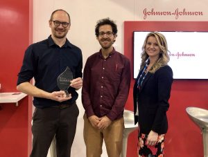
David Issadore (center) was announced as the awardee of the JPOD @ Philadelphia QuickFire Challenge by Katherine Merton (right), head of JLABS New York City, Boston, and Philadelphia, at last week’s BIO 2019 International convention. (Photo: Johnson & Johnson Innovation)
Chip Diagnostics is a Philadelphia-based device company founded in 2016 based on research from the lab of David Issadore, Assistant Professor of Bioengineering and Electrical and Systems Engineering in the School of Engineering and Applied Science. The startup combines microelectronics, microfluidics, and nanomaterials with the aim to better diagnose cancer. The company is developing technologies and digital assays for minimally-invasive early cancer detection and screening that can be done using mobile devices.
There has been a long interest in diagnosing cancer using blood tests by looking for proteins, cells, or DNA molecules shed by tumors, but these tests have not worked well for many cancers since the molecules shed tend to be either nonspecific or very rare.
Issadore’s group aims to target different particles called exosomes: Tiny particles shed by cells that contain similar proteins and RNA as the parent cancer cell. The problem, explains Issadore, is that because of the small size of the exosomes, conventional methods such as microscopy and flow cytometry wouldn’t work. “As an engineering lab, we saw an opportunity to build devices on a nanoscale that could specifically sort the cancer exosomes versus the background exosomes of other cells,” he explains.
After Issadore was approached by the IP group at PCI Ventures in the early stages of their research, Chip Diagnostics has since made huge strides as a company. Now, as the awardee of the JPOD @ Philadelphia QuickFire Challenge, Chip Diagnostics will receive $30,000 in grant funding to further develop the first-in-class, ultra-high-definition exosomal-based cancer diagnostic. The award also includes one year of residency at Pennovation Works as well as access to educational programs and mentoring provided by Johnson & Johnson Family of Companies global network of experts.
Joel Boerckel, Ph.D, Assistant Professor of Orthopaedic Surgery and Bioengineering
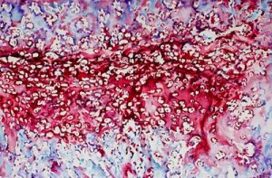
To treat large gaps in long bones, like the femur, which result from bone tumor removal or a shattering trauma, researchers at Penn Medicine and the University of Illinois at Chicago developed a process that partially recreates the bone growth process that occurs before birth. A bone defect of more than two centimeters is considered substantial, and current successful healing rates stand at 50% or less, with failure often resulting in amputation. The team hopes that their method, which they’ve developed in rodent models to mimic the process of rapid fetal bone growth, can substantially improve success rates. Their findings are published in Science Translational Medicine.
Watercolor- A watercolor image depicting the embryonic bone development process, endochondral ossification, featuring cartilage and bone. Credit: Joel Boerckel
“When bones are originally formed in the embryo, they’re first generated from cartilage, like a template,” says senior author Joel Boerckel, an assistant professor of orthopaedic surgery and bioengineering. “In order to regenerate bone within defects that otherwise won’t heal in grown people, we are seeking to recreate the embryonic bone development process.”
To do that, the researchers’ process begins with the delivery of specially engineered stem cells (called a condensation of mesenchymal cells) to the rodents’ bone defect, which sparks endochondral ossification, the specific term for embryonic bone development.
Bio-inspiration Informs New Football Helmet Design from IUPUI Students
Art, design, biology, and engineering all interact with each other in a recent design for a football helmet from two students— one of media arts and the other of engineering — at the Indiana University – Purdue University Indianapolis. Directed by Lecturer in Media Arts and Science Zebulun Wood, M.S., and Associate Professor of Mechanical and Energy Engineering and Assistant Professor of Biomedical Engineering Andres Tovar, Ph.D., the students found inspiration in biological structures like a pomelo peel, nautilus shell, and woodpecker skull to create energy-absorbing helmet liners. The resulting design took these natural concussion-reducing structures and created compliant mechanism lattice-based liners the replace the foam traditionally placed in between two harder shells of a typical helmet. Their work not only exemplifies the benefits of bio-inspiration, but demonstrates the way that several different domains of study can overlap in the innovation of a new product.
Study of Mechanical Properties of Hyaluronic Acid Could Help Inform Current Debates Over Treatment Regulation for Osteoarthritis
Arthritis is an extremely common condition, especially in older patients, in which inflammation of the joints can cause high amounts of stiffness and pain. Osteoarthritis in particular is the result of the degradation of flexible tissue between the bones of a joint, which increases friction in joint motion. A common treatment of this form of arthritis is the injection of hyaluronic acid, which is meant to provide joint lubrication, and decreases this friction between bones. Recently, however, there has been a debate over hyaluronic acid’s classification by the FDA and whether it should remain based on the knowledge of the mechanical actions of the acid in treatment for osteoarthritis or if potential chemical action of the acid should be considered as well.
Because of limited ways of testing the mechanical properties of the acid, many researchers felt that there could be more to hyaluronic acid’s role in pain relief for arthritic patients. But Lawrence Bonassar, Ph.D., the Daljit S. and Elaine Sarkaria Professor in Biomedical Engineering at the Meinig School of Bioengineering of Cornell University, had another idea. With his lab, he created a custom-made tribometer to measure the coefficient of friction of a given lubricant by rubbing a piece of cartilage back and forth across a smooth glass plate. The research demonstrated that hyaluronic acid’s ability to reduce the coefficient of friction aligned with patients’ pain relief. Bonassar and his team hope that these results will demonstrate the heavy contribution of mechanical action that hyaluronic acid has in osteoarthritis treatment, and help bring an end to the debate over its FDA classification.
A New Way of Mapping the Heart Could Lead to Better Understanding of Contractile Activity
Though reduced contractions in certain regions of the heart can be an indicator of a certain condition, there is currently no way to directly measure contractile activity. This is why Cristian Linte, Ph.D., an Associate Professor of Biomedical Engineering in the Kate Gleason College of Engineering at the Rochester Institute of Technology (RIT), hopes to create a map of the heart that can quantify contraction power. In collaboration with Niels Otani, Ph.D., an Associate Professor in the School of Mathematics at RIT, Linte plans to use an $850,000 grant from the National Science Foundation to achieve a more comprehensive understanding of the heart through both medical imaging and mechanical modeling. The group hopes that their approach will lead to not only a better way to diagnose certain heart conditions and diseases, but also open up understanding of active contraction, passive motion, and the stresses within the heart walls that underlie each.
Celebrity Cat Lil Bub Helps Penn and German Researchers Draw Public Attention to Genetics
Lil Bub’s unique appearance has garnered millions of online fans, and now, an avenue for researchers to talk about genetics. (Photo Courtesy of Mike Bridavsky)
In 2015, a group of curious researchers set out to sequence the genome of a celebrity cat named Lil Bub. They were hoping to understand the genetics behind Lil Bub’s extra toes and unique skeletal structure, which contribute to her heart-warming, kitten-like appearance. However, an equally important goal of their “LilBUBome” project was to invite the general public into the world of genetics.
Because of Lil Bub’s online fame, the project garnered attention from her fans and the media, all hoping to discover the secret to Lil Bub’s charm. As early as 2015, Gizmodo’s Kiona Smith-Strickland reported on the team’s intentions to sequence Lil Bub’s genome, and, since then, many have been awaiting the results of the LilBUBome.
The Alfred P. Sloan Foundation awarded a six-year grant to Barnard College and Columbia University’s School of Engineering and Applied Science to support graduate education for women in engineering. The funding will go towards a new five-year program that enables Barnard students to attain both a B.A. and M.S. in one year after their traditional four years of undergraduate education. The program will offer M.S. degrees in chemical engineering, biomedical engineering, and industrial engineering and operations research, and is one of the first of its kind for women’s colleges.
We would like to congratulate Jean Paul Allain, Ph.D., on being named the first head of the new Ken and Mary Alice Lindquist Department of Nuclear Engineering at Penn State. Allain, who is currently a Professor and head of graduate programs in the University of Illinois at Urbana-Champaign’s Department of Nuclear, Plasma, and Radiological Engineering, conducts research in models of particle-surface interactions. In addition to being head of the new department at Penn State, Allain will also hold a position as a Professor of Biomedical Engineering at the university.
We would also like to congratulate Andrew Douglas, Ph.D., on his appointment as the Vice Provost for Faculty Affairs at Johns Hopkins University. Douglas currently holds the position of Vice Dean for Faculty at the Whiting School of Engineering, and has joint appointments in Mechanical and Biomedical Engineering. Douglas’s research at Hopkins focuses on mechanical properties and responses of compliant biological tissue and on the nonlinear mechanics of solids, with a focus on soft tissues and organs like the heart and tongue.
Dan Huh, the Wilf Family Term Assistant Professor in the Department of Bioengineering, focuses his research on creating organs-on-chips: specially manufactured micro-devices with human cells that mimic the natural cellular processes of organs. Huh’s lab has engineered chips that approximate the functioning of placentas and lung disease, some of which were launched into space in May. Most recently, Huh published a review of organ-on-a-chip technology in the journal Science with graduate students Sunghee Estelle Park and Andrei Georgescu.
The June 2019 issue of Science is a special issue centered around the science of growing human organ models in the laboratory. Such in vitro organs are known as organoids; they grow and develop much like organs do in the body, as opposed to Huh’s organs-on-chips, in which cells from the relevant organs are grown within a fabricated device that imitates some of the organ’s functions and natural environment.
In a video accompanying the review article, Huh explains how organoid and organ-on-a-chip technologies differ and the advantages that accompany each approach:
Unlike Organ-on-a-Chip, which are heavily engineered man-made systems, organoids allow us to mimic the complex of the human body in a more natural way. So organoids represent a more realistic model, but they have problems because they develop in a highly variable fashion and it’s not very easy to control their environment. So we think that Organ-on-a-Chip Technology is a promising solution to many of these problems.
Read Huh, Park, and Georgescu’s review article at Science.
De la Fuente, who started at Penn earlier this year, was recognized because he “is pioneering the computerization of biological systems for the development of transformative biotechnologies designed to solve societal grand challenges such as antibiotic resistance.”
Vector Flow Imaging Helps Visualize Blood Flow in Pediatric Hearts
A group of biomedical engineers at the University of Arkansas used a new ultrasound-based imaging technique called vector flow imaging to help improve the diagnosis of congenital heart disease in pediatric patients. The study, led by associate professor of biomedical engineering Morten Jensen, Ph.D., collaborated with cardiologists at the local Children’s Hospital in Little Rock to produce images of the heart in infants to help potentially diagnose congenital heart defects. Though the use of vector flow imaging has yet to be developed for adult patients, this type of imaging could possibly provide more detail about the direction of blood flow through the heart than traditional techniques like echocardiography do. In the future, the use of both techniques could provide information about both the causes and larger effects of heart defects in patients.
Using Stem Cells to Improve Fertility in Leukemia Survivors
One of the more common side effects of leukemia treatment in female patients is infertility, but researchers at the University of Michigan want to change that. Led by associate professor of biomedical engineering Ariella Shikanov, Ph.D., researchers in her lab found ways of increasing ovarian follicle productivity in mice, which directly relates to the development of mature eggs. The project involves the use of adipose-derived stem cells, that can be found in human fat tissue, to surround the follicles in an ovary-like, three-dimensional scaffold. Because the radiation treatments for leukemia and some other cancers are harmful to follicles, increasing their survival rate with this stem cell method could reduce the rate of infertility in patients undergoing these treatments. Furthermore, this new approach is innovative in its use of a three-dimensional scaffold as opposed to a two-dimensional one, as it stimulates follicle growth in all directions and thus helps to increase the follicle survival rate.
Penn Engineers Look at How Stretching & Alignment of Collagen Fibers Help Cancer Cells Spread
Cancer has such a massive impact on people’s lives that it might be easy to forget that the disease originates at the cellular level. To spread and cause significant damage, individual cancer cells must navigate the fibrous extracellular environment that cells live in, an environment that Penn Engineer Vivek Shenoy has been investigating for years.
Shenoy’s most recent study on cancer’s mechanical environment was led by a postdoctoral researcher in his lab, Ehsan Ban. Paul Janmey, professor in Physiology and Bioengineering, and colleagues at Stanford University also contributed to the study. Shenoy also received the Heilmeier Award this March and delivered the Heilmeier Award Lecture in April.
Controlled Electrical Stimulation Can Prevent Joint Replacement Infections
Joint replacements are one of the most common kinds of surgery today, but they still require intense post-operative therapy and have a risk of infection from the replacement implant. These infections are usually due to the inflammatory response that the body has to any foreign object, and can become serious and life-threatening if left untreated. Researchers at the University of Buffalo Jacobs School of Medicine and Biomedical Sciences hope to offer a solution to preventing infections through the use of controlled electrical stimulation. Led by Mark Ehrensberger, Ph.D., Kenneth A. Krackow, M.D., and Anthony A. Campagnari, Ph.D., the treatment system uses the electrical signal to create an antibacterial environment at the interface of the body and the implant. While the signal does not prevent infections completely, these antibacterial properties will prevent infections from worsening to a more serious level. Patented as the Biofilm Disruption Device TM, the final product uses two electrode skin patches and a minimally invasive probe that delivers the electrical signal directly to the joint-body interface. The researchers behind the design hope that it can help create a more standard way of effectively treating joint replacement infections.
People and Places
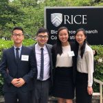
TBx: Gabriel Koo, Ethan Zhao, Daphne Cheung, and Shelly Teng
For their senior design project, four bioengineering seniors — Gabriel Koo, Ethan Zhao, Daphne Cheung, and Shelly Teng — created a low-cost tuberculosis diagnostic that they called TBx. Using their knowledge of the photoacoustic effect of certain dyes, the platform the group created can detect the presence of lipoarabinomannan in patient urine. The four seniors presented TBx at the Rice360 Design Competition in Houston, Texas this spring, which annually features student-designed low-cost global health technologies.
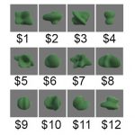
These 12 object-number value pairs were taught to the participants, who had to properly learn the associations to succeed in value judgement tests. The researchers investigated the differences in their brain activity patterns to see why some were faster learners than others.
Why do some people naturally excel at learning instruments, languages or technology while others take longer to pick up new knowledge? Learning requires the brain to encode information, changing its neural “wiring” and creating networks between brain regions.
Earlier research has suggested that part of what might slow down learners is over-thinking. A 2015 study led by Danielle Bassett, Eduardo D. Glandt Faculty Fellow and associate professor in the Department of Bioengineering, showed a correlation between slow learning and cognitive control: the brain’s ability to regulate itself by activating the necessary networks and inhibiting unnecessary activity. In that study, when people unnecessarily engaged parts of the brain linked to cognitive control, they were more likely to take longer to learn a simple task.
But beyond what might make an individual learn more slowly, the researchers want to know what sort of geometric patterns of brain activity make for better learning.
Evelyn Tang and Danielle Bassett
Their new study was led by Bassett and Evelyn Tang, who was an Africk Family Postdoctoral Fellow in Bassett’s Complex Systems Lab before starting at the Max Planck Institute this fall. Sharon Thompson-Schill, Christopher H. Browne Distinguished Professor and chair of Psychology, also contributed to the study.
SpaceX launched its 17th resupply mission to the International Space Station on May 4, with bioengineering professor Dan Huh’s organ-on-a-chip experiments in tow.
Dan Huh, the Wilf Family Term Assistant Professor in the Department of Bioengineering, researches human organs and the diseases that infect them by engineering devices made of living cells that act as stand-ins for organs. Huh’s lab has developed imitations of many organs, including the placenta and the eye, but it’s his lung-on-a-chip and his bone-marrow-on-a-chip that are reaching unprecedented heights as part of a new experiment taking place at the International Space Station (ISS).
On May 4, SpaceX launched a ISS-bound cargo capsule carrying Huh’s organ-on-a-chip experiments, which will remain in space for a month. Once back on Earth, the chips that spent time in space will be compared to control chips from Huh’s lab that are being monitored in parallel. Huh’s team is looking to see how being in space affects bacterial infections in lungs and white blood cell behavior in bone marrow. The researchers’ hope is that their studies will reveal important information about how human organs function both in space and on Earth.
Daniel Oberhaus of WIRED wrote an article describing the multiple organs-on-a-chip experiments being conducted at the ISS, including the two experiments headed by Huh:
Dan Huh is a bioengineer at the University of Pennsylvania and the lead researcher on the lung tissue chip headed to the ISS. This lung chip models a human airway and will be infected with Pseudomonas aeruginosa, a species of bacteria that had previously been found on the ISS. On Earth this bacteria is usually associated with respiratory infections, which are one of the leading types of illness on long-duration missions to the ISS.
Huh says scientists still know very little about why astronauts’ immune response seems to become suppressed in orbit, and the tissue chips are aimed at building a better understanding of the phenomenon.
When cells move throughout the body, they do so by dragging themselves, using molecular “arms” to pull themselves closer to where they need to be while unlatching themselves from the area they’re moving away from. In a recent study, Penn Engineers looked at a few mechanobiological factors that help regulate cells’ migration towards their destination, providing new insight into the gene expression feedback loops that keep them from getting stuck.
Joel Boerckel and Devon Mason
The research was led by Joel Boerckel, Assistant Professor of Orthopaedic Surgery in the Perelman School of Medicine and in Bioengineering in Penn Engineering, and bioengineering graduate student Devon Mason. Co-authors include bioengineering graduate student Joseph Collins and researchers from the University of Notre Dame, Indiana University and Purdue University.
Danielle Bassett, Eduardo D. Glandt Faculty Fellow and Associate Professor in theDepartment of Bioengineering, grew up in central Pennsylvania where she and her 10 siblings were homeschooled. Back then, Bassett had aspirations to become a professional pianist, a dream shattered by stress fractures in her arm at age 16.
Now, Bassett is a renowned physicist and MacArthur fellow who has pushed the field of network science, which studies connections and interactions between parts of a whole, to new realms. Bassett’s research focuses on brain function, including work on how brains of people with schizophrenia are organized, how brain communication changes with learning, and how the brain is able to switch between tasks.
Kelly Servick of Science sat down with Bassett to talk through her incredible journey from child pianist to leading network scientist:
““By 17, discouraged by her parents from attending college and disheartened at her loss of skill while away from the keys, she expected that responsibilities as a housewife and mother would soon eclipse any hopes of a career. ‘I wasn’t happy with that plan,’ she says.
Instead, Bassett catapulted herself into a life of research in a largely uncharted scientific field now known as network neuroscience. A Ph.D. physicist and a MacArthur fellow by age 32, she has pioneered the use of concepts from physics and math to describe the dynamic connections in the human brain. ‘She’s now the doyenne of network science,’ says theoretical neuroscientist Karl Friston of University College London. ‘She came from a formal physics background but was … confronted with some of the deepest questions in neuroscience.’”
Continue reading about Bassett’s career path and evolving research interests at Science.


 Art, design, biology, and engineering all interact with each other in a
 A group of biomedical engineers at the University of Arkansas used a