Penn Medicine researchers laud the early results for CAR T therapy in lupus patients, which point to broader horizons for the use of personalized cellular therapies.
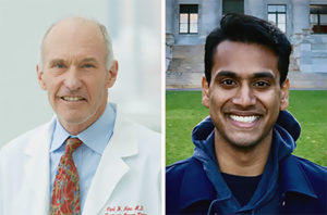
Penn Medicine’s Carl June and Daniel Baker.
Engineered immune cells, known as CAR T cells, have shown the world what personalized immunotherapies can do to fight blood cancers. Now, investigators have reported highly promising early results for CAR T therapy in a small set of patients with the autoimmune disease lupus. Penn Medicine CAR T pioneer Carl June and Daniel Baker, a doctoral student in cell and molecular biology in the Perelman School of Medicine, discuss this development in a commentary published in Cell.
“We’ve always known that in principle, CAR T therapies could have broad applications, and it’s very encouraging to see early evidence that this promise is now being realized,” says June, who is the Richard W. Vague Professor in Immunotherapy in the department of Pathology and Laboratory Medicine at Penn Medicine and director of the Center for Cellular Immunotherapies at the Abramson Cancer Center.
T cells are among the immune system’s most powerful weapons. They can bind to, and kill, other cells they recognize as valid targets, including virus-infected cells. CAR T cells are T cells that have been redirected, through genetic engineering, to efficiently kill specifically defined cell types.
CAR T therapies are created out of each patient’s own cells—collected from the patient’s blood, and then engineered and multiplied in the lab before being reinfused into the patient as a “living drug.” The first CAR T therapy, Kymriah, was developed by June and his team at Penn Medicine, and received Food & Drug Administration approval in 2017. There are now six FDA-approved CAR T cell therapies in the United States, for six different cancers.
From the start of CAR T research, experts believed that T cells could be engineered to fight many conditions other than B cell cancers. Dozens of research teams around the world, including teams at Penn Medicine and biotech spinoffs who are working to develop effective treatments from Penn-developed personalized cellular therapy constructs, are examining these potential new applications. Researchers say lupus is an obvious choice for CAR T therapy because it too is driven by B cells, and thus experimental CAR T therapies against it can employ existing anti-B-cell designs. B cells are the immune system’s antibody-producing cells, and, in lupus, B cells arise that attack the patient’s own organs and tissues.
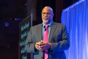
Penn Integrates Knowledge Professor Kevin Johnson takes the stage at 24th Engaging Minds. (Image: Ben Asen)
This past weekend in New York City, the University of Pennsylvania showcased its 24th Engaging Minds event, the first in person since 2019. It was hosted by Penn Alumni.
Three Penn Integrates Knowledge University Professors — Kevin Johnson, Lance Freeman and Dolores Albarracín, — each discussed their research. The audience, at least 600 in person and remote, heard about using city planning to promote racial equity, about how conspiracy theories come to life and propagate, and about the need for physicians to communicate effectively with patients and families.
Following brief remarks from Penn Alumni President Ann Reese, University President Liz Magill introduced the event. “As many of you know, I’ve been thinking a lot and speaking often about what makes Penn Penn,” she said. “What are our distinctive strengths? What are the unique contributions to society that we have made in the past and can make in the future? And where do we go from the extraordinary position we are in now?”
Magill went on to express gratitude for the speakers and invited the audience to think about how the researchers’ work and expertise furthered what she described as the “twin principles of truth and opportunity.”
He took the audience through his family history, education and training, pausing at a point on the timeline when he was a young physician-scientist who had just explained a new medical topic to a journalist. “I felt really good about the conversation — and then the article came out,” Johnson said.
In the piece, he had been cast as saying that the medical community was over-treating this condition, “which is not what I said.” He realized in that moment that as a physician, he had been taught to communicate what a study finds, not how to act based on those findings. That experience shifted his thinking on how to communicate scientific topics, and he has spent decades trying to move the needle on how others in his field perceive this.
“As scientists we face obstacles. We face the obstacle of scale, so, small projects that we’re asked to generalize. We face the issue of trust. And then we face the issue of values,” Johnson said. “I’ll add a fourth, which is format; the way we choose to reach specific audiences will be different.”
Read more about the 24th Engaging Minds at Penn Today.
Kevin Johnson is the David L. Cohen University of Pennsylvania Professor in the Departments of Biostatistics, Epidemiology and Informatics and Computer and Information Science. As a Penn Integrates Knowlegde (PIK) University Professor, Johnson also holds appointments in the Departments of Bioengineering and Pediatrics, as well as in the Annenberg School of Communication.
The Heilmeier Award honors a Penn Engineering faculty member whose work is scientifically meritorious and has high technological impact and visibility. It is named for the late George H. Heilmeier, a Penn Engineering alumnus and member of the School’s Board of Advisors, whose technological contributions include the development of liquid crystal displays and whose honors include the National Medal of Science and Kyoto Prize.
Bassett, who also holds appointments in Physics & Astronomy in Penn Arts & Sciences and in Neurology and Psychiatry in the Perelman School of Medicine, is a pioneer in the field of network neuroscience, an emerging subfield which incorporates elements of mathematics, physics, biology and systems engineering to better understand how the overall shape of connections between individual neurons influences cognitive traits. They lead the Complex Systems lab, which tackles problems at the intersection of science, engineering and medicine using systems-level approaches, exploring fields such as curiosity, dynamic networks in neuroscience, and psychiatric disease.
Bassett will deliver the 2022-23 Heilmeier Award Lecture in Spring 2023.
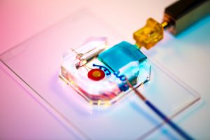
Dan Huh’s BIOLines Lab develops several different kinds of organ-on-a-chip systems, such as this blinking-eye-on-a-chip.
If you’d read about it in a science fiction novel, you might not have believed it. Human organs and organ systems — from lungs to blood vessels to blinking eyes — bio-miniaturized and stored on a plastic chip no larger than a matchbook.
But that’s the breathing, blinking reality at the Biologically Inspired Engineering Systems (BIOLines) Laboratory in the Department of Bioengineering in the School of Engineering and Applied Sciences at the University of Pennsylvania, a bona fide pioneer of what is now widely known as “organ-on-a-chip” technology. Proponents hope these devices can one day help scientists around the world learn more about the body’s inner workings and ultimately improve disease prevention and treatment.
“The century-old practice of cell culture is to grow living cells isolated from the human body in hard plastic dishes and keep them bathed in copious amounts of culture media under static conditions, and that is drastically different than the complex, dynamic environment of native tissues in which these cell reside,” said Dan Dongeun Huh, Ph.D., BIOLines’ principal investigator and an associate professor of Bioengineering in Penn’s School of Engineering and Applied Science. “What makes this organ-on-a-chip technology so unique and powerful is that it enables us to reverse-engineer living human tissues using microengineered devices and mimic their intricate biological interactions and physiological functions in ways that have not been possible using traditional cell culture techniques. This represents a major advance in our ability to model and understand the inner workings of complex physiological systems in the human body.”
Generally speaking, organ-on-a-chip devices are made of clear silicone rubber — the same material used to make contact lenses — and can vary in size and design. Embedded within are microfabricated three-dimensional chambers lined with different human cell types, arranged and propagated to ultimately form a structure complex enough to actually mimic the essential elements of a functioning organ.
With partners at the Perelman School of Medicine, BIOLines recently developed a newer variation of the organ-on-a-chip: one that replicates the interface between maternal tissue and the cells of the placenta at the critical moments in early pregnancy when the embryo is implanting in the uterus. Huh and Penn Medicine physicians led a study using the “implantation-on-a-chip” to observe things that would otherwise have been virtually unobservable.
Bella Mirro, a fourth year student in Bioengineering who also minors in Chemistry, spoke with 34th Street Magazine about her many roles at Penn, including being Co–President of Shelter Health Outreach Program (SHOP), a Research Assistant in lab of Michal A. Elovitz, the Hilarie L. Morgan and Mitchell L. Morgan President’s Distinguished Professor in Women’s Health at Penn Medicine, and a Penn Engineering Council Marketing Team Member. In this Q&A, she discusses her research in women’s health and her passions for accessible healthcare, serving Philadelphia’s homeless community, and good food.
Eight researchers from the Perelman School of Medicine have received research grants designed to invest in high-risk, high-reward projects.
Bushra Raj, Assistant Professor of Cell and Developmental Biology in the Perelman School of Medicine and member of the Penn Bioengineering Graduate Group, was one of three Penn winners of the NIH Director’s New Innovator Award for independent projects developed by early-career investigators. More additional Penn scientists who received NIH Director’s Transformative Research Award for a project focusing on cancer research.
Raj’s project focuses on “testing a novel technology that uses CRISPR/Cas gene-editing tools to genomically record inputs from two signaling pathways in the developing zebrafish brain.”
Established in 2009, the Transformative Research Award promotes cross-cutting, interdisciplinary science and is open to individuals and teams of investigators who propose research that could potentially create or challenge existing paradigms.
Each year, the the Department of Bioengineering seeks exceptional candidates to conduct summer research in bioengineering with the support of two scholarships: the Abraham Noordergraaf Student Summer Bioengineering Research Fund and the Blair Undergraduate Research Fund in the Department of Bioengineering. These scholarships provide a living stipend for students to conduct research on campus in a Penn research lab under the mentorship of a faculty member. The Abraham Noordergraaf Student Summer Bioengineering Research Fund provides financial support for undergraduate or graduate summer research opportunities in bioengineering with a preference for study in the area of cardiovascular systems. Dr. Noordergraaf, who died in 2014, was a founding member and first chair of Penn Bioengineering. The Blair Undergraduate Research Fund in the Department of Bioengineering supports three to five undergraduate research scholars each year with the support of Dr. James C. Blair II. After a competitive round of proposals, the following six scholars were chosen for the Summer 2022 semester. Keep reading below for the research abstracts and bios of the awardees.
The Blair Undergraduate Research Fund in the Department of Bioengineering (Blair Scholars)
Ella Atsavapranee
Student: Ella Atsavapranee (BE Class of 2023)
PI: Michael J. Mitchell, J. Peter and Geri Skirkanich Assistant Professor of Innovation, Bioengineering
“Lipid nanoparticle-mediated delivery of RAS protease to inhibit cancer cell growth”
Mutations in RAS, a family of proteins found in all human cells, drive a third of cancers, including many pancreatic, colorectal, and lung cancers. However, there are still no therapies that can effectively prevent RAS from causing tumor growth. Recently, a protease was engineered to specifically degrade active RAS, offering a promising new tool for treating these cancers. However, many protein-based therapies still cannot be effectively delivered to patients. Lipid nanoparticles (LNPs), which were used in the Pfizer-BioNTech and Moderna COVID-19 vaccines, have emerged as a promising platform for safe and effective delivery of both nucleic acids and proteins. We formulated a library of LNPs using different cationic lipids. We characterized the LNPs by size, charge, and pKa, and tested their ability to deliver fluorescently labeled protease. The LNPs were able to encapsulate and deliver a RAS protease, successfully reducing proliferation of colon cancer cells.
Ella is a senior from Maryland studying bioengineering and chemistry. She works in Dr. Michael Mitchell’s lab, developing lipid nanoparticles to deliver proteins that reduce cancer cell proliferation. She has also conducted research on early-stage cancer detection and therapy monitoring (at Stanford University) and drug delivery across the blood-brain barrier for neurodegenerative diseases (at University of Maryland). She is passionate about translational research, science communication, and promoting diversity in STEM.
Chiadika Eleh
Student: Chiadika Eleh (BE and CIS Class of 2024)
PI: Eric J. Brown, Associate Professor of Cancer Biology, Perelman School of Medicine
“Investigating Viability in ATR and WEE1 Inhibitor Treated Ovarian Cancer Cells”
High-grade serous ovarian cancers (HGSOCs) are an aggressive subtype of ovarian cancer, accounting for up to 80% of all ovarian cancer-related deaths. More than half of HGSOCs are homologous recombination deficient; thus, they lack a favorable response when treated with common chemotherapeutic trials. Therefore, new treatment strategies must be developed to increase the life expectancy and quality of life of HGSOC patients. To address the lack of effective treatment options, the Brown Lab is interested in combining ATR and WEE1 inhibition (ATRi/WEE1i) to target HGSOC cells. It has previously been shown that low-dose ATRi/WEE1i is an effective treatment strategy for CCNE1-amplified ovarian cancer-derived PDX tumors (Xu et al., 2021, Cell Reports Medicine). Therefore, the next step is to characterize the HGSOC-specific response to ATRi/WEE1i treatment. This project aims to characterize the viability phenotype of ovarian cancer (OVCAR3) cells in the presence of ATRi/WEE1i in both single and combination treatments. With further research, Eleh hopes to prove the hypothesis low-dose combination ATRi/WEE1i treatment will result in the synergistic loss of viability in OVCAR3 cells. This goal will be achieved through the treatment of OVCAR3 cells with ranging doses of ATRi and Wee1i over 24 and 48 hour time intervals. We hope that this data will help set a treatment baseline that can be used for all OVCAR30-based viability experiments in the future.
Chiadika Eleh is a Bioengineering and Computer Science junior and a member of Penn Engineering’s Rachleff Scholar program. As a Blair Scholar, she worked in Dr. Eric Brown’s cancer biology lab, where she studied cell cycle checkpoint inhibitors as a form of cancer treatment.
“Tbc1d2b regulates vascular formation during development and tissue repair after ischemia”
The mechanisms behind endothelial cells forming blood vessels remains unknown. We have identified Tbc1d2b as a protein that is integral to the regulation of vascular formation. In order to investigate the role of Tbc1d2b in tubule formation, fibrin gel bead assays will be conducted to evaluate how the presence of Tbc1d2b is required for angiogenesis. Fibrin gel bead assays simulate the extracellular matrix environment to support the in vitro development of vessels from human umbilical vein endothelial cells (HUVEC) coated on cytodex beads. In order to confirm the success of angiogenesis, immunostaining for Phalloidin and CD31 will be conducted. After confirmation that fibrin gel bead assays can produce in vitro tubules, sgRNA CRISPR knockout of Tbc1d2b will be performed on HUVEC cells which will then be used to conduct more fibrin gel bead assays. We hypothesize that HUVEC with the Tbc1d2b knockout phenotype will be unable to form tubules while wild type HUVEC will be able to.
Gloria Lee is a rising senior studying Bioengineering and Physics in the VIPER program from Denver, Colorado. Her research in Dr. Yi Fan’s lab focuses on the role that proteins play in cardiovascular tubule formation.
Abraham Noordergraaf Student Summer Bioengineering Research Fund (Noordergraaf Fellows)
Gary Lin
Student: Gary Lin (Master’s in MEAM Class of 2023)
PI: Michelle J. Johnson, Associate Professor in Physical Medicine and Rehabilitation, Perelman School of Medicine, and in Bioengineering
“Development and Integration of Dynamically Modulating Control Systems in the Rehabilitation Using Community-Based Affordable Robotic Exercise System (Rehab CARES)”
As the number of stroke patients requiring rehabilitative care continues to increase, strain is being put onto the US health infrastructure which already has a shortage of rehabilitation practitioners. To help alleviate this pressure, a cost-effective robotic rehabilitative platform was developed to increase access to rehabilitative care. The haptic TheraDrive, a one-degree of freedom actuated hand crank that can apply assistive and resistive forces, was modified to train pronation and supination at the elbow and pinching of the fingers in addition to flexion and extension of the elbow and shoulder. Two controllers were created including an open-loop force controller and a closed-loop proportional-integral (PI) with adaptive control gains based on subject performance in therapy-game tasks as well as galvanic skin response. Stroke subjects (n=11) with a range of cognitive and motor impairment completed 4 therapy games in both adaptive and non-adaptive versions of the controllers (n=8) while measuring force applied on the TheraDrive handle. Resulting normalized average power versus Upper Extremity Fugl-Meyer (UE-FM) and Montreal Cognitive Assessment (MoCA) correlation analyses showed that power was strongly correlated with UE-FM in 2 of the conditions and moderately correlated with the other 6 while MoCA was moderate correlated to 2 of the conditions and weakly correlated to the rest. Mann-Whitney U-tests between adaptive and non-adaptive versions of each therapy game showed no significant differences with regards to power between controller types (p<0.05).
Gary is a master’s student in the School of Engineering studying Mechanical Engineering and Applied Mechanics with a concentration in Robotic and Mechatronic systems. His research primarily focuses on developing affordable rehabilitation robotics for use in assessment and game-based therapies post neural injury. Many of his interests revolve around the design of mechatronic systems and the algorithms used to control them for use in healthcare spaces.
Priya Shah
Student: Priya Shah (BE Class of 2024)
PI: Alex J. Hughes, Assistant Professor in Bioengineering
“Optogenetic Control of Developing Kidney Cells for Future Treatment of End-Stage Renal Disease”
This project sought to build from prior research in the Hughes Lab on the geometric and mechanical consequences of kidney form on cell and tissue-scale function. While the developmental trajectory of the kidney is well understood, little is currently known about many factors affecting nephron progenitor differentiation rate. Insufficient differentiation of nephron progenitor cells during kidney formation can result in lower nephron number and glomerular density, which is a risk factor for progression to end-stage renal disease later in life. Prior studies indicated that the amount of nephron differentiation – and thus function of the adult kidney – is correlated to the packing of ureteric tubule tips present at the surface of the kidney. Building off of research conducted in the Bugaj Lab, we found that inserting an optogenetic construct into the genome of human embryonic kidney (HEK) cells allowed us to manipulate the contraction of those cells through exposing them to blue light. Manipulating the contraction of the cells allows for the manipulation of the packing of ureteric tubule tips at the kidney surface. We used a lentiviral vector to transduce HEK293 cells with the optogenetic construct and witnessed visible contraction of the cells when they were exposed to blue light. Future work will include using CRISPR-Cas9 to introduce the optogenetic construct into IPS cells.
Priya is a junior studying bioengineering and had the opportunity to work on manipulating developing kidney cells using an optogenetic construct in the Hughes Lab this summer. She is thrilled to continue this research throughout the coming school year. Outside of the lab, Priya is involved with the PENNaach dance team and the Society of Women Engineers, as well as other mentorship roles.
Cosette Tomita
Student: Cosette Tomita (Master’s in MEAM Class of 2023)
“Expression and Characterization of an Anti-Aβ42 scFv”
Background: Amyloid Beta (Aβ42) fibrils contribute to the pathology of Alzheimer’s Disease. Numerous monoclonal antibodies have been developed against Aβ42. In this study we have designed and expressed a short chain variable fragment specific to Aβ42 (Anti-Aβ42 scFv). To characterize our anti-Aβ42 scFv we have performed structural analysis using transmission electron microscopy (TEM) and binding kinetics using microscale thermophoresis (MST) compared to commercially available antibodies 6E10, Aducanumab, and an IgG isotype control. The goal of this study is to determine if labeling densities and binding constants for Aducanumab and anti-Aβ42 scFv are not significantly different.
Method: To characterize Aβ42 fibril associated antibodies we used negative stain TEM. Aβ42 fibrils were stained on a glow discharged copper grid, and incubated with gold conjugated anti-Aβ42 scFv, 6E10—which binds all Aβ species, aducanumab, or IgG isotype control. Labeling densities were calculated as the number of fibril-associated gold particles per 1 μm2 for each image. Next, we used microscale thermophoresis determine the binding kinetics. Antibodies or anti-Aβ42 scFv were labeled with Alexa Fluor-647 and unlabeled Aβ42 was titrated in a serial dilution over 16 capillaries. The average fluorescence intensity was plotted against the antibody or scFv concentration and the curves were analyzed using the GraphPad Prism software to calculate the dissociation constant (KD) values.
Results: We found a significant difference, tested with a one-way ANOVA (P <0.0001), in gold particle associated Aβ fibrils per 1 μm2 between anti-Aβ42 scFv, 6E10, aducanumab, and IgG isotype control. Further analysis of aducanumab and 6CO3 with unpaired student t-test indicates significant differences in fibril associated gold particles between aducanumab vs. 6E10 (P=0.0003), Aducanumab vs. Isotype control (P <0.0001), anti-Aβ42 scFv vs 6E10 (p=0.0072), and anti-Aβ42 scFv vs Isotype Control (P=0.0029) with no significant difference in labeling densities between Aducanumab and anti-Aβ42 scFv. The expected KD values from MST were 1.8μM for Aducanumab and anti-Aβ42 scFv, 10.3nM for 6E10 and no expected binding for the isotype control. The experimental KD values for anti-Aβ42 scFv and 6E10 are 0.1132μM and 1.467μM respectively. The KD value for Isotype control was undetermined, as expected, however, the KD for Aducanumab was undetermined due to suboptimal assay conditions. Due to confounding variables in the experimental set up such as the use of Aβ1-16 compared to Aβ42 and the use of different fluorophores—5-TAMRA, Alexa Fluor 647 or FITC— the experimental KD values were off by several orders of magnitude.
Conclusion: We have illustrated similar labeling densities between Aducanumab and our anti-Aβ42 scFv. In the future, we will further optimize the MST assay conditions and compare the KD values obtained by MST with other techniques such as surface plasma resonance.
Cosette was born and raised in Chicago land area. Go Sox! She attended University of Missouri where she majored in Chemistry and Biology. She synthesized sigma-2 radiotracers and developed advanced skills in biochemical techniques in Dr. Susan Lever’s lab. After graduation, she moved to NJ to work at Lantheus, a radiopharmaceutical company. She missed academia and the independence of program and project development, so she came to work at the Penn Cyclotron facility before entering the Bioengineering master’s program.
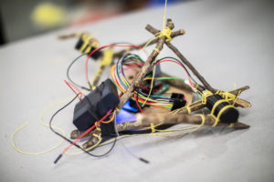
Devin Carroll, a doctoral candidate in the School of Engineering and Applied Sciences, is designing a modular robot called StickBot, which may be adapted for rehabilitation use in global public health settings.
StickBot in walking mode, using the sticks as legs to propel itself across the table.
In late summer, just as the leaves were starting to crisp and curl in the heat, Devin Carroll walked out of his apartment, looked on the ground, and picked up a couple of sticks that he thought might work for his robot. About half an inch thick and the length of an adult hand, he stripped the three sticks of their bark and lashed them with string to StickBot, a modular robot composed of circuitry, actuators, a microcontroller, and a motor driver.
Controlling the robot using an app he designed, Carroll shows how StickBot can pivot from using the sticks as legs in “crawler mode,” to using them as arms. In “grasper mode,” the sticks are attached to a controller plate on one side to form a hinge joint while moving with their free end to hold a cup upright.
Rather than a static, singular invention, StickBot is an idea, a flexible system that can be reconfigured in a variety of ways. A modular robot, StickBot’s components can be added, adjusted, and discarded as needed.
This article features quotes from Michelle Johnson, Associate Professor in Physical Medicine and Rehabilitation in the Perelman School of Medicine and in Bioengineering in the School of Engineering and Applied Sciences, and Director of the Rehabilitation Robotics Lab.
Neuroscientists frequently say that neural activity ‘represents’ certain phenomena, PIK Professor Konrad Kording and postdoc Ben Baker led a study that took a philosophical approach to tease out what the term means.
Neuroscientists use the word “represent” to encompass multifaceted relationships between brain activity, behavior, and the environment.
One of neuroscience’s greatest challenges is to bridge the gaps between the external environment, the brain’s internal electrical activity, and the abstract workings of behavior and cognition. Many neuroscientists rely on the word “representation” to connect these phenomena: A burst of neural activity in the visual cortex may represent the face of a friend or neurons in the brain’s memory centers may represent a childhood memory.
But with the many complex relationships between mind, brain, and environment, it’s not always clear what neuroscientists mean when they say neural activity “represents” something. Lack of clarity around this concept can lead to miscommunication, flawed conclusions, and unnecessary disagreements.
To tackle this issue, an interdisciplinary paper takes a philosophical approach to delineating the many aspects of the word “representation” in neuroscience. The work, published in Trends in Cognitive Sciences, comes from the lab of Konrad Kording, a Penn Integrates Knowledge University Professor and senior author on the study whose research lies at the intersection of neuroscience and machine learning.
“The term ‘representation’ is probably one of the most common words in all of neuroscience,” says Kording, who has appointments in the Perelman School of Medicine and School of Engineering and Applied Science. “But it might mean something very different from one professor to another.”
Also coauthor on the paper is Benjamin Lansdell, a data scientist in the Department of Developmental Neurobiology at St. Jude Children’s Hospital and former postdoctoral researcher in the Kording lab.
Funding for this study came from the National Institutes of Health (awards 1-R01-EB028162-01 and R01EY021579) and the University of Pennsylvania Office of the Vice Provost for Research.
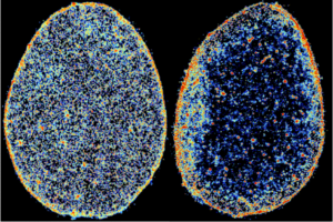
In these super-resolution images of tendon cell nuclei, the color coding represents chromatin density map, from low density in blue to high density in red. Comparing a healthy human tendon cell nucleus (left) to one diagnosed with tendinosis (right) shows that disease alters the spatial localization and compaction of chromatin.
In a recent study published in Nature Biomedical Engineering, the team detailed what they found when they closely observed the nucleus of cells inside connective tissues deteriorating as a result of tendinosis, which is the chronic condition that results from a tendon repeatedly suffering small injuries that don’t heal correctly. Using the latest super-resolution imaging techniques, they found that the tendon cells involved in maintaining the tissue’s structure in a diseased microenvironment improperly reorder their chromatin — the DNA-containing material that chromosomes are composed of — when attempting to repair.
This and other findings highlighted in the report point to the possibility of new treatments, such as small-molecule therapies, that could restore order to the affected cells.
“Interestingly, we were able to explain the role of mechanical forces on the 3-D organization of chromatin by developing a theory that integrates fundamental thermodynamic principles (physics) with the kinetics of epigenetic regulation (biology),” said study co-author and CEMB Director Vivek Shenoy in a news release from Penn Medicine News.
The CEMB, one of 18 active interdisciplinary research centers funded by the National Science Foundation’s Science and Technology Center (STC) program, brings together dozens of researchers from Penn Engineering and the Perelman School of Medicine, as well as others spread across campus and at partner institutions around the world.
With its funding recently renewed for another five years, the CEMB has entered into a new phase of its mission, centered on the nascent concept of “mechanointelligence,” which is exemplified by studies like this one. While mechanobiology is the study of the physical forces that govern the behavior of cells and their communication with their neighbors, mechanointelligence adds another layer of complexity: attempting to understand the forces that allow cells to sense, remember and adapt to their environments.
Ultimately, harnessing these forces would allow researchers to help multicellular organisms — plants, animals and humans — better adapt to their environments as well.
This story originally appeared in Penn Engineering Today.
Vivek Shenoy is Eduardo D. Glandt President’s Distinguished Professor in Materials Science and Engineering, Bioengineering, and in Mechanical Engineering and Applied Mechanics.