Week in BioE (March 14, 2018)

Nanotube Yarn Used for Neurological Applications

nanotubes
Microscopic image of carbon nanotube yarn.

Tapping into the autonomous nervous system – the control center for things like heartbeat and breathing – is a relatively new part of neurostimulation technologies to both record and direct organ function. Implants designed for stimulating peripheral nerves often fail because the protective tissue surrounding nerve bundles (the perineurium) is difficult to penetrate, and the body’s immune response often builds a scar around the implanted device.

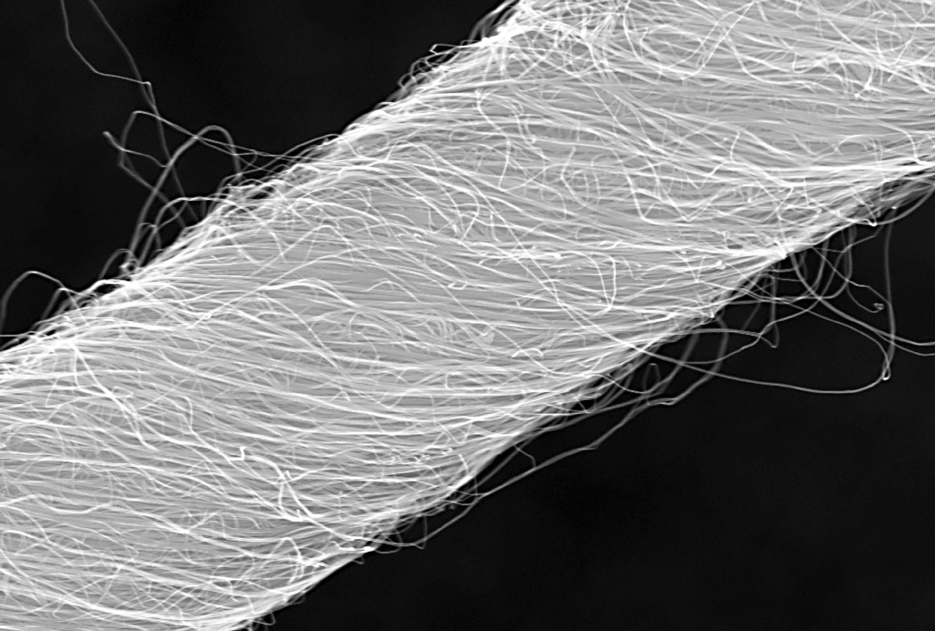
Now, a team of scientists from Case Western Reserve University (CWRUL) has used carbon nanotubes to overcome these obstacles, reporting their findings in Scientific Reports. The authors, led by Dominique M. Durand, Ph.D., Director of the Neural Engineering Center and El Lindseth Professor of Biomedical Engineering, Neurosciences, Physiology and Biophysics at CWRU, fabricated yarn made of carbon nanotubes that was 10 to 20 µm in diameter. The yarn was then used to create electrodes, which were implanted into rats to monitor activity of the glossopharyngeal and vagus nerves.

The authors found that they could use the implants to monitor nerve activity under conditions of hypoxia and stomach distention. They report that the success of their experiments likely derives from the similarity of the nanotube yarn to the actual neural tissue surrounding the implant. The implants are a long way from being tried in humans, but the large number of functions controlled by just these two nerves indicates that such implants could find use in an enormous number of diseases.

Better Screening of Nanoparticle Delivery

As we discussed last week, the development of gene-based therapies is hindered by the sheer size of the human genome. The immense volume of information involved can quickly become difficult to manage, so one way in which scientists “keep track” of genetic information during the process of introducing new genetic material into an organism is DNA barcoding. This process attaches a small piece of DNA to the gene being studied; if and when the gene causes cells to replicate, these cells will bear the barcode, thus allowing the observer to be certain of the gene identities the whole time.

Seeking to determine whether DNA barcoding of lipid nanoparticles for injection into living models would outperform in vitro testing, a team of investigators at Georgia Tech and Emory University conducted a comparison of the two techniques, reporting their findings in Nano Letters. The authors, led by James Dahlman, Ph.D., Assistant Professor of Biomedical Engineering at GT/Emory, found that in vitro testing did not predict in vivo delivery. Further, they were able to track several dozen barcodes delivered by nanoparticles to eight different cell lines.

The authors believe that their technique, which they call JOint Rapid DNA Analysis of Nanoparticles (JORDAN), is superior to in vitro screening of nanoparticles to predict successful transplantation. They are offering JORDAN online as open source software so other scientists can use the technology to more accurately screen nanomaterials.

Synthetic Biologists Create Gene Circuits

Among the many types of molecules that regulate genetic expression in the body are microRNAs, non-coding strands of RNA that are responsible for gene silencing and other forms of gene expression regulation. The ability to harness and control the functions of microRNAs could have important implications for disease prevention and treatment.

In a recent article in Systems Biology and Applications, researchers at the University of Texas, Dallas, report on their engineering of a microRNA-based genetic circuit and its deployment in living cells. They created the circuit using strands of RNA from a variety of organisms, including viruses and jellyfish. The authors, led by Leonidas Bleris, Ph.D., Associate Professor of Bioengineering at UT Dallas, used the circuits to better understand how microRNAs change gene expression under different conditions.

More importantly, the authors found that their circuit had the ability to outproduce types of gene expression, which decreased as the number of gene replications increased.  The authors believe that their discoveries could have applications in a number of genetic disorders.

Discouraging Smoking at the Level of the Brain

Cigarette smoking is the single greatest contributor to negative health outcomes in the population.  Nicotine addiction often appears during the teenage years, and aggressive advertising has been used for the last couple of decades to encourage people to quit smoking and younger people not to start. Despite the widespread use of advertising to change human behavior, remarkably little is known on how the brain responds to advertising messages.

Danielle S. Bassett, Ph.D., Eduardo D. Glandt Faculty Fellow and Associate Professor of Bioengineering at Penn, recently collaborated with faculty from Penn’s Annenberg School of Communication to determine the neuroscience underlying this outcome. The collaborators showed graphic warning labels to a cohort of smokers while they were subjected to functional magnetic resonance imaging, which images brain activity during specific tasks. They found that smokers whose brains showed greater coherence between regions in the valuation network were more likely to quit smoking. Determining why these brain regions acted as they did could yield even more effective smoking-cessation messaging.

Purdue Startup Working to Expand MRI

Engineers at Purdue, including Zhongming Liu, Ph.D., Assistant Professor of Biomedical Engineering and Electrical and Computer Engineering, have cofounded at startup company, called MR-Link, to develop and produce a coin-sized device that can be inserted into MRI machines, allowing them to perform multiple scans simultaneously.

The device could be useful in reducing the amount of electromagnetic force to which patients are exposed during an MRI scan. In addition, Dr. Liu and his colleagues believe the device will cost perhaps less than a tenth of what similar devices currently cost. Given the widespread use of MRI, the device could ultimately impact how a number of diseases and disorders are diagnosed and tracked.

New Faculty: Interview With Konrad Kording

Kording
Konrad Kording, PhD

This week, we present our interview with incoming faculty member Konrad Kording, who starts as a Penn Integrates Knowledge Professor in the Department of Bioengineering and the Department of Neuroscience in the Perelman School of Medicine. Konrad and Andrew Mathis discuss what neuroscience is and isn’t, the “C” word (consciousness), and what it’s like for a native of Germany to live in the United States.


PlayPlay

Uncertainty Investigated by Neuroscience

uncertainty


Uncertainty is part of life, but the underlying neuroscience of how we make decisions under conditions of uncertainty is only beginning to be understood. In a paper published Monday by Nature Human Behaviour, new Penn Bioengineering faculty member and Penn Integrates Knowledge Professor Konrad Kording, Ph.D., and his coauthor, Iris Vilares, Ph.D., of University College London, offer additional evidence that dopamine lies at the heart of how the brain operates when there is a lack of certainty.

Drs. Kording and Vilares devised a simple computerized test that examined the extent to which test takers relied on previous knowledge vs. what they saw at the present moment. They then administered the test to a cohort of patients with Parkinson’s disease, a condition associated with depleted dopamine levels. The patients were tested both while taking dopaminergic medication and while off it. They found that dopaminergic medication caused the patients to pay greater attention to sensory (i.e., visual) information — an effect that diminished as the patients learned. Ultimately, the study provided evidence that dopamine levels were related to the tendency to rely on new information, also called likelihood uncertainty.

“Scientists believe that understanding uncertainty is key to understanding how the brain computes,” Dr. Kording says. “There are many theories in this space. We provide fairly clean evidence for one of them, which is that dopamine encodes likelihood uncertainty. This information could change the way people think about the manner in which the brain deals with uncertainty.”

Mind Control and an Ethical Appeal

mind control brain
A “wiring diagram of the human brain,” produced using diffusion MRI scans of the brain.

A group of four scholars from the University of Pennsylvania, including Bioengineering professor Danielle Bassett, have issued a call in the journal Nature Human Behaviour for greater safeguards for patients as treatments in the field of neuroscience evolve and come ever closer to resembling “mind control.”

“While we don’t believe,” Bassett said, “that the science-fiction idea of mind control, totally overriding a person’s autonomy, will ever be possible, new brain-focused therapies are becoming more specific, targeted and effective at manipulating individuals’ mental states. As these techniques and technologies mature, we need systems in place to make sure they are applied such that they maximize beneficial effects and minimize unwanted side effects.”

Read more at the Penn News Web Site.

Chow Wins NIH Grant for Brain Study

Chow R01
Brian Chow, Ph.D.

The National Institutes of Health (NIH) has awarded a grant to Brian Chow, Ph.D., an assistant professor in the Department of Bioengineering, to study ultrafast genetically encoded voltage indicators (GEVIs). GEVIs are proteins that can detect changes in the electrical output of cells and report those changes by emitting different color light. His research seeks to create GEVIs that can report these changes much more rapidly – in fact, more than a million times more quickly than the velocity of the changes themselves – and apply these ultrafast GEVIs to the study of the brain.

The NIH-funded research will build on earlier research, employing de novo fluorescent proteins (dFPs) created in Dr. Chow’s lab. These dFPs, which are totally artificial and unrelated to natural proteins, report voltage changes in neurons by changing in brightness. Working with a team of investigators that includes faculty members from the Departments of Biochemistry & Biophysics and Neuroscience, Dr. Chow hopes to develop these ultrafast GEVIs.

“Monitoring thousands of neurons in parallel will shed new light on cognition, learning and memory, mood, and the physiological underpinnings of nervous system disorders,” he says.

Danielle Bassett on Social Networks, Brain Activity

danielle bassett
Danielle Bassett, PhD
Danielle Bassett, Eduardo D. Glandt Faculty Fellow and Associate Professor in the departments of Bioengineering and Electrical and Systems Engineering, recently collaborated with colleagues from the Annenberg School for Communication and elsewhere, applying her network science approach to the brain to a study of social networks.

When someone talks about using “your network” to find a job or answer a question, most people understand that to mean the interconnected web of your friends, family, and acquaintances. But we all have another key network that shapes our life in powerful ways: our brains.

In the brain, impulses whiz from one brain region to another, helping you formulate all of your thoughts and decisions. As science continues to unlock the complexities of the brain, a group of researchers has found evidence that brain networks and social networks actually influence and inform one another.

The study, published today in the Proceedings of the National Academy of Sciences looked at the brain’s response to social exclusion under fMRI, particularly in the mentalizing system, which includes separate regions of the brain that help us consider the views of others.

It found that people who show greater changes in connectivity in their mentalizing system during social exclusion compared to inclusion tend to have a less tightly knit social network — that is, their friends tend not to be friends with one another. By contrast, people with more close-knit social networks, in which many people in the network tend to know one another, showed less change in connectivity in their mentalizing regions.

Continue reading.

Konrad Kording: A Penn Integrates Knowledge Professor Coming to Penn BE

konrad kording
Konrad Kording, PhD

The Department of Bioengineering at the University of Pennsylvania is proud to announce that Konrad Kording, PhD, currently professor of physical medicine and rehabilitation, physiology, and applied mathematics at Northwestern University, will join the BE faculty in the fall.

Dr. Kording, a neuroscientist with advanced degrees in experimental physics and computational neuroscience, is a native of Germany. After earning his PhD in 2001 at the Swiss Federal Institute of Technology in Zurich, he held fellowships at University College, London, and MIT before arriving at Northwestern in 2006.

Kording’s groundbreaking interdisciplinary research uses data science to understand brain function, improve personalized medicine, collaborate with clinicians to diagnose diseases based on mobile phone data, and even understand the careers of professors.  Across many areas of biomedical research, his group analyzes large datasets to test new models and thus get closer to an understanding of complex problems in bioengineering, neuroscience, and beyond.

Dr. Kording’s appointment will be shared between the BE Department and the Department of Neuroscience in the Perelman School of Medicine.