David F. Meaney, Solomon R. Pollack Professor of Bioengineering, has been named the Senior Associate Dean of Penn Engineering, effective January 1, 2020. This newly created leadership position will have oversight responsibilities in budget, space and infrastructure planning; facilities and research services; and will create and cultivate new interschool partnerships that will expand Penn Engineering’s footprint on campus.
Meaney is well known not only for his scholarship and innovation in neuroengineering and concussion science, but also for his leadership during his highly successful tenure as Chair of the Department of Bioengineering.
“Dave’s strong connections to the health schools will help strengthen Penn Engineering’s initiatives throughout campus,” says Vijay Kumar, Nemirovsky Family Dean of Penn Engineering. “He will have oversight of Penn Health-Tech, the Center for Engineering MechanoBiology and other efforts between engineering and the health schools, and Dave brings his unique creativity, energy and leadership experience to these collaborative efforts.”
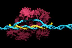
Positive results in first-in-U.S. trial of CRISPR-edited immune cells
3D render of the CRISPR-Cas9 genome editing system
Genetically editing a cancer patient’s immune cells using CRISPR/Cas9 technology, then infusing those cells back into the patient appears safe and feasible based on early data from the first-ever clinical trial to test the approach in humans in the United States. Researchers from the Abramson Cancer Center have infused three participants in the trial thus far—two with multiple myeloma and one with sarcoma—and have observed the edited T cells expand and bind to their tumor target with no serious side effects related to the investigational approach. Penn is conducting the ongoing study in cooperation with the Parker Institute for Cancer Immunotherapy and Tmunity Therapeutics.
“This trial is primarily concerned with three questions: Can we edit T cells in this specific way? Are the resulting T cells functional? And are these cells safe to infuse into a patient? This early data suggests that the answer to all three questions may be yes,” says the study’s principal investigator Edward A. Stadtmauer, section chief of Hematologic Malignancies at Penn. Stadtmauer will present the findings next month at the 61st American Society of Hematology Annual Meeting and Exposition.
Tulane researchers join NIH HEAL initiative for research into opioid crisis
A Tulane University professor and researcher of biomedical engineering will join fellow researchers from over 40 other institutions in the National Institute of Health’s Help to End Addiction Long-Term (HEAL) Initiative. Of the $945 million that make up the project, Michael J. Moore, Ph.D. will receive a share of $1.2 million to advance research in modeling human pain through computer chips, with the help of fellow Tulane researchers Jeffrey Tasker, Ph.D., and James Zadina, Ph.D., each with backgrounds in neuroscience.
Because of the opioid epidemic sweeping the nation, Moore notes that there’s a rapid search going on to develop non-addictive painkiller options. However, he also sees a gap in adequate models to test those new drugs before human clinical trials are allowed to take place. Here is where he hopes to step in and bring some innovation to the field, by integrating living human cells into a computer chip for modeling pain mechanisms. Through his research, Moore wants to better understand not only how some drugs can induce pain, but also how patients can grow tolerant to some drugs over time. If successful, Moore’s work will lead to a more rapid and less expensive screening option for experimental drug advancements.
New machine learning-assisted microscope yields improved diagnostics
Researchers at Duke University recently developed a microscope that uses machine learning to adapt its lighting angles, colors, and patterns for diagnostic tests as needed. Most microscopes have lighting tailored to human vision, with an equal distribution of light that’s optimized for human eyes. But by prioritizing the computer’s vision in this new microscope, researchers enable it to see aspects of samples that humans simply can’t, allowing for a more accurate and efficient diagnostic approach.
Led by Roarke W. Horstmeyer, Ph.D., the computer-assisted microscope will diffuse light through a bowl-shaped source, allowing for a much wider range of illumination angles than traditional microscopes. With the help of convolutional neural networks — a special kind of machine learning algorithm — Horstmeyer and his team were able to tailor the microscope to accurately diagnose malaria in red blood cell samples. Where human physicians typically perform similar diagnostics with a rate of 75 percent accuracy, this new microscope can do the same work with 90 percent accuracy, making the diagnostic process for many diseases much more efficient.
Case Western Reserve University researchers create first-ever holographic map of brain
A Case Western Reserve University team of researchers recently spearheaded a project in creating an interactive holographic mapping system of the human brain. The design, which is believed to be the first of its kind, involves the use of the Microsoft HoloLens mixed reality platform. Lead researcher Cameron McIntyre, Ph.D., sees this mapping system as a better way of creating holographic navigational routes for deep brain stimulation. Recent beta tests with the map by clinicians give McIntyre hope that the holographic representation will help them better understand some of the uncertainties behind targeted brain surgeries.
More than merely providing a useful tool, McIntyre’s project also brings together decades’ worth of neurological data that has not yet been seriously studied together in one system. The three-dimensional atlas, called “HoloDBS” by his lab, provides a way of finally seeing the way all of existing neuro-anatomical data relates to each other, allowing clinicians who use the tool to better understand the brain on both an analytical and visual basis.
Implantable cancer traps reduce biopsy incidence and improve diagnostic
Biopsies are one of the most common procedures used for cancer diagnostics, involving a painful and invasive surgery. Researchers at the University of Michigan are trying to change that. Lonnie Shea, Ph.D., a professor of biomedical engineering at the university, worked with his lab to develop implants with the ability to attract any cancer cells within the body. The implant can be inserted through a scaffold placed under the patient’s skin, making it a more ideal option than biopsy for inaccessible organs like lungs.
The lab’s latest work on the project, published in Cancer Research, details its ability to capture metastatic breast cancer cells in vivo. Instead of needing to take biopsies from areas deeper within the body, the implant allows for a much simpler surgical procedure, as biopsies can be taken from the implant itself. Beyond its initial diagnostic advantages, the implant also has the ability to attract immune cells with tumor cells. By studying both types of cells, the implant can give information about the current state of cancer in a patient’s body and about how it might progress. Finally, by attracting tumor and immune cells, the implant has the ability to draw them away from the area of concern, acting in some ways as a treatment for cancer itself.
People and Places
Cesar de la Fuente-Nunez, PhD
The Philadelphia Inquirer recently published an article detailing the research of Penn’s Presidential Assistant Professor in Psychiatry, Microbiology, and Bioengineering, Cesar de la Fuente, Ph.D. In response to a growing level of worldwide deaths due to antibiotic-resistant bacteria, de la Fuente and his lab use synthetic biology, computation, and artificial intelligence to test hundreds of millions of variations in bacteria-killing proteins in the same experiment. Through his research, de la Fuente opens the door to new ways of finding and testing future antibiotics that might be the only viable options in a world with an increasing level of drug-resistant bacteria
Emily Eastburn, a Ph.D. candidate in Bioengineering at Penn and a member of the Boerckel lab of the McKay Orthopaedic Research Laboratory, recently won the Ashton fellowship. The Ashton fellowship is an award for postdoctoral students in any field of engineering that are under the age of 25, third-generation American citizens, and residents of either Pennsylvania or New Jersey. A new member of the Boerckel lab, having joined earlier this fall, Eastburn will have the opportunity to conduct research throughout her Ph.D. program in the developmental mechanobiology and regeneration that the Boerckel lab focuses on.
A Q&A with neuroscientist Konrad Kording on how connections between minds and machines are portrayed in popular culture, and what the future holds for this reality-defying technology.
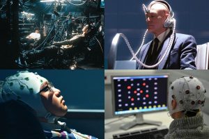
Science fiction and superhero films portray brain-machine interfaces as malevolent robots that plug into human brains for fuel in The Matrix (top left) or as power-enhancing devices in X-Men (top right). In reality, they can help patients use artificial limbs or directly connect to computers. (Image credits, from top left to bottom right: Warner Brothers, 20th Century Fox, Intelligent Films, AFP Photo/Jean-Pierre Clatot)
For the many superheroes that use high-powered gadgets to save the day, there’s an equal number of villains who use technology nefariously. From robots that plug into human brains for fuel in “The Matrix” to the memory-warping devices seen in “Men in Black,” “Captain Marvel,” and “Total Recall,” technology that can control people’s minds is one of the most terrifying examples of technology gone wrong in science fiction and superhero films.
Now, progress made on brain-machine interfaces, technology that provides a direct communication link between a brain and an external device, is bringing us closer to a world that feels like science fiction. Elon Musk’s company NeuraLink is working on a device to let people control computers with their minds, while Facebook’s “mind-reading initiative” can decode speech from brain activity. Is this progress a glimpse into a dark future, or are there more empowering ways in which brain-machine interfaces could become a force for good?
Q: What are the main challenges in connecting brains to devices?
The key problem is that you need to get a lot of information out of brains. Today’s prosthetic devices are very slow, and if we want to go faster it’s a tradeoff: I can go slower and then I am more precise, or I can go faster and be more noisy. We need to get more data out of brains, and we want to do it electrically, meaning we need to get more electrodes into brains.
So what do you need? You need a way of getting electrodes into the brain without making your brain into a pulp, you want the electrodes to be flexible so they can stay in longer, and then you want the system to be wireless. You don’t want to have a big connector on the top of your head.
It’s primarily a hardware problem. We can get electrodes into brains, but they deteriorate quickly because they are too thick. We can have plugs on people’s heads, but it’s ruling out any real-world usage. All these factors hold us back at the moment.
That’s why the Neuralink announcement was very interesting. They get a rather large number of electrodes into brains using well-engineered approaches that make that possible. What makes the difference is that Neuralink takes the best ideas in all the different domains and puts them together.
Q: Most examples in pop culture of connecting brains to machines have villainous or nefarious ends. Does that match up with how brain-machine interfaces are currently being developed?
Let’s say you’ve had a stroke, you can’t talk, but there’s a prosthetic device that allows you to talk again. Or if you lost your arm, and you get a new one that’s as good as the original—that’s absolutely a force for good.
It’s not a dark, ugly future thing, it’s a beautiful step forward for medicine. I want to make massive progress in these diseases. I want patients who had a stroke to talk again; I want vets to have prosthetic devices that are as good as the real thing. I think short-term this is what’s going to happen, but we are starting to worry about the dark sides.
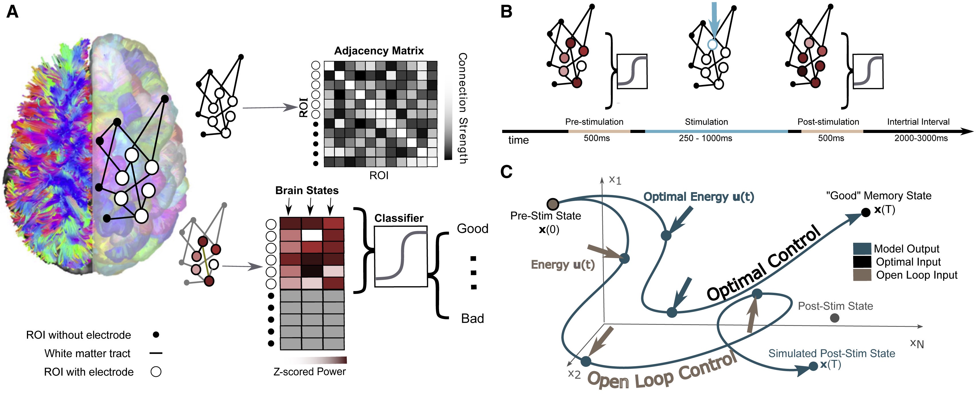
The researchers’ model involves mapping the connections between different regions of an individual’s brain while they performed a basic memory task, then using that data to predict how electrical stimulation in one region would affect activity throughout the network. Individuals’ improved performance on the same memory task after stimulation suggests the model could eventually be generalized toward a variety of stimulation therapies.
Brain stimulation, where targeted electrical impulses are directly applied to a patient’s brain, is already an effective therapy for depression, epilepsy, Parkinson’s and other neurological disorders, but many more applications are on the horizon. Clinicians and researchers believe the technique could be used to restore or improve memory and motor function after an injury, for example, but progress is hampered by how difficult it is to predict how the entire brain will respond to stimulation at a given region.
In an effort to better personalize and optimize this type of therapy, researchers from the University of Pennsylvania’s School of Engineering and Applied Science and Perelman School of Medicine, as well as Thomas Jefferson University Hospital and the University of California, Riverside, have developed a way to model how a given patient’s brain activity will change in response to targeted stimulation.
To test the accuracy of their model, they recruited a group of study participants who were undergoing an unrelated treatment for severe epilepsy, and thus had a series of electrodes already implanted in their brains. Using each individual’s brain activity data as inputs for their model, the researchers made predictions about how to best stimulate that participant’s brain to improve their performance on a basic memory test.
The participants’ brain activity before and after stimulation suggest the researchers’ models have meaningful predictive power and offer a first step towards a more generalizable approach to specific stimulation therapies.
Danielle Bassett and Jennifer Stiso.
The study, published in the journal Cell Reports, was led by Danielle Bassett, J. Peter Skirkanich Professor in Penn Engineering’s Department of Bioengineering, and Jennifer Stiso, a neuroscience graduate student in Penn Medicine and a member of Bassett’s Complex Systems Lab.
These 12 object-number value pairs were taught to the participants, who had to properly learn the associations to succeed in value judgement tests. The researchers investigated the differences in their brain activity patterns to see why some were faster learners than others.
Why do some people naturally excel at learning instruments, languages or technology while others take longer to pick up new knowledge? Learning requires the brain to encode information, changing its neural “wiring” and creating networks between brain regions.
Earlier research has suggested that part of what might slow down learners is over-thinking. A 2015 study led by Danielle Bassett, Eduardo D. Glandt Faculty Fellow and associate professor in the Department of Bioengineering, showed a correlation between slow learning and cognitive control: the brain’s ability to regulate itself by activating the necessary networks and inhibiting unnecessary activity. In that study, when people unnecessarily engaged parts of the brain linked to cognitive control, they were more likely to take longer to learn a simple task.
But beyond what might make an individual learn more slowly, the researchers want to know what sort of geometric patterns of brain activity make for better learning.
Evelyn Tang and Danielle Bassett
Their new study was led by Bassett and Evelyn Tang, who was an Africk Family Postdoctoral Fellow in Bassett’s Complex Systems Lab before starting at the Max Planck Institute this fall. Sharon Thompson-Schill, Christopher H. Browne Distinguished Professor and chair of Psychology, also contributed to the study.
New Studies in Mechanobiology Could Open Doors for Cellular Disease Treatment
When we think of treatments at the cellular level, we most often think of biochemical applications. But what if we began to consider more biomechanical-oriented approaches in the regulation of cellular life and death? Under a grant from the National Science Foundation (NSF),Worcester Polytechnic Institute’s (WPI) Head of the Department of Biomedical Engineering Kristen Billiar, Ph.D., performs research that looks at the way mechanical stimuli can affect and trigger programmed cell death.
Billiar, who received his M.S.E. and Ph.D. from Penn, began his research by first noticing the way that cells typically respond to the mechanical stimuli in their everyday environment, such as pressure or stretching, with behaviors like migration, proliferation, or contraction. He and his research team hope to find a way to eventually predict and control cellular responses to their environment, which they hope could open doors to more forms of treatment for disorders like heart disease or cancer, where cellular behavior is directly linked to the cause of the disease.
Self-Learning Algorithm Could Help Improve Robotic Leg Functionality
Obviously, one of the biggest challenges in the field of prosthetics is the extreme difficulty in creating a device that perfectly mimics whatever the device replaces for its user. Particularly with more complex designs that involve user-controlled motion for joints in the limbs or hands, the electrical circuits implemented are by no means a perfect replacement of the neural connections in the human body from brain to muscle. But recently at the University of Southern California Viterbi School of Engineering, a team of researchers led by Francisco J. Valero-Cuevas, Ph. D., developed an algorithm with the ability to learn new walking tasks and adapt to others without any additional programming.
The algorithm will hopefully help to speed the progress of robotic interactions with the world, and thus allow for more adaptive technology in prosthetics, that responds to and learns with their users. The algorithm Valero-Cuevas and his team created takes inspiration from the cognition involved with babies and toddlers as they slowly learn how to walk, first through random free play and then from pulling on relevant prior experience. In a prosthetic leg, the algorithm could help the device adjust to its user’s habits and gait preferences, more closely mimicking the behavior of an actual human leg.
Neurofeedback Can Improve Behavioral Performance in High-Stress Situations
We’re all familiar with the concept of being “in the zone,” or the feeling of extraordinary focus that we can sometimes have in situations of high-stress. But how can we understand this shift in mindset on a neuroengineering level? Using the principal of the Yerkes-Dodson law, which says that there is a state of brain arousal that is optimal for behavioral performance, a team of biomedical engineering researchers at Columbia University hope to find ways of applying neurofeedback to improving this performance in demanding high-stress tasks.
Led by Paul Sajda, Ph.D., who received his doctoral degree from Penn, the researchers used a brain-computer interface to collect electroencephalography (EEG) signals from users immersed in virtual reality aerial navigation tasks of varying difficulty levels. In doing so, they were able to make connections between stressful situations and brain activity as transmitted through the EEG data, adding to the understanding of how the Yerkes-Dodson law actually operates in the human body and eventually demonstrating that the use of neurofeedback reduced the neural state of arousal in patients. The hope is that neurofeedback may be used in the future to help treat emotional conditions like post-traumatic stress disorder (PTSD).
Ultrasound Stimulation Could Lead to New Treatments for Inflammatory Arthritis
Arthritis, an autoimmune disease that causes painful inflammation in the joints, is one of the more common diseases among older patients, with more than 3 million diagnosed cases in the United States every year. Though extreme measures like joint replacement surgery are one solution, most patients simply treat the pain with nonsteroidal anti-inflammatory drugs or the adoption of gentle exercise routines like yoga. Recently however, researchers at the University of Minnesota led by Daniel Zachs, M.S.E., in the Sensory Optimization and Neural Implant Coding Lab used ultrasound stimulation treatment as a way to reduce arthritic pain in mice. In collaboration with Medtronic, Zachs and his team found that this noninvasive ultrasound stimulation greatly decreased joint swelling in mice who received the treatment as opposed to those that did not. They hope that in the future, similar methods of noninvasive treatment will be able to be used for arthritic patients, who otherwise have to rely on surgical remedies for serious pain.
People and Places
Leadership and Inspiration: EDAB’s Blueprint for Engineering Student Life
To undergraduates at a large university, the administration can seem like a mysterious, all-powerful entity, creating policy that affects their lives but doesn’t always take into account the reality of their day-to-day experience. The Engineering Deans’ Advisory Board (EDAB) was designed to bridge that gap and give students a platform to communicate with key decision makers.
The 13-member board meets once per week for 60 to 90 minutes. The executive board, comprised of four members, also meets weekly to plan out action items and brainstorm. Throughout his interactions with the group, board president Jonathan Chen, (ENG ‘19, W ‘19), has found a real kinship with his fellow board members, who he says work hard and enjoy one another’s company in equal measure.
Bioengineering major Daphne Cheung (ENG’19) joined the board as a first-year student because she saw an opportunity to develop professional skills outside of the classroom. “For me, it was about trying to build a different kind of aptitude in areas such as project management, and learning how to work with different kinds of people, including students and faculty, and of course, the deans,” she says.
Purdue University College of Engineering and Indiana University School of Medicine Team Up in New Engineering-Medicine Partnership
The Purdue University College of Engineering and the Indiana University School of Medicine recently announced a new Engineering-Medicine partnership, that seeks to formalize ongoing and future collaborations in research between the two schools. One highlight of the partnership is the establishment of a new M.D./M.S. degree program in biomedical engineering that will allow medical students at Indiana University to receive M.S.-level training in engineering technologies as they apply to clinical practice. The goal of this new level of collaboration is to further involve Purdue’s engineering program in the medical field, and to exhibit the benefits that developing an engineering mindset can have for medical students. The leadership of this new partnership includes
A new approach uses “mechanoceuticals” to treat pain.
Drugs are commonly injected directly into an injury site to speed healing. For chronic pain, clinicians can inject drugs to reduce inflammation in painful joints, or can inject nerve blockers to block the nerve signals that cause pain. In a recent study, a group from UCLA developed a technique to deform a material surrounding nerve fibers to trigger a response in the fibers that would relieve pain. The combination of mechanics and treatment – i.e., ‘mechanoceuticals’ – is a clever way to trick fibers and reverse painful symptoms. Done without any injections and simply controlling magnetic fields outside the body, this approach can be reused as necessary.
The design of this mechanoceutical was completed by Dino Di Carlo, PhD, Professor of Bioengineering, and his team at UCLA’s Sameuli School of Engineering. By encasing tiny, magnetic nanoparticles within a biocompatible hydrogel, the group used magnetic force to stimulate nerve fibers and cause a corresponding decrease in pain signals. This promising development opens up a new approach to pain management, one which can be created with different biomaterials to suit different conditions, and delivered “on demand” without worrying about injections or, for that matter, any prescription drugs.
Understanding the Adolescent Brain
It’s no surprise that adults and adolescents often struggle to understand one another, but the work of neurologists and other researchers provides a possible physical reason for why that might be. Magnetic resonance elastrography (MRE) is a tool used in biomedical imaging to estimate the mechanical properties, or stiffness, of tissue throughout the body. Unexpectedly, a recent study suggests that brain stiffness correlates with cognitive ability, suggesting MRE may provide insight into patients’ behavior, psychology, and psychiatric state.
A new paper in Developmental Cognitive Neuroscience published the results of a study using MRE to track the relative “stiffness” vs. “softness” of adult and adolescent brains. The University of Delaware team, led by Biomedical Engineering Assistant Professor Curtis Johnson, PhD, and his doctoral student Grace McIlvain, sampled 40 living subjects (aged 12-14) and compared the properties to healthy adult brains.
The study found that children and adolescent brains are softer than those of adults, correlating to the overall malleability of childhood development. The team hopes to continue their studies with younger and older children, looking to demonstrate exactly when and how the change from softness to stiffness takes place, and how these properties correspond to individual qualities such as risk-taking or the onset of puberty. Eventually, establishing a larger database of measurements in the pediatric brain will help further studies into neurological and cognitive disorders in children, helping to understand conditions such as multiple sclerosis, autism, and cerebral palsy.
Can Nanoparticles Replace Stents?
Researchers and clinicians have made amazing advances in heart surgery. Stents, in particular, have become quite sophisticated: they are used to both prop open clogged arteries as well as deliver blood-thinning medication slowly over days to weeks in the area of the stent. However, the risk of blood clotting increases with stents and the blood vessels can constrict over time after the stent is placed in the vessel.
A recent NIH grant will support the design of a stent-free solution to unclog blood vessels. Led by Shaoqin Gong, PhD, Vilas Distinguished Professor of Biomedical Engineering at UW-Madison, the team used nanoparticles (or nanoclusters) to directly target the affected blood vessels and prevent regrowth of the cells post-surgery, eliminating the need for a stent to keep the pathways open. These nanoclusters are injected through an intravenous line, further reducing the risks introduced by the presence of the stent. As heart disease affects millions of people worldwide, this new material has far-reaching consequences. Their study is published in the September edition of Biomaterials.
NIST Grant Supports
The National Institute of Standards and Technology (NIST) awarded a $30 million grant to Johns Hopkins University, Binghamton University, and Morgan State University as part of their Professional Research Experience Program (PREP). Over five years, this award will support the collaboration of academics from all levels (faculty, postdoc, graduate, and undergraduate) across the three universities, enabling them to conduct research and attend NIST conferences.
The principal investigator for Binghamton U. is Professor and Chair of the Biomedical Engineering Department, Kaiming Ye, PhD. Dr. Ye is also the Director of the Center of Biomanufacturing for Regenerative Medicine (CBRM), which will participate in this collaborative new enterprise. Dr. Ye hopes that this grant will create opportunities for academics and researchers to network with each other as well as to more precisely define the standards for the fields of regenerative medicine and biomaterial manufacturing.
The gift honors the late A. James Clark, former CEO of Clark Enterprises and Clark Construction Group LLC, one of the country’s largest privately-held general building contractors. It is designed to prepare future engineering and business leaders, with an emphasis on low income families and first-generation college students. Clark never forgot that his business successes began with an engineering scholarship. This has guided the Clark family’s longstanding investments in engineering education and reflects its commitment to ensure college remains accessible and affordable to high-potential students with financial need.
We are proud to say that three incoming Clark Scholars from the Freshman Class of 2022 will be part of the Bioengineering Department here at Penn.
And finally, our congratulations to the new Dean of the School of Engineering at the University of Mississippi: David A. Puleo, PhD. Dr. Puleo earned his bachelor’s degree and doctorate in Biomedical Engineering from Rensselaer Polytechnic Institute. Most recently he served as Professor of Biomedical Engineering and Associate Dean for Research and Graduate Studies at the University of Kentucky’s College of Engineering. Building on his research in regenerative biomaterials, he also founded Regenera Materials, LLC in 2014. Over the course of his career so far, Dr. Puleo received multiple teaching awards and oversaw much departmental growth within his previous institution, and looks poised to do the same for “Ole Miss.”
Like many other fields, biomedical research is experiencing a data explosion. Some estimates suggest that the amount of data generated from the health sciences is now doubling every eighteen months, and experts expect it to double every seventy-three days by 2020. One challenge that researchers face is how to meaningfully analyze this data tsunami.
The challenge of interpreting data occurs at all scales, and a recent collaboration shows how new approaches can allow us to handle the volumes of data emerging at the level of individual cells to infer more about how biology “works” at this level. Wharton Statistics Department researchers Mo Huang and Jingshu Wang (PhD Student and Postdoctoral Researcher, respectively) collaborated with Arjun Raj’s lab in Bioengineering and published their findings in recent issues of Nature Methods and Proceedings of the National Academy of Sciences. One study focused on a de-noising technique called SAVER to provide more precise data from single cell experiments and significantly improves the ability to detect trends in a dataset, similar to how increasing sample size helps improve the ability to determine differences between experimental groups. The second method, termed DESCEND, creates more accurate characterization of gene expression that occur in individual cells. Together these two methods will improve data collection for biologists and medical professionals working to diagnose, monitor, and treat diseased cells.
Dr. Raj’s team contributed data to the cause and acted as consultants on the biological aspects of this research. Further collaboration involved Mingyao Li, PhD, Professor of Biostatistics at the Perelman School of Medicine, and Nancy Zhang, PD, Professor Statistics at the Wharton School. “We are so happy to have had the chance to work with Nancy and Mingyao on analyzing single cell data,” said Dr. Raj. “The things they were able to do with our data are pretty amazing and important for the field.”
Advancements in Biomaterials
This blog features many new biomaterials techniques and substances, and there are several exciting new developments to report this week. First, the journal of Nature Biomedical Engineering published a study announcing a new therapy to treat or even eliminate lung infections, such as those acquired while in hospital or as the result of cystic fibrosis, which are highly common and dangerous. Researchers identified and designed viruses to target and kill the bacteria which causes these infections, but the difficulty of administering them to patients has proven prohibitive. This new therapy, developed by researchers at the Georgia Institute of Technology, is administered as a dry powder directly to the lungs and bypasses many of the delivery problems appearing in past treatments. Further research on the safety of this method is required before clinical trials can begin.
A team at Harvard University published another recent study in Nature Biomedical Engineering announcing their creation of a tissue-engineered scale model of the left human heart ventricle. This model is made from degradable fibers that simulate the natural fibers of heart tissue. Lead investigator Professor Kevin Kit Parker, PhD, and his team eventually hope to build specific models culled from patient stem cells to replicate the features of that patient’s heart, complete with the patient’s unique DNA and any heart defects or diseases. This replica would allow researchers and clinicians to study and test various treatments before applying them to a specific patient.
Lastly, researchers at the Tufts University School of Engineering published in the Proceedings of the National Academy of Sciences on their creation of flexible magnetic composites that respond to light. This material is capable of macroscale motion and is extremely flexible, allowing its adaptation into a variety of substances such as sponges, film, and hydrogels. Author and graduate student Meg Li explained that this material differs from similar substances in its complexity; for example, in the ability for engineers to dictate specific movements, such as toward or away from the light source. Co-author Fiorenzo Omenetto, PhD, suggests that with further research, these movements could be controlled at even more specific and detailed levels.
CFPS: Getting Closer to “On Demand” Medicine
A recent and growing trend in medicine is the move towards personalized or “on demand” medicine, allowing for treatment customized to specific patients’ needs and situations. One leading method is Cell-Free Protein Synthesis (CFPS), a way of engineering cellular biology without using actual cells. CFPS is used to make substances such as medicine, vaccines, and chemicals in a sustainable and portable way. One instance if the rapid manufacture of insulin to treat diabetic patients. Given that many clinics most in need of such substances are found in remote and under-served locations far away from well-equipped hospitals and urban infrastructure, the ability to safely and quickly create and transport these vital substances to patients is vitally important.
The biggest limiting factor to CFPS is difficulty of replicating Glycosylation, a complex modification that most proteins undergo. Glycosylation is important for proteins to exert their biological function, and is very difficult to synthetically duplicate. Previously, achieving successful Glycosylation was a key barrier in CFPS. Fortunately, Matthew DeLisa, PhD, the Williams L. Lewis Professor of Engineering at Cornell University and Michael Jewett, PD, Associate Professor of Chemical and Biological Engineering at Northwestern University, have created a “single-pot” glycoprotein biosynthesis that allows them to make these critical molecules very quickly. The full study was recently published in NatureCommunications. With this new method, medicine is one step closer to being fully “on demand.”
People and Places
The Institute of Electrical and Electronics Engineers (IEEE) interviewed our own Penn faculty member Danielle Bassett, PhD, the Edwardo D. Glandt Faculty Fellow and Associate Professor in Bioengineering, for their website. Dr. Bassett, who shares a joint appointment with Electrical Systems Engineering (ESE) at Penn, has published groundbreaking research in Network Neuroscience, Complex Systems, and more. In the video interview (below), Dr. Bassett discusses current research trends in neuroscience and their applications in medicine.
Finally, a new partnership between Case Western Reserve University and Cleveland Clinic seeks to promote education and research in biomedical engineering in the Cleveland area. Cleveland Clinic Lerner Research Institute‘s Chair of Biomedical Engineering, Geoff Vince, PhD, sees this as an opportunity to capitalize on the renown of both institutions, building on the region’s already stellar reputation in the field of BME. Dozens of researchers from both institutions will have the opportunity to collaborate in a variety of disciplines and projects. In addition to professional academics and medical doctors, the leaders of this new partnership hope to create an atmosphere that can benefit all levels of education, all the way down to high school students.
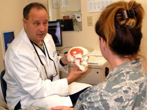
TMD is a common condition affecting movement of the jaw
Medical researchers have long been baffled by the need to find safe and effective treatment for a common condition called temporomandibular joint dysfunction (TMD). Affecting around twenty-five percent of the adult population worldwide, TMD appears overwhelmingly in adolescent, premenopausal women. Many different factors such as injury, arthritis, or grinding of the teeth can lead to the disintegration of or damage to the temporomandibular joint (TMJ), which leads to TMD, although the root cause is not always clear. A type of temporomandibular disorder, TMD can result in chronic pain in the jaw and ears, create difficulty eating and talking, and even cause occasional locking of the joint, making it difficult to open or close one’s mouth. Surgery is often considered a last resort because the results are often short-lasting or even dangerous.
The state of TMD treatment may change with the publication of a study in Science Translational Medicine. With contributions from researchers at the University of California, Irvine (UCI), UC Davis, and the University of Texas School of Dentistry at Houston, this new study has successfully implanted engineered discs made from rib cartilage cells into a TMJ model. The biological properties of the discs are similar enough to native TMJ cells to more fully reduce further degeneration of the joint as well as potentially pave the way for regeneration of joints with TMD.
Senior author Kyriacos Athanasiou, PhD, Distinguished Professor of Biomedical Engineering at UCI, states the next steps for the team of researchers include a long-term study to ensure ongoing effectiveness and safety of the implants followed by eventual clinical trials. In the long run, this technique may also prove useful and relevant to the treatment of other types of arthritis and joint dysfunction.
Advances in Autism Research
Currently, diagnosis of autism spectrum disorders (ASD) has been limited entirely to clinical observation and examination by medical professionals. This makes the early identification and treatment of ASD difficult as most children cannot be accurately diagnosed until around the age of four, delaying the treatment they might receive. A recent study published in the journal of Bioengineering & Translational Medicine, however, suggests that new blood tests may be able to identify ASD with a high level of accuracy, increasing the early identification that is key to helping autistic children and their families. The researchers, led by Juergen Hahn, PhD, Professor and Department Head of Biomedical Engineering at the Rensselaer Polytechnic Institute, hope that after clinical trials this blood test will become commercially available.
In addition to work that shows methods to detect autism earlier, the most recent issue of Nature Biomedical Engineering includes a study to understand the possible causes of autism and, in turn, develop treatments for the disease. The breakthrough technology of Cas9 enzymes allowed researchers to edit the genome, correcting for symptoms that appeared in mice which resembled autism, including exaggerated and repetitive behaviors. This advance comes from a team at the University of California, Berkeley, which developed the gene-editing technique known as CRISPR-Gold to treat symptoms of ASD by injecting the Cas9 enzyme into the brain without the need for viral delivery. The UC Berkeley researchers suggest in the article’s abstract that these safe gene-editing technologies “may revolutionize the treatment of neurological diseases and the understanding of brain function.” These treatments may have practical benefits for the understanding and treatment of such diverse conditions as addiction and epilepsy as well as ASD.
Penn Professor’s Groundbreaking Bioengineering Technology
Our own D. Kacy Cullen, PhD, was recently featured in Penn Today for his groundbreaking research which has led to the first implantable tissue-engineered brain pathways. This technology could lead to the reversal of certain neurodegenerative disorders, such as Parkinson’s disease.
With three patents, at least eight published papers, $3.3 million in funding, and a productive go with the Penn Center for Innovation’s I-Corps program this past fall, Dr. Cullen is ready to take this project’s findings to the next level with the creation of a brand new startup company: Innervace. “It’s really surreal to think that I’ve been working on this project, this approach, for 10 years now,” he says. “It really was doggedness to just keep pushing in the lab, despite the challenges in getting extramural funding, despite the skepticism of peer reviewers. But we’ve shown that we’re able to do it, and that this is a viable technology.” Several Penn bioengineering students are involved in the research conducted in Dr. Cullen’s lab, including doctoral candidate Laura Struzyna and recent graduate Kate Panzer, who worked in the lab all four years of her undergraduate career.
In addition to his appointment as a Research Associate Professor of Neurosurgery at the Perelman School of Medicine at the University of Pennsylvania, Dr. Cullen also serves as a member of Penn’s Department of Bioengineering Graduate Group Faculty, and will teach the graduate course BE 502 (From Lab to Market Place) for the BE Department this fall 2018 semester. He also serves as the director for the Center of Neurotrauma, Neurodegeneration, and Restoration at the VA Medical Center.
New Prosthetics Will Have the Ability to Feel Pain
New research from the Department of Biomedical Engineering at Johns Hopkins University (JHU) has found a way to address one of the difficult aspects of amputation: the inability for prosthetic limbs to feel. This innovative electronic dermis is worn over the prosthetic, and can detect sensations (such as pain or even a light touch), which are conveyed to the user’s nervous system, closing mimicking skin. The findings of this study were recently published in the journal ScienceRobotics.
While one might wonder at the value of feeling pain, both researchers and amputees verify that physical sensory reception is important both for the desired realism of the prosthetic or bionic limb, and also to alert the wearer of any potential harm or damage, the same way that heat can remind a person to remove her hand from a hot surface, preventing a potential burn. Professor Nitish Thakor, PhD, and his team hope to make this exciting new technology readily available to amputees.
People and Places
Women are still vastly outnumbered in STEM, making up only twenty percent of the field, and given the need for diversification, researchers, educators, and companies are brainstorming ways to proactively solve this problem by promoting STEM subjects to young women. One current initiative has been spearheaded by GE Healthcare and Milwaukee School of Engineering University (MSOE) who are partnering to give middle school girls access to programs in engineering during their summer break at the MSOE Summer STEM Camp, hoping to reduce the stigma of these subjects for young women. GE Girls also hosts STEM programs with a number of institutions across the U.S.
The National Science Policy Network (NSPN) “works to provide a collaborative resource portal for early-career scientists and engineers involved in science policy, diplomacy, and advocacy.” The NSPN offers platforms and support including grant funding, internships, and competitions. Chaired and led by emerging researchers and professors from around the country, including biomedical engineering PhD student Michaela Rikard of the University of Virginia, the NSPN seeks to provide a network for young scientists in the current political climate in which scientific issues and the very importance of the sciences as a whole are hotly contested and debated by politicians and the public. The NSPN looks to provide a way for scientists to have a voice in policy-making. This new initiative was recently featured in the Scientific American.
Upon its original founding in 2000, the Bill and Melinda Gates Foundation has included the eradication of malaria as part of its mission, pledging around $2 billion to the cause in the years since. One of its most recent initiatives is the funding of a bioengineering project which targets the type of mosquitoes which carry the deadly disease. Engineered mosquitoes (so-called “Friendly Mosquitoes”) would mate in the wild, passing on a mosquito-killing gene to their female offspring (only females bite humans) before they reach maturity. While previous versions of “Friendly Mosquitoes” have been met with success, concerns have been raised about the potential long-term ecological effects to the mosquito population. UK-based partner Oxitec expects to have the new group ready for trials in two years.
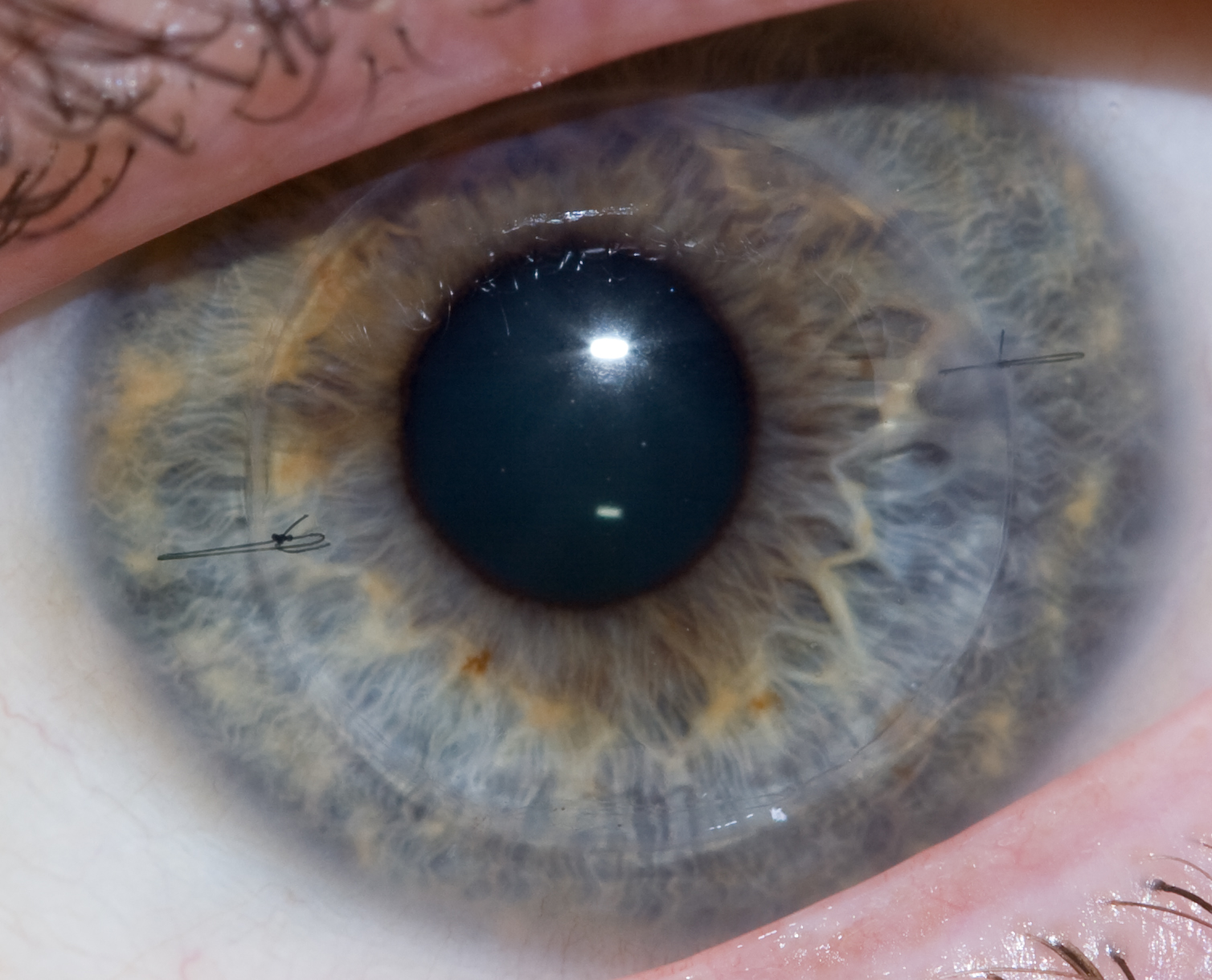
A human eye that received a cornea transplant one year postoperatively.
Disorders of or damage to the cornea — the clear covering over the lens of the eye — can be threatening to vision, and for the last century, corneal transplantation has been a cornerstone of treatment for these conditions. However, corneal transplants are complicated by two key facts: first, as with virtually all transplant procedures, donor organs are in short supply; and second, rejection is common, and recipients of transplants face repeated procedures or a lifetime of steroid eyedrops to prevent rejection.
One way of obviating these issues is the use of synthetic materials, which can now be manufactured with three-dimensional printing. In a new study from scientists at the Institute of Genetic Medicine at Newcastle University in the UK, to be published this summer in Experimental Eye Research, synthetic corneal tissue was 3D printed using a bioink loaded with encapsulated keratocytes (corneal cells), in combination with computer modeling based on actual corneas. The study is only proof to show that printing a biological replicate of the cornea is possible, but it lays the groundwork for future studies in animals.
Engineering Brain Recovery
One of the reasons why stroke is such a damaging event is the inability of damaged brain tissue to regenerate. Angiogenesis, the growth of new blood vessels, can help to regenerate brain tissue but properly guiding the process of angiogenesis is rather difficult.
However, a new report in Nature Materials indicates success using an injectable biogel for this purpose. In the report, a team led by Tatiana Segura, PhD, Professor of Biomedical Engineering at Duke with colleagues at UCLA, details its engineering of an injectable gel using nanoparticles consisting of heparin (a blood-thinning agent to prevent unwanted blood clotting) and vascular endothelial growth factor (VEGF) to stimulate brain regeneration. After injecting the gel in a mouse model of stroke, the mice showed a significant improvement in recovery compared to animals not receiving the engineered nanomaterial.
Here at Penn, D. Kacy Cullen, PhD, Research Associate Professor of Neurosurgery in the Perelman School of Medicine, has been investigating the use of implantable tissue-engineered brain pathways to treat and perhaps reverse the effects of neurodegnerative diseases like Parkinson’s disease. Penn Today has the story, with video of Dr. Cullen and photos and quotes from several of our own Bioengineering students.
Streamlining Environmental Bioengineering
Outside of the health sciences, bioengineering has applications in diverse fields, including energy development and environmental protection. Biofuels are one application for bioengineering that received a major boost recently. In an article published in NPJ Systems Biology and Applications, engineers from the US Department of Energy’s Lawrence Berkeley National Laboratory describe how they used machine learning to better predict the ability of engineered microbes to produce biofuel. With this information, they can then better adjust fuel-producing microbial pathways to maximize production. The machine learning model is a significant improvement over earlier, traditionally algorithmic approaches requiring complex differential equations. The time saved could, over generations of adjustments, result in a significant increase in output.
More on Pilots
Last week, we discussed how the cognitive load borne by airline pilots differs between simulated and real flight. Other scientists, it turns out, are looking at ways that pilots — in particular, fighter pilots — can overcome fatigue. With more than $1 million in grants from the US Department of Defense, Merhavan Singh, PhD, Dean of the Graduate School of Biomedical Sciences at the University of North Texas Health Science Center, and Kai Shen, PhD, Associate Professor in the Department of Chemistry and Forensic Science at Savannah State University in Georgia, are investigating compounds targeting the sigma 1 receptor, which the scientists believe could combat fatigue and also have neuroprotective effects if activated. This is particularly important among fighter pilots serving in conflict, who are often sleep deprived but must remain alert during missions.
People and Places
Having achieved success in its mission, the University of Alabama at Birmingham’s PREP Scholars Program, which supports underrepresented minority students in pursuing graduate study in bioengineering and biomedical engineering, has received an additional $1.8 million in support from the National Institutes of Health. The money will enable the funding of 40 students over the next five years.
Jeffrey Collins Wolchok, PhD, and Kartik Balachandran, PhD, both associate professors in the Department of Biomedical Engineering at the University of Arkansas, have received a $375,000 grant from the National Science Foundation to study the long-term effects of multiple concussions on the brain. With the increased emphasis in the scientific community and media on traumatic brain injury and chronic traumatic encephalopathy, including among former athletes, the two scientists will develop brain on a chip technology to examine the issue.
Finally, this week, the Best College Reviews website published its Top 10 list of online Master’s programs in biomedical engineering. Purdue University’s program finished in first place, with appearances on the list by Colorado State, UC Riverside, Stevens Tech, and Worcester Tech.


 When we think of treatments at the cellular level, we most often think of biochemical applications. But what if we began to consider more biomechanical-oriented approaches in the regulation of cellular life and death? Under a grant from the National Science Foundation (NSF),Worcester Polytechnic Institute’s (WPI) Head of the Department of Biomedical Engineering