Rooting Out Systemic Bias in Neuroscience Publishing

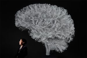
An interdisciplinary research team has found statistical evidence of women being under-cited in academic literature. They are now studying similar effects along racial lines.

By Izzy Lopez

Danielle Bassett, Jordan Dworkin and Perry Zurn are leading efforts to analyze systemic bias in neuroscience citations, and have suggestions for combatting it.

Scientific papers are the backbone of a research community and the citation of those papers sparks conversation in a given field. This cycle of publication and citation leads to new knowledge, but what happens when implicit discrimination in a field leads to papers by minority scholars being cited less often than their counterparts? A new team of researchers has come together to ask this question and dig into the numbers of gender and racial bias in neuroscience.

The team members include physicist and neuroscientist Danielle Bassett, J. Peter Skirkanich Professor of Bioengineering at the University of Pennsylvania, with secondary appointments in the Departments of Neurology and Psychiatry in Penn’s Perelman School of Medicine, statistician Jordan Dworkin, then a graduate student in Penn Medicine’s Department of Biostatistics, Epidemiology and Bioinformatics, and ethicist Perry Zurn, an Assistant Professor of Philosophy at American University.

Their study on gender bias, which recently appeared in Nature Neuroscience, reports on the extent and drivers of gender imbalance in neuroscience reference lists. The team has also published a perspective paper in Neuron that makes practical recommendations for improving awareness of this issue and correcting for biases.

They are now working on a second study, led by Maxwell Bertolero, a postdoctoral researcher in Bassett’s lab, that considers the extent and drivers of racial imbalances in neuroscience reference lists.

Together, Bassett, Dworkin and Zurn are using their combined research strengths to uncover the under-citation of women or otherwise minority-led papers in neuroscience and to assess its significance. This research is fundamental in highlighting a true gap in representation in research paper citations, which can have detrimental effects for women and other minorities leading science. In addition, they provide actionable steps to address the problem and build a more equitable future.

Your research team is a distinctive one. How did you come together for a study about gender discrimination in neuroscience citations?

As it turns out, there is really strong literature on issues of diversity and citation in science. Some disciplines have done field-specific investigations, such as the foundational studies in political science, international relations, and economics, but there wasn’t yet any research in neuroscience. Since biomedical sciences often have different approaches to citation, it seemed that it would be worth doing a deeper neuroscience-specific investigation to give quantitative backing to the issue of gender bias in neuroscience research.

Danielle Bassett: When Jordan and I started working together on this project, I knew it was important. To do it right, we needed to present the information in a way that made it actionable, with clear recommendations about how each of us as scientists can help address the issue. We also needed to add someone to the team with expertise in gender theory and research ethics. We especially wanted to make sure we were discussing gender bias in a way that was informed by recent advances in gender studies. That’s when we brought Perry in.

Perry Zurn: I’m a philosopher by training, with a focus on ethics and politics. Citations are both an ethical and a political issue. Citations reflect whose questions and whose contributions are recognized as important in the scholarly conversation. As such, citations can either bring in marginalized voices, voices that have been historically excluded from a conversation, or they can simply replicate that exclusion. My own field of philosophy has just as much of a problem with gender and racial diversity as STEM fields, something Dani and I have been talking about for a long time. This work seemed like a natural point of collaboration.

Describe this study and what it means for promoting gender diversity in neuroscience.

Dworkin: To understand the role of gender in citation practices, we looked at the authors and reference lists of articles published in five top neuroscience journals since 1995. We accounted for self-citations, and various potentially relevant characteristics of papers, and we found that women-led papers are under-cited relative to what would be expected if gender was not a consideration in citation behavior. Importantly, we also found that the under-citation of women-led papers is driven largely by the citation behavior of men-led teams. We also found that this trend is getting worse over time, because the field is getting more diverse while citation rates are generally staying the same.

For a very simple example, if there were 10 women and 90 men neuroscientists in 1980, then citing 10% women would be roughly proportional. But with a diversifying field, say there are now 200 women and 200 men neuroscientists and citations are still 10% women. Sure, the percentage of women cited didn’t go down, but that percentage is now vastly lower than the true percentage in the field. That’s a dramatic example, but it shows you that if we’re going to call for equality in scientific citation, the number of women-led papers on a given reference list should reflect, or even exceed, the number of available and relevant women-led papers in a field, and our work found that it does not.

Bassett: This under-citation of women scientists is a key issue because the gaps in the amount of engagement that women’s work receives could have detrimental downstream effects on conference invitations, grant and fellowship awards, tenure and promotion, inclusion in syllabi, and even student evaluations. As a result, understanding and eliminating gender bias in citation practices is vital for addressing gender imbalances in a field.

Why are citations important to gender representation in neuroscience?

Bassett: There are a lot of underrepresented scholars who have fantastic ideas and write really interesting papers but they’re not being acknowledged — and cited — in the way they deserve. And there are great role models for all the young women who are thinking about going into science, but unless the older women scientists are being cited, the younger ones will never see them. Without serious changes in the field, and a deep commitment to gender and racial diversity, many young women and minority scientists won’t stick with it, they won’t be hired, they won’t be promoted, and they won’t be put in the textbooks.

Zurn: Exactly. I think it’s important not only to think about who we’re citing as leading scientists, but also what sorts of people we’re representing as scientists at all. If you are looking at neuroscience as a field and you see predominantly white cisgender men in the research labs and the reference lists, then you begin to think that is what a neuroscientist looks like. But this homogeneity is neither representative of an increasingly diverse field like neuroscience, nor supportive of continuing efforts to diversify STEM in general. We need to expand what a scientist looks like and citations are one way to do that.

Read the full interview on the Penn Engineering blog.

Danielle Bassett also has appointments in Penn Engineering’s Department of Electrical and Systems Engineering and Penn Arts & Sciences Department of Physics and Astronomy.

Jordan Dworkin is now an Assistant Professor of Clinical Biostatistics in the Department of Psychiatry at Columbia University.

Kristin Linn, Assistant Professor of Biostatics, Russell Shinohara, Associate Professor of Biostatistics, and Erin Teich, a postdoctoral researcher in Bassett’s lab, also contributed to the study published in Nature Neuroscience. It was supported by the National Institute of Neurological Disorders and Stroke through grants R01 NS085211 and R01 NS060910, the John D. and Catherine T. MacArthur Foundation, the Alfred P. Sloan Foundation, and the National Science Foundation through CAREER Award PHY-1554488.

What do ‘Bohemian Rhapsody,’ ‘Macbeth,’ and a list of Facebook Friends All Have in Common?

New research finds that works of literature, musical pieces, and social networks have a similar underlying structure that allows them to share large amounts of information efficiently.

Examples of statistical network analysis of characters in two of Shakespeare’s tragedies. Two characters are connected by a line, or edge, if they appear in the same scene. The size of the circles that represent these characters, called nodes, indicate how many other characters one is connected to. The network’s density relates to how complete the graph is, with 100% density meaning that it has all of the characters are connected. (Image: Martin Grandjean)


By Erica K. Brockmeier

To an English scholar or avid reader, the Shakespeare Canon represents some of the greatest literary works of the English language. To a network scientist, Shakespeare’s 37 plays and the 884,421 words they contain also represent a massively complex communication network. Network scientists, who employ math, physics, and computer science to study vast and interconnected systems, are tasked with using statistically rigorous approaches to understand how complex networks, like all of Shakespeare, convey information to the human brain.

New research published in Nature Physics uses tools from network science to explain how complex communication networks can efficiently convey large amounts of information to the human brain. Conducted by postdoc Christopher Lynn, graduate students Ari Kahn and Lia Papadopoulos, and professor Danielle S. Bassett, the study found that different types of networks, including those found in works of literature, musical pieces, and social connections, have a similar underlying structure that allows them to share information rapidly and efficiently.

Technically speaking, a network is simply a statistical and graphical representation of connections, known as edges, between different endpoints, called nodes. In pieces of literature, for example, a node can be a word, and an edge can connect words when they appear next to each other (“my” — “kingdom” — “for” — “a” — “horse”) or when they convey similar ideas or concepts (“yellow” — “orange” — “red”).

The advantage of using network science to study things like languages, says Lynn, is that once relationships are defined on a small scale, researchers can use those connections to make inferences about a network’s structure on a much larger scale. “Once you define the nodes and edges, you can zoom out and start to ask about what the structure of this whole object looks like and why it has that specific structure,” says Lynn.

Building on the group’s recent study that models how the brain processes complex information, the researchers developed a new analytical framework for determining how much information a network conveys and how efficient it is in conveying that information. “In order to calculate the efficiency of the communication, you need a model of how humans receive the information,” he says.

Continue reading at Penn Today.

To Err is Human, to Learn, Divine

Researchers develop a new model for how the brain processes complex information: by striking a balance between accuracy and simplicity while making mistakes along the way.

By Erica K. Brockmeier

New research finds that the human brain detects patterns in complex networks by striking a balance between simplicity and complexity, much like how a pointillist painting can be viewed up close to see the finer details or from a distance to see its overall structure.

The human brain is a highly advanced information processor composed of more than 86 billion neurons. Humans are adept at recognizing patterns from complex networks, such as languages, without any formal instruction. Previously, cognitive scientists tried to explain this ability by depicting the brain as a highly optimized computer, but there is now discussion among neuroscientists that this model might not accurately reflect how the brain works.

Now, Penn researchers have developed a different model for how the brain interprets patterns from complex networks. Published in Nature Communications, this new model shows that the ability to detect patterns stems in part from the brain’s goal to represent things in the simplest way possible. Their model depicts the brain as constantly balancing accuracy with simplicity when making decisions. The work was conducted by physics Ph.D. student Christopher Lynn, neuroscience Ph.D. student Ari Kahn, and Danielle Bassett, J. Peter Skirkanich Professor in the departments of Bioengineering and Electrical and Systems Engineering.

This new model is built upon the idea that people make mistakes while trying to make sense of patterns, and these errors are essential to get a glimpse of the bigger picture. “If you look at a pointillist painting up close, you can correctly identify every dot. If you step back 20 feet, the details get fuzzy, but you’ll gain a better sense of the overall structure,” says Lynn.

To test their hypothesis, the researchers ran a set of experiments similar to a previous study by Kahn. That study found that when participants were shown repeating elements in a sequence, such as A-B-C-B, etc., they were automatically sensitive to certain patterns without being explicitly aware that the patterns existed. “If you experience a sequence of information, such as listening to speech, you can pick up on certain statistics between elements without being aware of what those statistics are,” says Kahn.

To understand how the brain automatically understands such complex associations within sequences, 360 study participants were shown a computer screen with five gray squares corresponding to five keys on a keyboard. As two of the five squares changed from gray to red, the participants had to strike the computer keys that corresponded to the changing squares. For the participants, the pattern of color-changing squares was random, but the sequences were actually generated using two kinds of networks.

The researchers found that the structure of the network impacted how quickly the participants could respond to the stimuli, an indication of their expectations of the underlying patterns. Responses were quicker when participants were shown sequences that were generated using a modular network compared to sequences coming from a lattice network.

Continue reading on Penn Today.

This paper was also profiled on the website Big Think.

Penn Postdoctoral Researcher David Lydon-Staley Appointed Assistant Professor in Annenberg School for Communication

by Sophie Burkholder

A Penn Bioengineer will soon join the Annenberg School for Communication as an Assistant Professor of Communication. David Lydon-Staley, Ph.D., recently completed two years as a Postdoctoral Researcher in Penn’s Complex Systems Lab, led by Danielle Bassett, Ph.D., the J. Peter Skirkanich Professor of Bioengineering and Electrical and Systems Engineering.

David Lydon-Staley, Ph.D.

Lydon-Staley started out studying English and Psychology in his undergraduate education, going on to pursue a Ph.D. from Penn State University in Human Development and Family Studies. What brought him to Bassett’s lab was his interest in using cognitive neuroscience to understand the brain patterns and behaviors behind substance abuse and addiction. There, Lydon-Staley examined networks of nicotine withdrawal behaviors, how those behaviors impact each other, and what information they might hold about how to help smokers in their quit attempts. “David’s breadth of interest is only rivalled by his expansive expertise and bottomless enthusiasm,” says Bassett. “I feel incredibly lucky to have had the chance to work with him.”

In his new role at Annenberg, Lydon-Staley will launch the Addiction, Health, and Adolescence Lab, or “AHA!” for short. “My recent work examines engagement with new media during the course of daily life, and how the information sought and encountered relates to both curiosity and substance use,” he says. Lydon-Staley’s new lab will use methods like experience-sampling and functional Magnetic Resonance Imaging to understand brain and behavior, while drawing on theories and tools from  communication, psychology, cognitive neuroscience, network science, and more.

Even though Lydon-Staley will be working out of a new school at Penn, he still has plans to continue collaborating with the Bassett Lab. One ongoing project he has with the lab involves studying how curiosity works in everyday life, and another looks at moment-to-moment patterns of cigarette withdrawal in daily smokers. “Working in the Bassett Lab gave me the confidence and ability to stretch my wings, chase ideas across traditional disciplinary lines, learn new skills, and collaborate with creative and capable scientists every day,” says Lydon-Staley. Those are opportunities he hopes to keep chasing and fostering in his new position.

Beyond continuing his prior research from a communication-based angle, Lydon-Staley is also excited to develop new classes in the Annenberg School. “Annenberg is a very special place. It is an active school, with frequent seminars and many vibrant research centers,” he says. Informed and inspired by the breadth of research from Annenberg scholars, Lydon-Staley hopes that he can create classes that focus on the psychology of time and timing in everyday life—topics that he spends a lot of time thinking about himself.

Above all, Lydon-Staley is excited by the opportunity to stay at Penn and continue the kind of versatile and multi-faceted studies that have been the bedrock of his research so far. He hopes to continue expanding his previous work with not only the Engineering School, but the School of Medicine and the Graduate School of Education as well. “The opportunities for interdisciplinary collaboration at Penn are unrivaled, and I am constantly in awe of the quality of students here.”

Bioengineering News Round-Up (April 2020)

by Sophie Burkholder

How to Heal Chronic Wounds with “Smart” Bandages

Some medical conditions, like diabetes or limb amputation, have the potential to result in wounds that never heal, affecting patients for the rest of their lives. Though normal wound-healing processes are relatively understood by medical professionals, the complications that can lead to chronic non-healing wounds are often varied and complex, creating a gap in successful treatments. But biomedical engineering faculty from the University of Connecticut want to change that.

Ali Tamayol, Ph.D., an Associate Professor in UConn’s Biomedical Engineering Department, developed what he’s calling a “smart” bandage in collaboration with researchers from the University of Nebraska-Lincoln and Harvard Medical School. The bandage, paired with a smartphone platform, has the ability to deliver medications to the wound via wirelessly controlled mini needles. The minimally invasive device thus allows doctors to control medication dosages for wounds without the patient even having to come in for an appointment. Early tests of the device on mice showed success in wound-healing processes, and Tamayol hopes that soon, the technology will be able to do the same for humans.

A New Patch Could Fix Broken Hearts

Heart disease is by far one of the most common medical conditions in the world, and has a high risk of morbidity. While some efforts in tissue engineering have sought to resolve cardiac tissue damage, they often require the use of existing heart cells, which can introduce a variety of complications to its integration into the human body. So, a group of bioengineers at Trinity College in Dublin sought to eliminate the need for cells by creating a patch that mimics both the mechanical and electrical properties of cardiac tissue.

Using thermoelastic polymers, the engineers, led by Ussher Assistant Professor in Biomedical Engineering Michael Monaghan, Ph.D., created a patch that could withstand multiple rounds of stretching and exhibited elasticity: two of the biggest challenges in designing synthetic cardiac tissues. With the desired mechanical properties working, the team then coated the patches with an electroconductive polymer that would allow for the necessary electrical signaling of cardiac tissue without decreasing cell compatibility in the patch. So far, the patch has demonstrated success in both mechanical and electrical behaviors in ex vivo models, suggesting promise that it might be able to work in the human body, too.

3-D Printing a New Tissue Engineering Scaffold

While successful tissue engineering innovations often hold tremendous promise for advances in personalized medicine and regeneration, creating the right scaffold for cells to grow on either before or after implantation into the body can be tricky. One common approach is to use 3-D printers to extrude scaffolds into customizable shapes. But the problem is that not all scaffold materials that are best for the body will hold up their structure in the 3-D printing process.

A team of biomedical engineers at Rutgers University led by Chair of Biomedical Engineering David I. Schreiber, Ph.D., hopes to apply the use of hyaluronic acid — a common natural molecule throughout the human body — in conjunction with polyethylene glycol to create a gel-like scaffold. The hope is that the polyethylene glycol will improve the scaffold’s durability, as using hyaluronic acid alone creates a substance that is often too weak for tissue engineering use. Envisioning this gel-like scaffold as a sort of ink cartridge, the engineers hope that they can create a platform that’s customizable for a variety of different cells that require different mechanical properties to survive. Notably, this new approach can specifically control both the stiffness and the ligands of the scaffold, tailoring it to a number of tissue engineering applications.

A New Portable Chip Can Track Wide Ranges of Brain Activity

Understanding the workings of the human brain is no small feat, and neuroscience still has a long way to go. While recent technology in brain probes and imaging allows for better understanding of the organ than ever before, that technology often requires immense amounts of wires and stationary attachments, limiting the scope of brain activity that can be studied. The answer to this problem? Figure out a way to implant a portable probe into the brain to monitor its everyday signaling pathways.

That’s exactly what researchers from the University of Arizona, George Washington University, and Northwestern University set out to do. Together, they created a small, wireless, and battery-free device that can monitor brain activity by using light. The light-sensing works by first tinting some neurons with a dye that can change its brightness according to neuronal activity levels. Instead of using a battery, the device relies on energy from oscillating magnetic fields that it can pick up with a miniature antenna. Led in part by the University of Arizona’s Gutruf Lab, the new device holds promise for better understanding how complex brain conditions like Alzheimer’s and Parkinson’s might work, as well as what the mechanisms of some mental health conditions look like, too.

People & Places

Each year, the National Academy of Engineering (NAE) elects new members in what is considered one of the highest professional honors in engineering. This year, NAE elected 87 new members and 18 international members, including a former Penn faculty member and alumna Susan S. Margulies, Ph.D. Now a professor of Biomedical Engineering at Georgia Tech and Emory University, Margulies was recognized by the NAE for her contributions to “elaborating the traumatic injury thresholds of brain and lung in terms of structure-function mechanisms.” Congratulations, Dr. Margulies!

Nimmi Ramanujam, Ph.D., a Distinguished Professor of Bioengineering at Duke University, was recently announced as having one of the highest-scoring proposals for the MacArthur Foundation’s 100&Change competition for her proposal “Women-Inspired Strategies for Health (WISH): A Revolution Against Cervical Cancer.” Dr. Ramanujam’s proposal, which will enter the next round of competition for the grant, focuses on closing the cervical cancer inequity gap by creating a new model of women-centered healthcare.

Penn Bioengineering and COVID-19

A message from Penn Bioengineering Professor and Chair Ravi Radhakrishnan:

In response to the unprecedented challenges presented by the global outbreak of the novel coronavirus SARS-CoV-2, Penn Bioengineering’s faculty, students, and staff are finding innovative ways of pivoting their research and academic projects to contribute to the fight against COVID-19. Though these projects are all works in progress, I think it is vitally important to keep those in our broader communities informed of the critical contributions our people are making. Whether adapting current research to focus on COVID-19, investing time, technology, and equipment to help health care infrastructure, or creating new outreach and educational programs for students, I am incredibly proud of the way Penn Bioengineering is making a difference. I invite you to read more about our ongoing projects below.

RESEARCH

Novel Chest X-Ray Contrast

David Cormode, Associate Professor of Radiology and Bioengineering

Nanomedicine and Molecular Imaging Lab

Peter Noel, Assistant Professor of Radiology and BE Graduate Group Member

Laboratory for Advanced Computed Tomography Imaging

The Cormode and Noel labs are working to develop dark-field X-ray imaging, which may prove very helpful for COVID patients. It involves fabricating diffusers that incorporate gold nanoparticles to modify the X-ray beam. This method gives excellent images of lung structure. Chest X-ray is being used on the front lines for COVID patients, and this could potentially be an easy to implement modification of existing X-ray systems. The additional data give insight into the health state of the microstructures (alveoli) in the lung. This new contrast mechanics could be an early insight into the disease status of COVID-19 patients. For more on this research, see Cormode and Noel’s chapter in the forthcoming volume Spectral, Photon Counting Computed Tomography: Technology and Applications, edited by Katsuyuki Taguchi, Ira Blevis, and Krzysztof Iniewski (Routledge 2020).

Immunotherapy

Michael J. Mitchell, Skirkanich Assistant Professor of Innovation in Bioengineering

Mitchell Lab

Mike Mitchell is working with Saar Gill (Penn Medicine) on engineering drug delivery technologies for COVID-19 mRNA vaccination. He is also developing inhalable drug delivery technologies to block COVID-19 internalization into the lungs. These new technologies are adaptations of prior research published Volume 20 of Nano Letters (“Ionizable Lipid Nanoparticle-Mediated mRNA Delivery for Human CAR T Cell Engineering” January 2020) and discussed in Volume 18 of Nature Reviews Drug Discovery (“Delivery Technologies for Cancer Immunotherapy” January 2019).

Respiratory Distress Therapy Modeling

Ravi Radhakrishnan, Professor, and Chair of Bioengineering and Professor of Chemical and Biomolecular Engineering

Radhakrishnan Lab

Computational Models for Targeting Acute Respiratory Distress Syndrome (ARDS). The severe forms of COVID-19 infections resulting in death proceeds by the propagation of the acute respiratory distress syndrome or ARDS. In ARDS, the lungs fill up with fluid preventing oxygenation and effective delivery of therapeutics through the inhalation route. To overcome this major limitation, delivery of antiinflammatory drugs through the vasculature (IV injection) is a better approach; however, the high injected dose required can lead to toxicity. A group of undergraduate and postdoctoral researchers in the Radhakrishnan Lab (Emma Glass, Christina Eng, Samaneh Farokhirad, and Sreeja Kandy) are developing a computational model that can design drug-filled nanoparticles and target them to the inflamed lung regions. The model combines different length-scales, (namely, pharmacodynamic factors at the organ scale, hydrodynamic and transport factors in the tissue scale, and nanoparticle-cell interaction at the subcellular scale), into one integrated framework. This targeted approach can significantly decrease the required dose for combating ARDS. This project is done in collaboration with Clinical Scientist Dr. Jacob Brenner, who is an attending ER Physician in Penn Medicine. This research is adapted from prior findings published in Volume 13, Issue 4 of Nanomedicine: Nanotechnology, Biology and Medicine: “Mechanisms that determine nanocarrier targeting to healthy versus inflamed lung regions” (May 2017).

Diagnostics

Sydney Shaffer, Assistant Professor of Bioengineering and Pathology and Laboratory Medicine

Syd Shaffer Lab

Arjun Raj, Professor of Bioengineering

Raj Lab for Systems Biology

David Issadore, Associate Professor of Bioengineering and Electrical and Systems Engineering

Issadore Lab

Arjun Raj, David Issadore, and Sydney Shaffer are working on developing an integrated, rapid point-of-care diagnostic for SARS-CoV-2 using single molecule RNA FISH. The platform currently in development uses sequence specific fluorescent probes that bind to the viral RNA when it is present. The fluorescent probes are detected using a iPhone compatible point-of-care reader device that determines whether the specimen is infected or uninfected. As the entire assay takes less than 10 minutes and can be performed with minimal equipment, we envision that this platform could ultimately be used for screening for active COVID19 at doctors’ offices and testing sites. Support for this project will come from a recently-announced IRM Collaborative Research Grant from the Institute of Regenerative Medicine with matching funding provided by the Departments of Bioengineering and Pathology and Laboratory Medicine in the Perelman School of Medicine (PSOM) (PI’s: Sydney Shaffer, Sara Cherry, Ophir Shalem, Arjun Raj). This research is adapted from findings published in the journal Lab on a Chip: “Multiplexed detection of viral infections using rapid in situ RNA analysis on a chip” (Issue 15, 2015). See also United States Provisional Patent Application Serial No. 14/900,494 (2014): “Methods for rapid ribonucleic acid fluorescence in situ hybridization” (Inventors: Raj A., Shaffer S.M., Issadore D.).

HEALTH CARE INFRASTRUCTURE

Penn Health-Tech Coronavirus COVID-19 Collaborations

Brian Litt, Professor of Bioengineering, Neurology, and Neurosurgery

Litt Lab

In his role as one of the faculty directors for Penn Health-Tech, Professor Brian Litt is working closely with me to facilitate all the rapid response team initiatives, and in helping to garner support the center and remove obstacles. These projects include ramping up ventilator capacity and fabrication of ventilator parts, the creation of point-of-care ultrasounds and diagnostic testing, evaluating processes of PPE decontamination, and more. Visit the Penn Health-Tech coronavirus website to learn more, get involved with an existing team, or submit a new idea.

BE Labs COVID-19 Efforts

BE Educational Labs Director Sevile Mannickarottu & Staff

BE Educational Labs staff members Dana Abulez (BE ’19, Master’s BE ’20) and Matthew Zwimpfer (MSE ’18, Master’s MSE ’19) take shifts to laser-cut face shields.

The George H. Stephenson Foundation Educational Laboratory & Bio-MakerSpace staff have donated their PPE to Penn Medicine. Two staff members (Dana Abulez, BE ’19, Master’s BE ’20 and Matthew Zwimpfer, MSE ’18, Master’s MSE ’19) took shifts to laser-cut face shields in collaboration with Penn Health-Tech. Dana and Matthew are also working with Dr. Matthew Maltese on his low-cost ventilator project (details below).

Low-Cost Ventilator

Matthew Maltese, Adjunct Professor of Medical Devices and BE Graduate Group Member

Children’s Hospital of Philadelphia Center for Injury Research and Prevention (CIRP)

Dr. Maltese is rapidly developing a low-cost ventilator that could be deployed in Penn Medicine for the expected surge, and any surge in subsequent waves. This design is currently under consideration by the FDA for Emergency Use Authorization (EUA). This example is one of several designs considered by Penn Medicine in dealing with the patient surge.

Face Shields

David F. Meaney, Solomon R. Pollack Professor of Bioengineering and Senior Associate Dean

Molecular Neuroengineering Lab

Led by David Meaney, Kevin Turner, Peter Bruno and Mark Yim, the face shield team at Penn Health-Tech is working on developing thousands of rapidly producible shields to protect and prolong the usage of Personal Protective Equipment (PPE). Learn more about Penn Health-Tech’s initiatives and apply to get involved here.

Update 4/29/20: The Penn Engineering community has sprung into action over the course of the past few weeks in response to COVID-19. Dr. Meaney shared his perspective on those efforts and the ones that will come online as the pandemic continues to unfold. Read the full post on the Penn Engineering blog.

OUTREACH & EDUCATION

Student Community Building

Yale Cohen, Professor of Otorhinolaryngology, Department of Psychology, BE Graduate Group Member, and BE Graduate Chair

Auditory Research Laboratory

Yale Cohen, and Penn Bioengineering’s Graduate Chair, is working with Penn faculty and peer institutions across the country to identify intellectually engaging and/or community-building activities for Bioengineering students. While those ideas are in progress, he has also worked with BE Department Chair Ravi Radhakrishnan and Undergraduate Chair Andrew Tsourkas to set up a dedicated Penn Bioengineering slack channel open to all Penn Bioengineering Undergrads, Master’s and Doctoral Students, and Postdocs as well as faculty and staff. It has already become an enjoyable place for the Penn BE community to connect and share ideas, articles, and funny memes.

Undergraduate Course: Biotechnology, Immunology, Vaccines and COVID-19 (ENGR 35)

Daniel A. Hammer, Alfred G. and Meta A. Ennis Professor of Bioengineering and Chemical and Biomolecular Engineering

The Hammer Lab

This Summer Session II, Professor Dan Hammer and CBE Senior Lecturer Miriam R. Wattenbarger will teach a brand-new course introducing Penn undergraduates to a basic understanding of biological systems, immunology, viruses, and vaccines. This course will start with the fundamentals of biotechnology, and no prior knowledge of biotechnology is necessary. Some chemistry is needed to understand how biological systems work. The course will cover basic concepts in biotechnology, including DNA, RNA, the Central Dogma, proteins, recombinant DNA technology, polymerase chain reaction, DNA sequencing, the functioning of the immune system, acquired vs. innate immunity, viruses (including HIV, influenza, adenovirus, and coronavirus), gene therapy, CRISPR-Cas9 editing, drug discovery, types of pharmaceuticals (including small molecule inhibitors and monoclonal antibodies), vaccines, clinical trials. Some quantitative principles will be used to quantifying the strength of binding, calculate the dynamics of enzymes, writing and solving simple epidemiological models, methods for making and purifying drugs and vaccines. The course will end with specific case study of coronavirus pandemic, types of drugs proposed and their mechanism of action, and vaccine development.
Update 4/29/20: Read the Penn Engineering blog post on this course published April 27, 2020.

Neuromatch Conference

Konrad Kording, Penn Integrates Knowledge University Professor of Bioengineering, Neuroscience, and Computer and Information Science

Kording Lab

Dr. Kording facilitated Neuromatch 2020, a large virtual neurosciences conferences consisting of over 3,000 registrants. All of the conference talk videos are archived on the conference website and Dr. Kording has blogged about what he learned in the course of running a large  conference entirely online. Based on the success of Neuromatch 1.0, the team are now working on planning Neuromatch 2.0, which will take place in May 2020. Dr. Kording is also working on facilitating the transition of neuroscience communication into the online space, including a weekly social (#neurodrinking) with both US and EU versions.

Neuromatch Academy

Konrad Kording, Penn Integrates Knowledge University Professor of Bioengineering, Neuroscience, and Computer and Information Science

Kording Lab

Dr. Kording is working to launch the Neuromatch Academy, an open, online, 3-week intensive tutorial-based computational neuroscience training event (July 13-31, 2020). Participants from undergraduate to professors as well as industry are welcome. The Neuromatch Academy will introduce traditional and emerging computational neuroscience tools, their complementarity, and what they can tell us about the brain. A main focus is not just on using the techniques, but on understanding how they relate to biological questions. The school will be Python-based making use of Google Colab. The Academy will also include professional development / meta-science, model interpretation, and networking sessions. The goal is to give participants the computational background needed to do research in neuroscience. Interested participants can learn more and apply here.

Journal of Biomedical Engineering Call for Review Articles

Beth Winkelstein, Vice Provost for Education and Eduardo D. Glandt President’s Distinguished Professor of Bioengineering

Spine Pain Research Lab

The American Society of Medical Engineers’ (ASME) Journal of Biomechanical Engineering (JBME), of which Dr. Winkelstein is an Editor, has put out a call for review articles by trainees for a special issue of the journal. The call was made in March 2020 when many labs were ramping down, and trainees began refocusing on review articles and remote work. This call continues the JBME’s long history of supporting junior faculty and trainees and promoting their intellectual contributions during challenging times.
Update 4/29/20: CFP for the special 2021 issue here.

Are you a Penn Bioengineering community member involved in a coronavirus-related project? Let us know! Please reach out to ksas@seas.upenn.edu.


Penn Bioengineering Junior Shreya Parchure Named Goldwater Scholar

Shreya Parchure (BSE ’21)

Shreya Parchure is one of four juniors at the University of Pennsylvania who have been selected as Goldwater Scholars by the Barry Goldwater Scholarship & Excellence in Education Foundation, which provides scholarships of as much as $7,500 to undergraduate students interested in pursuing research careers in the natural sciences, mathematics, or engineering. Each year Penn’s Center for Undergraduate Research and Fellowships (CURF) nominates four students for the award and provides advising.

Shreya Parchure, from Fremont, California, is a bioengineering major who has been working with Roy Hamilton, the director of the Laboratory for Cognition and Neural Stimulation in the Perelman School of Medicine, characterizing a form of non-invasive brain stimulation for use in neurorehabilitation after stroke. The work with Hamilton is through a Faculty Mentoring Undergraduate Research grant. She also is creating a cardiac surgical device with support from Penn Health-Tech. She is a Rachleff Scholar, and a recipient of a Vagelos Undergraduate Research Grant. As a United Nations Millennium Fellow, Parchure led a social-impact initiative expanding her work with Penn’s Intercultural Leadership Program. She serves as a CURF Research Peer Advisor and as co-editor-in-chief of the Penn Bioethics Journal. She intends to pursue an M.D./Ph.D. in neuroengineering and conduct medical research.

Originally posted on the Penn Engineering blog. Read about Penn’s other Goldwater Scholars at Penn Today.

Listen: Danielle Bassett Uses Network Science to Find Links in Human Curiosity

Danielle Bassett, Ph.D.

Danielle Bassett, J. Peter Skirkanich Professor of Bioengineering and Electrical and Systems Engineering, is a curious scientist.

Featured on a recent episode of “Choosing to be Curious” on WERA 96.7 Radio Arlington, Bassett discussed her work in studying curiosity and the potential neural mechanisms behind it. In her work, Bassett strives to re-conceptualize curiosity itself, defining it as not just seeking new bits information, but striving to understand the path through which those bits are connected.

Bassett is a pioneering researcher in the field of network science and how its tools can be applied to understand the brain. Now, Bassett and her research team are using the tools of network science and complex systems theory to uncover what common styles of curiosity people share and how individual styles differ. In addition, the team is exploring if there are canonical types of curiosity among humans or if each person’s curiosity architecture is unique.

This isn’t the first time Bassett has combined the tools of disparate fields to pursue her research. For as long as she can remember, Bassett has been insatiably curious and, while she was homeschooled as a child, she often wandered from one subject to the next and let her own interest guide her path. For Bassett, studying curiosity with the tools of physical, biology, and engineering is a natural step in her research journey.

In her interview with host Lynn Borton, Bassett says:

“What took me to curiosity is the observation that there’s a problem in defining the ways in which we search for knowledge. And that perhaps the understanding of curiosity could be benefitted by a scientific and mathematical approach. And that maybe the tools and conceptions that we have in mathematics and physics and other areas of science are useful for understanding curiosity. Which most people would consider to be more in the world of the humanities than the sciences….“Part of what I’m hoping to do is to illustrate that there are connections between disciplines that seem completely separate. Sometimes some of the best ideas in science are inspired not by a scientific result but by something else.”

To hear more about Bassett’s research on curiosity, listen to the full episode of Choosing to Be Curious.

Originally posted on the Penn Engineering blog.

Bioengineering Round-Up (January 2020)

by Sophie Burkholder

University of Washington Researchers Engineer a New Way to Study Circulatory Obstruction

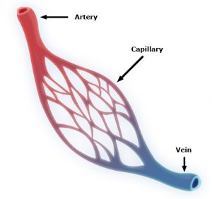
Capillaries are one of the most important forms of vasculature in our body, as they allow our blood to transfer nutrients to other parts of our body. But for how much effect capillary functionality can have on our health, their small size makes them extremely difficult to engineer into models for a variety of diseases. Now, researchers at the University of Washington led by Ying Zheng, Ph. D., engineered a three-dimensional microvessel model with living cells to study the mechanisms of microcirculatory obstruction involved with malaria.

Rather than just achieving a physical model of capillaries, these researchers created a model that allowed them to study typical flow and motion through capillaries, before comparing it to deficiencies in this behavior involved with diseases like malaria. The shape of the engineered model is similar to that of an hourglass, allowing the researchers to study instances where red blood cell transit may encounter bottlenecks between the capillaries and other vessels. Using multiphoton technology, Zheng and her team created 100mm capillary models with etched-in channels and a collagen base, to closely model the typical size and rigidity of the vessels. Tested with malaria-infected blood cells, the model showed similar circulatory obstructive behavior to that which occurs in patients, giving hope that this model can be transferred to other diseases involving such obstruction, like sickle cell anemia, diabetes, and cardiovascular conditions.

Understanding a Cell Membrane Protein Could Be the Key to New Cancer Treatments

Almost every cell in the body has integrins, a form of proteins, on its membrane, allowing cells to sense biological information from beyond their membranes while also using this feedback information to initiate signals within cells themselves. Bioengineers at the Imperial College of London recently looked at the way another membrane protein, called syndecan-4, interacts with integrins as a potential form of future cancer treatment. Referred to as “cellular hands” by lead researcher of the study Armando del Rio Hernandez, Ph.D., syndecan-4 sometimes controls the  development of diseases or conditions like cancer and fibrosis. Hernandez and his team specifically studied the ties of syndecan-4 to yes-associated protein (YAP) and enzyme called P13K, both of which are affiliated with qualities of cancer progression like halted apoptosis or cell stiffening. Knowing this, Hernandez and his team hope to continue research into understanding the mechanisms of syndecan-4 throughout the cell, in search of new mechanisms and targets to focus on with future developments of cancer treatments.

A New Medical Device Could Improve Nerve Functionality After Severe Damage

Serious nerve damage remains difficult to repair surgically, often involving the stretching of nerves for localized damage, or the transfer of healthy nerve cells from another part of the body to fill larger gaps in nerve damage. But these imperfect solutions limit the return of full nerve function and movement to the damaged part of the body, and in more serious cases with large areas of nerve damage, can also risk damage in other areas of the body that healthy nerves are borrowed from for treatment. A new study from the University of Pittsburgh published in Science Translational Medicine led by Kacey Marra, Ph. D., has successfully repaired nerve damage in mice and monkeys using a biodegradable tube that releases growth factors called glial-cell-derived neurotrophic factors over time.

Marra and her team showed that this new device restored nerve function up to 80% in nonhuman primates, where current methods of nerve replacement often only achieve 50-60% functionality restoration. The device might have an easier time getting FDA-approval, since it doesn’t involve the use of stem cells in its repair mechanisms. Hoping to start human clinical trials in 2021, Marra and her team hope that the device will help both injured veterans and typical patients with nerve damage, and see potential future applications in facial nerve damage as well.

A New Computational Model Could Improve Treatments for Cancer, HIV, and Autoimmune Diseases

With cancer, HIV, and other autoimmune diseases, the best treatment options for patients are often determined with trial-and-error methods, leading to prolonged instances of ineffective approaches and sometimes unnecessary side effects. A group of researchers led by Wesley Errington, Ph.D., at the University of Minnesota decided to take a computational approach this problem, in an effort to more quickly and efficiently determine the most appropriate treatment for a given patient. Based on parameters controlling interactions between molecules with multiple binding sites, the team’s new model looks primarily at binding strength, linkage rigidity, and size of linkage arrays. Because diseases can often involve issues in molecular binding, the model aimed to model the 78 unique binding configurations for cases of when interacting molecules only have three binding sites, which are often difficult to observe experimentally. This new approach will allow for faster and easier determination of treatments for patients with diseases involving these molecular interactions.

Improved Drug Screening for Glioblastoma Patients

A new microfluidic brain chip from researchers at the University of Houston could help improve treatment evaluations for brain tumors. Glioblastoma patients, who have a five-year survival rate of a little over 5%, are some of the most common patients suffering from malignant brain tumors. This new chip, developed by the lab of Yasemin Akay, Ph.D., can quickly determine cancer drug effectiveness by analyzing a piece of cultured tumor biopsy from a patient by incorporating different chemotherapy treatments through the microfluidic vessels. Overall, Akay and her team found that this new chip holds hope as a future efficient and inexpensive form of drug screening for glioblastoma patients.

People and Places

The brain constructs maps to guide people, not just of physical spaces but also to connect stimuli around them, like conversations and other people. It’s long been known that the brain area responsible for this spatial navigation—the medial temporal lobe—is also involved in recalling memories.

Michael Kahana (left) is principal investigator in the Defense Advanced Research Projects Agency’s RAM program and a professor in the Department of Psychology. Ethan Solomon is an M.D./Ph.D. student in the Department of Bioengineering of the School of Engineering and Applied Science and in the Perelman School of Medicine.

Now, neuroscientists at the University of Pennsylvania have discovered that the signals the brain produces during spatial navigation and episodic memory recall look similar. Low-frequency brain waves called the theta rhythm appear as people jump from one memory to the next, as many prior studies looking only at human navigation have shown. The new findings, which suggest that the brain structures responsible for helping people navigate the world may also “navigate” a mental map of prior experiences, appear in the Proceedings of the National Academy of Sciences.

Read the rest of this story featuring Penn Bioengineering’s Graduate Group member Michael Kahana and M.D./Ph.D. student Ethan Solomon on Penn Today.

The Florida Institute of Technology recently announced plans to start construction in spring 2020 on a new Health Sciences Research Center, set to further establish biomedical engineering and pre-medical coursework and research at the institute. With plans to open the new center in 2022, Florida Tech anticipates increased enrollment in the two programs, and hopes that the center will offer more opportunities in a growing professional field.

Anson Ong, Ph.D., the Associate Dean of Administration and Graduate Programs at the University of Texas at San Antonio, was recently elected to the International College of Fellows of Biomaterials Science and Engineering. With a focus on research in biomaterial implants for orthopaedic applications, Ong’s election to the college honors his advancement and contribution to the field of biomaterials research.

Danielle Bassett Among Science’s ‘Favorite Photos of 2019’

MATTHEW BENDER/JAMES BARTOLOZZI

Among shots of a towering thunderstorm reaching into the stratosphere, the moon Daphnis peeking through Saturn’s rings, and an extremely close-up of a highly-endangered pangolin in Mozambique, one of Science’s favorite photos of 2019 was taken in Penn Engineering’s Raisler Lounge.

There, Danielle Bassett, J. Peter Skirkanich Professor in the Department of Bioengineering, poses underneath a giant visualization of the brain’s structural connections, projected on the wall behind her. Bassett’s research combines elements of physics, mathematics, engineering and neuroscience to provide a new look at how brain function arises from these networks of neurons.

Kelly Servick of Science profiled Bassett last year, revealing how a child whose parents discouraged her from attending college went on to become a pioneer in a highly interdisciplinary way of understanding the brain.

Read Bassett’s profile in Science here, and see the rest of the journal’s favorite photos of the year here.

Originally posted on the Penn Engineering blog. Media contact Evan Lerner.