The introduction of morphine in the 19th century to alleviate pain revolutionized medicine in a way few innovations do, but it brought with it a grave unintended consequence: addiction. In today’s society, opioid addiction is creating the biggest health crisis of the last half century. Affecting nearly 1 in 100 people, opioid addiction occurs more than type 1 diabetes, multiple sclerosis, or a number of other diseases. The addiction crisis also appears in global affairs and impacts our national security: heroin production in Afghanistan over the last 40 years has been critical to funding military actions by insurgent groups against both the US and, in the past, the Soviet Union.
However, bioengineers at Stanford have begun to tackle the issue of production and might have begun to tackle the issue of addiction. In the lab of Christine Smolke, Ph.D., Professor of Bioengineering at Stanford, they have been genetically engineering yeast to produce opioids. They described the process in a 2015 article from Science. Now, in a recent interview in Fast Company, Dr. Smolke discusses the possibility of using the yeast producing method she pioneered to produce opioids without addiction potential. These alternative drugs are very expensive to produce, and Dr. Smolke’s process could provide safer, less addictive compounds to people in need.
Breaking the Barrier
Among the challenges faced by bioengineers working on therapies for brain disease is the blood-brain barrier (BBB), a tightly regulated boundary between the circulatory system and the brain that prevents all but the tiniest molecules from getting into the brain. The poor permeability of the BBB to many molecules means that one needs to use higher drug dosages to reach the brain, which is one of the reasons why most psychiatric medications have a broad array of side effects.
One way of circumventing this issue is to deliver the drugs directly to the brain, rather than using oral or intravenous delivery methods that need to cross the BB. Here, the challenge is one of size — unless a needle used to administer such a drug is very small, it will invariably damage brain tissue, which can have devastating consequences. Answering this call has been Robert Langer, Ph.D., David H. Koch Institute Professor at MIT, whose lab has successfully microfabricated delivery cannulas as small as 30 microns, about one-third the diameter of human hair. As they report in Science Translational Medicine, the new cannula can target brain areas as small as 1 cubic millimeter.
Dr. Langer and his colleagues used the new cannula to create an implantable device, called the miniaturized neural drug delivery system (MiNDS), that they subsequently tested in rats and rhesus monkeys. They found that the device could modulate neuronal activity in both animals. In addition, MiNDS could also record and transmit information from the treatment site to enable feedback control. Going forward, the study authors envision the use of non-metallic materials to fashion cannulas and hydrogel coatings to facilitate MR imaging and increase biocompatibility.
Unlocking the Mystery of IPF
Idiopathic pulmonary fibrosis (IPF) is a lung disease that causes permanent scarring of the lung tissue. The disease affects around five million people worldwide, mainly people 50 or older, and the five-year mortality rate is very high. Although risk factors, such as cigarette smoking, have been identified, as the word “idiopathic” implies, the cause is unknown, making it difficult to create effective therapies other than ones that merely slow the progression of the disease.
However, thanks to a new discovery, we might be closer to effective treatments. In an article in the Journal of Clinical Investigation Insight, a team of scientists from Yale University report that the tissue lesions that constitute IPF are made up of roughly one-fifth pericytes — a type of contractile cell that plays an important role in the proper function of capillaries, including those in the lungs.
The study authors, led by Anjelica Gonzalez, Ph.D., Donna L. Dubinsky Associate Professor of Biomedical Engineering at Yale, found that IPF caused pericytes to take on the properties of myofibroblasts, a cell type that is important to the wound-healing process. They found further that treatment of these myofibroblast-like pericytes with nintedanib, a drug approved for IPF treatment, reversed this effect. Armed with this knowledge, we come a step closer to designing and producing more effective therapies for IPF, as well as for diseases with similar effects.
People and Places
Washington University in St. Louis has announced it will launch a Ph.D. program in imaging science, to enroll its first cohort this fall. The program will be headed by Mark Anastasio, Ph.D., Professor of Biomedical Engineering and a 1993 recipient of an MSE from Penn. WashU’s program is only the second such program in the country, following the program at the Rochester Institute of Technology.
Closer to home, Johns Hopkins is the recipient of a $50 million donation from the United Arab Emirates. The money will be used to create the Sheikh Khalifa Stroke Institute, which will unite faculty members from biomedical engineering, neurology, and rehabilitation medicine to advance research into stroke.
George H.W. Bush refused to eat it, but maybe he should start. It turns out that broccoli, combined with bioengineered yogurt, could provide effect cancer prevention. We’ve known for some time that compounds in certain fresh vegetables can increase chemoprevention, but the levels are usually too low to be effective, or they can’t be assimilated optimally by the body. However, scientists in Singapore found that engineered bacteria, when ingested by mice with colorectal cancer, had anticancer effects. The bacteria caused the secretion of an enzyme by the cancer cells that transformed glucosinolates — compounds found in vegetables — into molecules with anticancer efficacy. The scientists report their findings in Nature Biomedical Engineering.
The authors programmed an E. coli cell line to bind to heparan sulfate proteoglycan, a cell surface protein that occurs in colorectal cancer cells. Once the engineered bacteria bound to the cancer cells, the bacteria secreted myrosinase, an enzyme that commonly occurs in many plants to defend them against aphids. In the cell model employed by the authors, myrosinase caused the conversion of glucosinolates into sulforaphane, which in turn could inhibit cancer cell growth.
The scientists then applied their system in a mouse model of colorectal cancer, feeding the mice yogurt infused with the engineered bacteria. They found that the mice fed broccoli plus the yogurt developed fewer and smaller tumors than mice fed broccoli alone. Additional testing is necessary, of course, but the study authors believe that their engineered bacteria could be used both as a preventive tool in high-risk patients and as a supplement for cancer patients after surgery to remove their tumors.
The Gates of CRISPR
About two years ago, software giant Microsoft unveiled Azimuth, a gene-editing tool for CRISPR/Casa9 that it had developed in collaboration with scientists at the Broad Institute. Now, in response to concerns that CRIPR may edit more of the genome than a bioengineer wants, the team has introduced a tool called Elevation. A new article in Nature Biomedical Engineering discusses the new tool.
In the article, the team, co-led by John C. Doench, Ph.D., Institute Scientist at the Broad Institute, describes how it developed Azimuth and Elevation, both of which are machine learning models, and deployed the tools to compare their ability to predict off-target editing with the ability of other approaches. The Elevation model outperformed the other methods. In addition, the team has implemented a cloud-based service for end-to-end RNA design, which should alleviate some of the time and resource handicaps that scientists face in using CRISPR.
Reducing Infant Mortality With an App
Among the challenges still faced in the developing world with regard to health care is high infant mortality, with the most common cause being perinatal asphyxia, or lack of oxygen reaching the infant during delivery. In response, Nigerian graduate student Charles C. Onu, a Master’s student in the computer science lab of Doina Precup, Ph.D., at McGill University in Montreal, founded a company called Ubenwa, an Igbo word that means “baby’s cry.”
With Ubenwa and scientists from McGill, Onu developed a smartphone app and a wearable that apply machine learning to instantly diagnose birth asphyxia based on the sound of a baby’s cry. In initial testing, the device performed well, with sensitivity of more than 86% and specificity of more than 89%. You can read more about the development and testing of Ubenwa at Arxiv.
People and Places
Several universities have announced that they are introducing new centers for research in bioengineering. Purdue University secured $27 million in funding from Semiconductor Research Corp. for its Center for Brain-inspired Computing Enabling Autonomous Intelligence, or C-BRIC, which opened last month. The center will develop, among other technologies, robotics that can operate without human intervention.
In Atlanta, Emory University received a $400 million pledge from the Robert W. Woodruff Foundation for two new centers — the Winship Cancer Institute Tower and a new Health Sciences Research Building. The latter will host five research teams, including one specializing in biomedical engineering. Further north in Richmond, Virginia Commonwealth University announced that it will begin construction on a new $92 million Engineering Research Building in the fall. The uppermost floors of the new building will include labs for the college’s Department of Biomedical Engineering.
Finally, North Carolina’s Elon College will introduce a bachelor’s degree program in engineering in the fall. The program will offer concentrations in biomedical engineering and computer engineering. Sirena Hargrove-Leak, Ph.D., is director of the program.
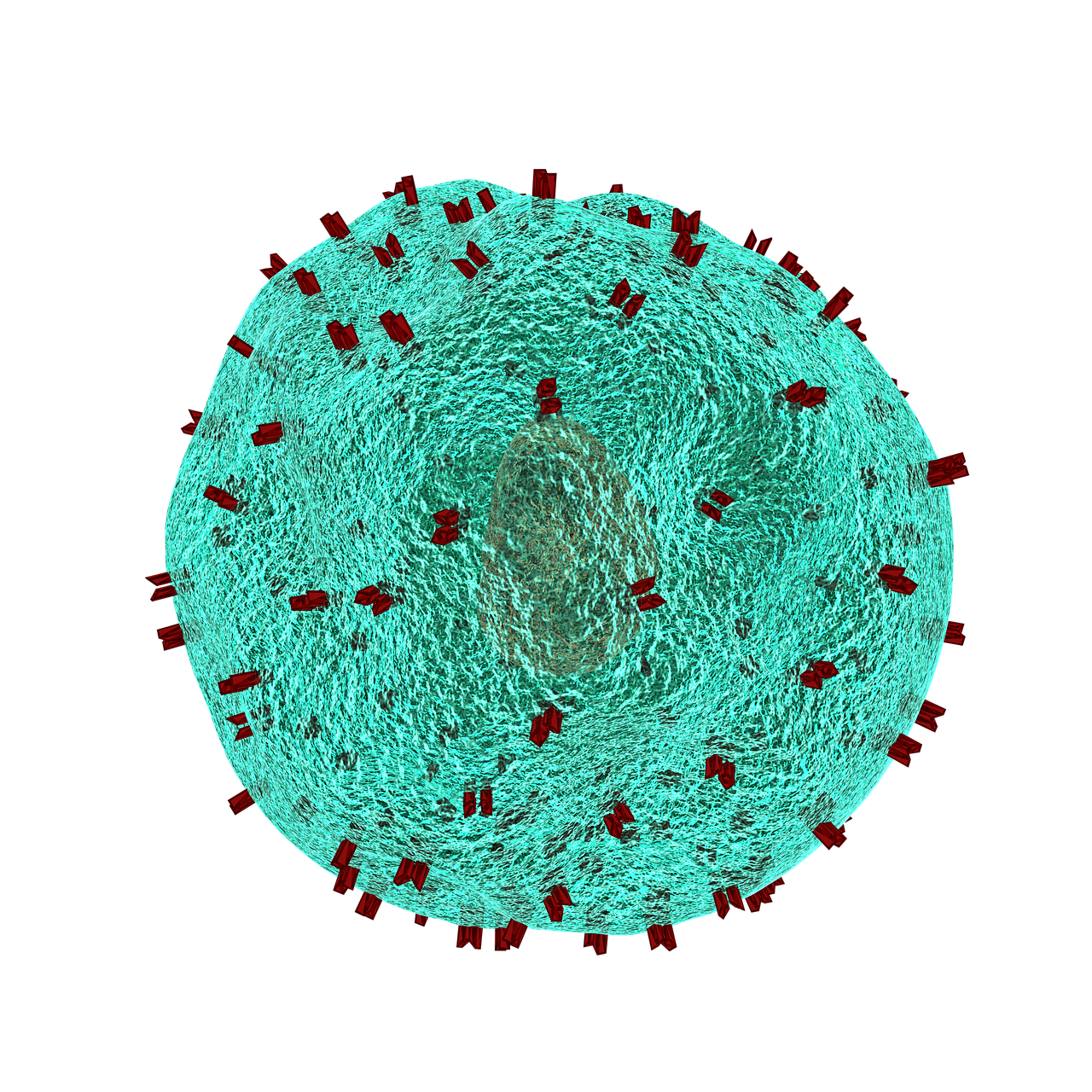
T lymphocytes in the immune system play a vital role in the body to recognize invasion by an outside element. When foreign bacteria enter the body, receptors on the T cell surface detect antigens associated with the bacteria and send a signal deploying phagocytes to attack and defeat the invading bacteria. While evolution and vaccination make the immune system very efficient, the inability of T cell receptors (TCRs) to detect cancer makes normal T cells relatively ineffective in resisting cancer. One of the ways to overcome this limitation of the immune system is to better understand how the TCRs respond to antigens. Analyses of the proteins involved in TSR responses are useful but limited by several factors, including the dizzying amount of data involved. Data analysis techniques have been helpful but have offered little information about the general reactions of TSRs, rather than how they react to specific antigens.
A possible solution to this obstacle is ImmunoMap, developed by scientists collaborating between Johns Hopkins University and Memorial Sloan Kettering Cancer Center. In a study recently published in Cancer Immunology Research, the authors, led by Jonathan P. Schneck, M.D., Ph.D., a professor of pathology at Johns Hopkins associated with the university’s Institute for Cell Engineering and Institute for Nanobiotechnology, describe their creation and deployment of ImmunoMap, a group of artificial intelligence algorithms that use machine learning to process large amounts of sequencing data and compare data from different antigens with each other.
The authors trained ImmunoMap initially using data from a mouse model of melanoma, in which the algorithm demonstrated significantly better performance than traditional methods. Subsequently, ImmunoMap was applied to patient response data from a melanoma clinical trial of the chemotherapy agent nivolumab. The algorithm discovered a new group of patients that would respond positively to nivolumab treatment — a finding missed by popular past methods. More testing of ImmunoMap is necessary, but the technology could make significant contributions to the monitoring of cancer patients receiving chemotherapy. In addition, it could to help to better predict response in patients before they begin specific chemotherapy regimes.
Wearables Improving Health
Among the most troubling health disparities related to global wealth inequality is the higher rate of mortality among children suffering from cancer. Fever is a common symptom of children undergoing cancer treatment, and this symptom may indicate more serious health issues that require the attention of a doctor. However, continuously monitoring skin temperature in children from low resource settings is difficult. Seeking to help remedy this problem, undergraduate engineering students at Harvard collaborated with the Dana-Farber/Boston Children’s Cancer & Blood Disorders Center’s Global Health Initiative to develop tools for earlier fever detection and treatment.
In a course taught by David Mooney, Ph.D., Robert P. Pinkas Family Professor of Bioengineering at Harvard, students developed a wearable device that sounds an alarm when the wearer needs medical help. The app can send patients’ recorded messages to their doctors, who can then review the temperature data and messages from the children before responding. Fashioned like a wristwatch, the extra-durable and waterproof device will next move into pilot testing among a larger patient population.
Meanwhile, at Northwestern, John A. Rogers, Ph.D., the Louis Simpson and Kimberly Querrey Professor of Materials Science and Engineering, Biomedical Engineering, and Neurological Surgery in Northwestern’s McCormick School of Engineering, has partnered with cosmetics giant L’Oréal to create the world’s smallest wearable. The device, which is smaller than an adult fingernail, measures UV sun exposure for the wearer and can tell when they should go back inside instead of risking overexposure. Unsurprisingly, it’s solar powered, and it was demonstrated a couple of weeks ago at a consumer electronics show in Las Vegas.
Growing Hydrogels Like Human Tissue
Scientists at Carnegie Mellon University and Nanyang Technological University in Singapore have collaborated in a process to create polyacrylamide gels that grow in a manner resembling natural tissue. K. Jimmy Hsia, Ph.D., Professor of Biomedical Engineering at Carnegie Mellon, is co-lead author of a new study in PNAS describing this new growth mode.
In the study, Dr. Hsia and his coauthors report that, in the same way that growth factors secreted by a living organism affect the generation of new tissue, oxygen can be modulated to control how hydrogels grow. Moreover, while growth is under way, the process could be continued to efficiently manage the mass transfer of nutrients from cell to cell. Finally, the authors detail the mechanical processes that help to shape the final product. With this new process, the ability to design and create materials for applications such as robotics and tissue engineering comes a step closer to resembling living tissue as closely as possible.
People and Places
Engineers at Virginia Tech have been awarded a $1.1 million grant from the Virginia Research Investment Committee to develop a device that uses low-energy electric fields for the treatment of brain tumors. Rafael Davalos, Ph.D., L. Preston Wade Professor of Biomedical Engineering and Mechanics, is the chief investigator on the grant.
The Department of Biomedical Engineering has announced the appointment of Kam W. Leong, Ph.D., as the Samuel Y. Sheng Professor of Biomedical Engineering. Dr. Leong earned his Ph.D. in chemical engineering from the University of Pennsylvania and taught at Duke and Johns Hopkins before arriving at Columbia in 2006. He was previously the James B. Duke Professor of Biomedical Engineering at Duke. Congratulations to Dr. Leong!
The sheer complexity of the human brain means that, despite the tremendous advances made in neuroscience, there is still much we don’t know about what goes on inside our heads and how it goes awry in mental disorders. Even with the most advanced techniques, much of what we’ve learned about the brain is descriptive — telling that something is different between health and unhealthy function — but not why that something is different or how we could change it.
Rat microglia and neurons stained for different proteins
Among the approaches that have provided important insights into these questions is network science, which seeks to understand the brain as a complex system of multiple interacting components. Now, in a review published recently in Neuron, Danielle Bassett, Ph.D., Eduardo D. Glandt Faculty Fellow and Associate Professor of Bioengineering, and Richard Betzel, Ph.D., a postdoc in Dr. Bassett’s lab, have collaborated with scientists from the University of Heidelberg in Germany. The review covers a broad range of discoveries and innovations, moving from earlier, two-dimensional approaches to understanding the brain, such as graph theory, to newer approaches including multilayer networks, generative network models, and network control theory.
“Stating what is different in brain networks of individuals with disorders of mental health is not the same as identifying why” says Bassett. “Here we propose that emerging tools from network science can be used to identify true mechanisms of mental health disorders, and bridge molecular and genetic mechanisms through brain physiology, thus informing interventions in the form of pharmacological manipulations and brain stimulation.”
The developing human brain contains a cacophony of electrical and chemical signals from which emerge the powerful adult capacities for decision-making, strategizing, and critical thinking. These signals support the trafficking of information across brain regions, in patterns that share many similarities with traffic patterns in railway and airline transportation systems. Yet while air traffic is guided by airport control towers, and railway routes are guided by signal control rooms, it remains a mystery how the information traffic in the brain is guided and how that guidance changes as kids grow.
In part, this mystery has been complicated by the fact that, unlike transportation systems, the brain is not hooked up to external controllers. Control must happen internally. The problem becomes even more complicated when we think about the sheer number of routes that must exist in the brain to support the full range of human cognitive capabilities. Thus, the controllers would need to produce a large set of control signals or use different control strategies. Where internal controllers might be, how they produce large variations in routing, and whether those controllers and their function change with age are important open questions.
A recent paper published in Nature Communications – a product of collaboration among the Departments of Bioengineering and Electrical & Systems Engineering at the University of Pennsylvania and the Department of Psychiatry of Penn’s Perelman School of Medicine – offers some interesting answers. In their article, Danielle Bassett, Ph.D., Eduardo D. Glandt Faculty Fellow and Associate Professor in the Penn BE Department, Theodore D. Satterthwaite, M.D., Assistant Professor in the Penn Psychiatry Department, postdoctoral fellow Evelyn Tang, and their colleagues suggest that control in the human brain works in a similar way to control in man-made robotic and other mechanical systems. Specifically, controllers exist inside each human brain, each region of the brain can perform multiple types of control, and this control grows as children grow.
As part of this study, the authors applied network control theory — an emerging area of systems engineering – to explain how the pattern of connections (or network) between brain areas directly informs the brain’s control functions. For example, hubs of the brain’s information trafficking system (like Grand Central Station in New York City) show quite different capacities for and sensitivities to control than non-hubs (like Newton Station, Kansas). Applying these ideas to a large set of brain imaging data from 882 youths in the Philadelphia area between the ages of 8 and 22 years old, the authors found that the brain’s predicted capacity for control increases over development. Older youths have a greater predicted capacity to push their brains into nearby mental states, as well as into distant mental states, indicating a greater potential for diversity of mental operations than in younger youths.
The investigators then asked whether the principles of network control could explain the specific manner in which connections in the brain change as youths age. They used tools from evolutionary game theory – traditionally used to study Darwinian competition and evolving populations in biology – to ‘evolve’ brain networks in silico from their 8-year old state to their 22-year-old state. The results demonstrated that the optimization of network control is a principle that explains the observed changes in brain connectivity as youths develop over childhood and adolescence. “One of the observations that I think is particularly striking about this study,” Bassett says, “is that the principles of network controllability are sufficient to explain the observed evolution in development, suggesting that we have identified a quintessential rule of developmental rewiring.”
This research informs many possible future directions in scientific research. “Showing that network control properties evolve during adolescence also suggests that abnormalities of this developmental process could be related to cognitive deficits that are present in many neuropsychiatric disorders,” says Satterthwaite. The discovery that the brain optimizes certain network control functions over time could have important implications for better understanding of neuroplasticity, skill acquisition, and developmental psychopathology.
Late last semester, Penn Bioengineering Department chair David Meaney and senior lecturer LeAnn Dourte held a second roundtable with BE undergrads Eric Helfgott, Joseph Maggiore, Kayla Prezelski, and Margaret Schroeder. They picked up on topics from the last roundtable, extending the topics to balancing an engineering workload and other commitments.
One of the key processes in embryonic development and growth through childhood and adolescence is that of how tissue folds into the specific shapes required for them to function in the body. For instance, mesenchymal stem cells, which form a variety of tissues including bones, muscles, and fat, are required to “know” what shapes to take on as they form organ systems and other structures. Therefore, a big concern in tissue engineering is determining how to control these processes of tissue folding.
Just in time for his arrival at Penn Bioengineering, Dr. Alex Hughes, a new assistant professor in the department, is the lead author on a new paper in Developmental Cell that explores this concern. The study was coauthored with Dr. Zev Gartner of the University of California, Berkeley, where Dr. Hughes just finished a postdoctoral fellowship. In the paper, the authors used three-dimensional cell-patterning techniques, embryonic tissue explants, and finite element modeling to determine that the folding process involves the interaction of a protein called myosin II with the extracellular matrix, itself the molecular material that provides a structural framework for developing tissues. With the knowledge gained in the initial experiments, the authors were then able to reproduce the tissue folding process in the lab.
“Bioengineers are currently thinking about building tissues,” Dr. Hughes says, “not just at the level of organoids, but at the level of organs in the body. One of my interests at Penn is to harness developmental principles that link these length scales, allowing us to design medically relevant scaffolds and machines.”
Equipped with the knowledge gleaned from this research, future studies could contribute further to the ability to generate tissues and even organ systems in laboratories. Ultimately, this knowledge could revolutionize transplant medicine, as well as variety of other fields.
Dr. Konrad Kording, a University of Pennsylvania PIK Professor in Bioengineering and Neuroscience, has been named an associate fellow by the Canadian Institute for Advanced Research (CIFAR), an advanced study institute headquartered in Toronto and partially funded by the government of Canada. Dr. Kording’s fellowship is in the institute’s Learning in Machines & Brains area, which has been one of CIFAR’s 14 interdisciplinary study fields since 2004. He joins 32 other fellows currently supported by the institute for their work in this area.
“The CIFAR program in Learning in Machines & Brains brings together many of the world’s leading deep learning scientists,” Dr. Kording says. “I look forward to collaborate with them to figure out how the brain learns.”
CIFAR was founded in 1982. Over the last 35 years, the institute has supported the work of scientists in 133 countries, including 18 Nobel Prize laureates.
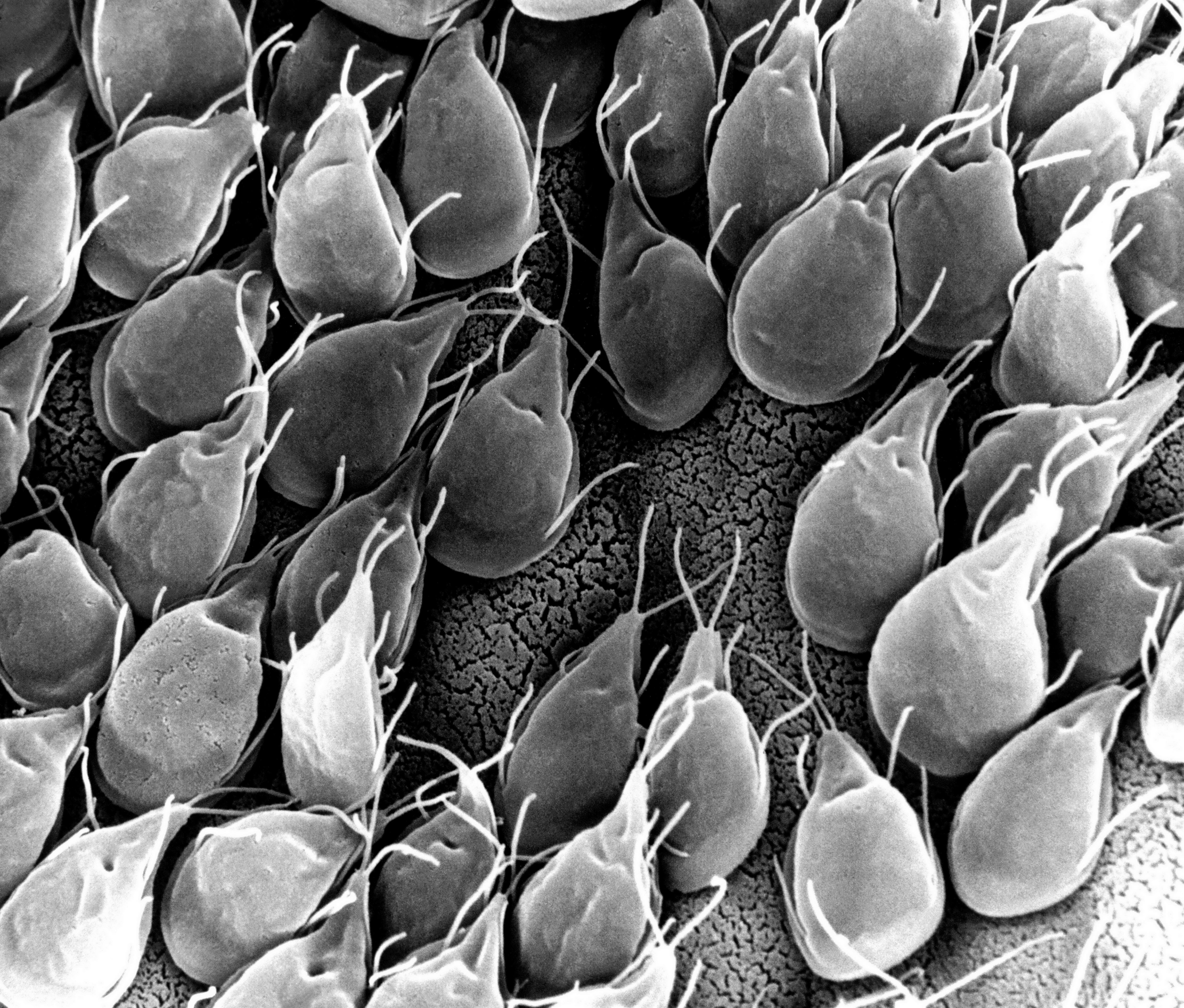
Small intestinal mucosa infested with Giardia lamblia parasites
Diseases of the small intestine, including Crohn’s disease and microbial infections, impose a huge burden on health. However, finding treatments for these diseases is challenged by the lack of optimal models for studying disease. Animal models are only so close to human disease states, and laboratory models using cell lines do not completely mimic the environment inside the gut.
However, these limitations might be overcome soon thanks to the research of scientists at Tufts University. In an article recently published in PLOS ONE, a team led by David L. Kaplan, Ph.D., of the Tufts Department of Biomedical Engineering, describes how they used donor stem cells and a compartmentalized biomimetic scaffold to model and generate small intestine cells that could differentiate into the broad variety of cell types common to that organ.
The study team tested the response of its cell model to E. coli, a common pathogen. At the genetic level, the model matched the reaction of the human small intestine when exposed to this bacterium. The success of the model could translate into its use in the near future to better understand the digestive system’s response to infection, as well as to test individualized treatments for inflammatory bowel diseases such as Crohn’s.
Saving Battle-wounded Eyes
The increase in combat survival rates has led to a higher incidence of veterans with permanent vision loss due to catastrophic damage to the eye. Globe injuries will recover of some vision, if caught in time. However, combat care for eye injuries often occurs hundreds or thousands of miles away from emergency rooms with attending ophthalmologists. With this unavoidable delay in treatment, people with globe injuries suffer blindness and often enucleation.
However, battle medics might soon have something in their arsenals to prevent such blinding injuries immediately in the combat theater. As reported recently in Science Translational Medicine, engineers at the University of Southern California (USC) and ophthalmologists from USC’s Roski Eye Institute have collaborated in creating a new material for temporary sealing of globe injuries. The study authors, led by John J. Whalen, III, Ph.D., used a gel called poly(N-isopropylacrylamide) (PNIPAM), already under investigation for treating retinal injuries. PNIPAM is a thermoresponsive sealant, meaning it is a liquid at cooler temperature but an adhesive gel at warmer temperature. These interesting properties mean PNIPAM can be applied as a liquid and then solidifies quickly on the eye. The authors manipulated PNIPAM chemically to make it more stable at body temperature. As envisioned, the gel, when used with globe injuries, could be applied by medics and then removed with cold water just before the eye is treated.
The study team has tested the gel in rabbits, where it showed statistically significant improvement in wound sealing and no negative effects on the eyes or overall health of the rabbits. The authors believe the material will be ready for human testing in 2019.
Predicting Seizures in Epilepsy
Epilepsy is a central nervous system disorder characterized by seizure activity that can range in severity from mild to debilitating. Many patients with epilepsy experience adequate control of seizures with medications; however, about a third of epileptic patients have intractable cases requiring surgery or other invasive procedures.
In what could be a breakthrough in the treatment of refractive epilepsy, scientists from Australia in collaboration with IBM Research-Australia have used big data from epilepsy patients to develop a computer model that can predict when seizures will occur. So far, the technology predicts 69% of seizures in patients. While it’s still short of a range of accuracy making it feasible for use in patients outside of experimental settings, the acquisition of ever-increasing amounts of data will render the model more accurately.
The Art of Genetic Engineering
Among the techniques used in genetic engineering is protein folding, which is one of the naturally occurring processes that DNA undergoes as it takes on three dimensions. Among the major developments in genetic engineering was the discovery of the ability to fold DNA strands artificially, in a process called DNA origami.
Now, as suggested by the name “origami,” some people have begun using the process in quasi-artistic fashion. In an article recently published in Nature, bioengineers at CalTech led by Lulu Qian, Ph.D., assistant professor of bioengineering, showed they were able to produce a variety of shapes and designs using DNA origami, including a nanoscale replica of Leonardo da Vinci’s Mona Lisa.
DNA now also has another unique artistic application — tattoos, although people’s opinions of whether tattooing constitutes art might vary. Edith Mathiowitz, Ph.D., of Brown University’s Center for Biomedical Engineering, is among the patenters of Everence, a technology that takes DNA provided by a customer and incorporates it into tattoo ink. Potential tattooees can now have the DNA of loved ones incorporated into their bodies permanently, if they should so wish.
People and Places
The University of Washington has launched its new Institute for Nano-engineered Systems, cutting the ribbon on the building on December 4. The center will house facilities dedicated to scalable nanomanufacturing and integrated photonics, among others. Meanwhile, at the University of Chicago, Rama Ranganathan, M.D., Ph.D., a professor in the Department of Biochemistry and Molecular Biology and the Institute for Molecular Engineering, will lead that college’s new Center for Physics of Evolving Systems. Congratulations!
This interview with Dr. LeAnn Dourte is a collaboration between Double Shelix and the University of Pennsylvania Department of Bioengineering! Thanks to Kayla and Sally for conducingt this interview! If there’s someone else at UPenn BioE (or elsewhere!) that you think they should feature, let them know!

 The introduction of morphine in the 19th century to alleviate pain revolutionized medicine in a way few innovations do, but it brought with it a grave unintended consequence: addiction. In today’s society, opioid addiction is creating the biggest health crisis of the last half century. Affecting nearly 1 in 100 people, opioid addiction occurs more than type 1 diabetes, multiple sclerosis, or a number of other diseases. The addiction crisis also appears in global affairs and impacts our national security: heroin production in Afghanistan over the last 40 years has been critical to funding military actions by insurgent groups against both the US and, in the past, the Soviet Union.