The annual Bioengineering Graduate Symposium provides an opportunity for current bioengineering graduate students to showcase their research to faculty and peers through poster sessions and short talks.
Date: January 8th
Location: Wu & Chen Auditorium and Levine Hall Lobby
Time: Check-in opens 12:45, event starts 1 pm.
Keynote Speaker: Dr. Sujata Bhatia,
Professor of Chemical and Bimolecular Engineering, faculty director, McNair Scholars Program,
University of Delaware
“Bioengineering to Alleviate the Global Burden of Disease”
Biochemical and biomedical engineers face an unprecedented opportunity to improve and save the lives of millions worldwide. Both high-income and low-income countries are experiencing an explosion in the incidence of chronic diseases, including coronary artery disease, stroke, diabetes, and cancer. At the same time, low-income countries continue to be plagued by infectious diseases, including HIV, tuberculosis, diarrheal disease, and pneumonia; low-income countries are often said to experience a dual burden of chronic and infectious diseases. Innovative biomedical materials will only reach the clinic if these technologies solve pressing clinical problems. This talk will describe opportunities for bioengineers to alleviate the global burden of disease. The talk will additionally highlight specific examples of unmet clinical needs. Bioengineers in industry, academia, and government can all make a difference, not only by designing novel biomedical products, but also by training the next generation of bioengineers and shaping the future direction of research and development.
Michael Mitchell, PhD, Skirkanich Assistant Professor of Innovation in the Department of Bioengineering at Penn, has been honored with a Rising Star Award in Cellular and Molecular Bioengineering from the Biomedical Engineering Society (BMES). According to the BMES website, “The BMES Cellular and Molecular Bioengineering Special Interest Group brings together researchers with diverse scientific and clinical interests with a common goal of understanding and engineering molecules, cells, their interactions and microenvironments in the pursuit of controlling biological processes and improving the practice of medicine.” Dr. Mitchell received the award and delivered a lecture at the 2019 Cellular and Molecular Bioengineering Conference in San Diego, California in January, 2019.
One of six early-stage investigators from across the nation to receive the honor, Dr. Mitchell was recognized for his work on engineering delivery technologies for cancer gene therapy and immunotherapy, which is helping to lay the foundation for a new class of therapeutic strategies against hematologic cancers such as multiple myeloma and leukemia. In 2018, Dr. Mitchell was awarded the NIH Director’s New Innovator Award for this research, and received the Burroughs Wellcome Fund Career Award at the Scientific Interface) in 2016. He joined the Penn faculty in January 2018 after completing an NIH NCI postdoctoral fellowship with Dr. Robert Langer at the Koch Institute for Integrative Cancer Research at MIT.
The BE Seminar Series continues this week. We hope to see you there!
Speaker: Alison Pouch, Ph.D.
Research Associate, Penn Image Computing & Science Lab, Department of Radiology
Gorman Cardiovascular Research Group, Department of Surgery, University of Pennsylvania
Date: November 29, 2018
Time: 12:00 pm
Location: Room 337, Towne Building
“Shaping Innovation in Heart Valve Surgery with Image-Based Modeling”
A number of challenging questions routinely arise before heart valve surgery. Should a regurgitant valve be repaired or replaced? If repaired, which techniques and maneuvers should be employed? When is the optimal time to perform surgery? This is just the beginning of a series of questions whose answers rely on an individualized assessment of heart valve morphology and function. With advanced imaging technology like 3D echocardiography already in the operating room, we have a window into patient-specific disease characteristics that cannot otherwise be appreciated immediately before surgery. Unfortunately, imaging resources are often not tapped to their full potential, which contributes to delays in surgical innovation. This presentation introduces an image-based modeling approach to creating enhanced visualizations and quantification of heart valves from real-time 3D echocardiography. The goal of this approach is to fundamentally change the way that surgeons interact with and utilize images in the operating room. Pre-operative image-based heart valve models can be used to characterize mechanisms of disease prior to cardiopulmonary bypass and to identify connections between pre-operative image features and clinical outcomes. Image-based modeling provides a means for surgeons to innovate and master new surgical techniques like bicuspid aortic valve repair and to devise new standardized approaches for surgical treatment of ischemic mitral regurgitation. Image analysis and surgical innovation are linked within and beyond the cardiovascular domain, and this work aims to optimize the potential of imaging and shape modeling for pre-surgical planning.
Scientific claims are a foundation of scientific discourse. Extracting claims from scientific articles is primarily done by researchers during literature review and discussions. The enormous growth in scientific articles makes this ever more challenging and time-consuming. Here, we develop a deep neural network architecture to solve the problem. Our model an F1 score of 0.704 on a large corpus of expertly annotated claims within abstracts. Our results suggest that we can use a small dataset of annotated resources to achieve high-accuracy claim detection. We release a tool for discourse and claim detection, and a novel dataset annotated by experts. We discuss further applications beyond Biomedical literature.
Multiple Sclerosis Lesion Segmentation with Joint Label Fusion Evaluated on OASIS and CNN
Scientific claims are a foundation of scientific discourse. Extracting claims from scientific articles is primarily done by researchers during literature review and discussions. The enormous growth in scientific articles makes this ever more challenging and time-consuming. Here, we develop a deep neural network architecture to solve the problem. Our model an F1 score of 0.704 on a large corpus of expertly annotated claims within abstracts. Our results suggest that we can use a small dataset of annotated resources to achieve high-accuracy claim detection. We release a tool for discourse and claim detection, and a novel dataset annotated by experts. We discuss further applications beyond Biomedical literature.
Michael Mitchell, Skirkanich Assistant Professor of Innovation in Penn Engineering’s Department of Bioengineering, is drawing on a variety of fields — biomaterials engineering, data science, gene therapy and machine learning — to tailor the next generation of drug delivery vehicles with this level of precision.
His work in this field has earned him a $2.4 million NIH Director’s New Innovator Award, which is part of the NIH Common Fund’s High-Risk, High-Reward Research program. The High-Risk, High-Reward Research program supports innovative research proposals that might not prove successful in the conventional peer-review process despite their potential to advance medicine.
The BE Seminar Series continues next week with three lectures delivered by our current PhD Students. We hope to see you there!
“Magnetic Susceptibility of Hemorrhagic Myocardial Infarction”
Brianna Moon, PhD Candidate
Speaker: Brianna Moon, PhD Candidate
Research Advisor: Walter Witschey, PhD
Date: Thursday, September 20, 2018
Time: 12:05pm-12:25pm
Location: Room 337 Towne Building
Abstract:
Hemorrhagic myocardial infarction (MI) has been reported in 41% and 54% of ST-elevated MI patients after primary percutaneous coronary intervention. These patients are at high risk for adverse left ventricle (LV) remodeling, impaired LV function and increased risk of fatal arrhythmias. Relaxation time MRI such as T2*-maps are sensitive to hemorrhagic infarct iron content, but are also affected by myocardial edema and fibrosis. Quantitative Susceptibility Mapping (QSM), which uses the MR signal phase to quantify tissue magnetic susceptibility, may be a more specific and sensitive marker of hemorrhagic MI. The objective of this study was to develop and validate cardiac QSM in a large animal model of myocardial infarction, investigate the association of magnetic susceptibility with iron content and infarct pathophysiology, and compare QSM to relaxation time mapping, susceptibility-weighted imaging (SWI), and late-gadolinium enhanced (LGE) MRI.
“Role of ACTG2 Mutations in Visceral Myopathy”
Sohaib Hashmi, MD/PhD Student
Speaker: Sohaib Hashmi, MD/PhD Student
Research Advisor: Robert O. Heuckeroth, MD, PhD
Date: Thursday, September 20, 2018
Time: 12:30-12:50pm
Location: Room 337 Towne Building
Abstract:
Visceral myopathy is a debilitating chronic medical condition in which smooth muscle of the bowel, bladder, and uterus is weak or dysfunctional. When the bowel muscle is weak and unable to efficiently contract, the bowel becomes distended which causes pain, bilious vomiting, growth failure, and nutritional deficiencies. The abdominal distension can become life-threatening. Patients often become dependent on intravenous nutrition and undergo multiple rounds of abdominal surgery, which only partially alleviates symptoms. Recently, rare mutations in gamma smooth muscle actin (ACTG2) have been shown to be responsible for a large subset of visceral myopathies. ACTG2 is a critical protein in the smooth muscle contractile apparatus. However, we have only limited knowledge of how ACTG2 mutations may cause human disease. To improve our understanding of the pathophysiology of ACTG2 mutations, my work has the following specific aims:
1)Determine how pathogenic ACTG2 mutations affect actin structure and function in primary human intestinal smooth muscle cells (HISMCs).
2) Examine the effects of ACTG2 mutations on differentiation of human pluripotent stem cells into smooth muscle cells (SMCs).
These studies will allow us to elucidate the mechanisms through which ACTG2 mutations impair normal visceral smooth muscle development and function. I am examining the effects of ACTG2 mutations on actin filament organization in fixed cells and actin dynamics in live cells using fluorescent probes. I will investigate changes in contractile force generation using traction force microscopy. I am also developing a novel differentiation method to convert pluripotent stem cells into cells closely resembling visceral SMCs. We will use this method with CRISPR/Cas9 gene editing to study the effects of the mutations on SMC differentiation. We are currently generating pluripotent stem cell lines containing ACTG2 mutations and performing assays to investigate actin organization and dynamics. We are using these systems to identify robust phenotypes highlighting the mechanisms through which ACTG2 mutations cause disease. We hope to leverage this information in the selection of targets for high-throughput drug screening, which may eventually lead to novel treatment strategies for visceral myopathies.
“Distinct Patterns of Longitudinal Cortical Thinking and Perfusion in Pathological Subtypes of Behavioral Variant Frontotemporal Degeneration”
Christopher Olm, PhD Student
Speaker: Christopher Olm, PhD Student
Research Advisor: Murray Grossman, MD, EdD
Date: Thursday, September 20, 2018
Time: 12:55-1:15pm
Location: Room 337 Towne Building
Abstract:
Two main sources of pathology have been identified in the behavioral variant of frontotemporal dementia (bvFTD): tau inclusions (FTLD-tau) and TDP-43 aggregates (FTLD-TDP). With therapies emerging that target these proteins, exploring distinct trajectories of degeneration can be extremely helpful for tracking progression in clinical trials and improve prognosis estimation. We hypothesized that longitudinal cortical thinning (CT) would identify areas of extant disease progression in bvFTD subgroups and longitudinal hypoperfusion would identify distinct regions of anticipated neurodegeneration. We included N=47 patients with probable or definite bvFTD and two MRI scanning sessions including T1-weighted and arterial spin labeling (ASL) scans, recruited through the Penn Frontotemporal Degeneration Center. Neuropathology, genetic mutations, or CSF protein markers (phosphorylated tau (p-tau)/Ab1-42<.09 for likely FTLD; p-tau<8.75 for FTLD-TDP) were used to identify bvFTD with likely FTLD-tau (n=28, mean age=63.1 years, mean disease duration=3.89 years) or likely FTLD-TDP (n=19, mean age=61.9, mean disease duration=3.06). Voxel-wise cortical thickness and cerebral blood flow estimates were generated for each T1 and ASL scan, respectively, using longitudinal pipelines in ANTs. We created annual change images by subtracting follow-up images from baseline and dividing by inter-scan interval. In whole brain voxel-wise comparisons, FTLD-tau showed significantly greater right orbitofrontal CT and longitudinal hypoperfusion in right middle temporal and angular cortex relative to FTLD-TDP. FTLD-TDP displayed greater progressive CT in left superior and middle frontal cortex, precentral gyrus, and right temporal cortex, and longitudinal hypoperfusion in medial prefrontal cortex relative to FTLD-tau. In conclusion, FTLD-tau and FTLD-TDP show distinct patterns of longitudinal CT and hypoperfusion. Structural and functional MRI contribute independent information potentially useful for characterizing disease progression in vivo for clinical trials.
Please join us for the first of our seminar lectures this year!
Rosalind Picard, ScD, FIEEE
Director of Affective Computing Research
Faculty Chair, MIT Mind+Hand+Heart
MIT Media Lab
“What Can We Discover About Emotions and the Brain from Noninvasive Measures?”
Date: Thursday, September 13, 2018
Time: 12:00PM-1:00 PM
Location: 337 Towne Building
Abstract:
Years ago, our team at MIT created wearable as well as non-contact imaging technology and machine learning algorithms to detect changes in human emotion. As we shrunk the sensors and made them able to comfortably collect data 24/7, we started to discover several surprising findings, such as that autonomic activity measured through a sweat response was more specific than 100 years of studies had assumed. While we originally thought this signal of “arousal” or “stress” was quite generally related to overall activation, we learned it could peak even when a patient’s EEG showed a lack of cortical brain activity. This talk will highlight some of the most surprising findings along the journey of measuring emotion “in the wild”with implications for anxiety, depression, sleep-memory consolidation, epilepsy, autism, pain studies, and more. What is the grand challenge we aim to solve next?
Bio:
Rosalind Picard, ScD, FIEEE, is founder and director of the Affective Computing Research Group at the MIT Media Laboratory, co-founder and Chief Scientist of Empatica, improving lives with clinical quality wearable sensors and analytics, and co-founder of Affectiva, providing tools for Emotion AI. Picard is the author of over two hundred fifty peer-reviewed scientific articles and of the book, Affective Computing, which helped launch that field. Picard’s lab at MIT develops technologies to better measure, understand, forecast, and regulate emotion, including personalized machine-learning analytics that work with wearables and your smartphone.
A new approach uses “mechanoceuticals” to treat pain.
Drugs are commonly injected directly into an injury site to speed healing. For chronic pain, clinicians can inject drugs to reduce inflammation in painful joints, or can inject nerve blockers to block the nerve signals that cause pain. In a recent study, a group from UCLA developed a technique to deform a material surrounding nerve fibers to trigger a response in the fibers that would relieve pain. The combination of mechanics and treatment – i.e., ‘mechanoceuticals’ – is a clever way to trick fibers and reverse painful symptoms. Done without any injections and simply controlling magnetic fields outside the body, this approach can be reused as necessary.
The design of this mechanoceutical was completed by Dino Di Carlo, PhD, Professor of Bioengineering, and his team at UCLA’s Sameuli School of Engineering. By encasing tiny, magnetic nanoparticles within a biocompatible hydrogel, the group used magnetic force to stimulate nerve fibers and cause a corresponding decrease in pain signals. This promising development opens up a new approach to pain management, one which can be created with different biomaterials to suit different conditions, and delivered “on demand” without worrying about injections or, for that matter, any prescription drugs.
Understanding the Adolescent Brain
It’s no surprise that adults and adolescents often struggle to understand one another, but the work of neurologists and other researchers provides a possible physical reason for why that might be. Magnetic resonance elastrography (MRE) is a tool used in biomedical imaging to estimate the mechanical properties, or stiffness, of tissue throughout the body. Unexpectedly, a recent study suggests that brain stiffness correlates with cognitive ability, suggesting MRE may provide insight into patients’ behavior, psychology, and psychiatric state.
A new paper in Developmental Cognitive Neuroscience published the results of a study using MRE to track the relative “stiffness” vs. “softness” of adult and adolescent brains. The University of Delaware team, led by Biomedical Engineering Assistant Professor Curtis Johnson, PhD, and his doctoral student Grace McIlvain, sampled 40 living subjects (aged 12-14) and compared the properties to healthy adult brains.
The study found that children and adolescent brains are softer than those of adults, correlating to the overall malleability of childhood development. The team hopes to continue their studies with younger and older children, looking to demonstrate exactly when and how the change from softness to stiffness takes place, and how these properties correspond to individual qualities such as risk-taking or the onset of puberty. Eventually, establishing a larger database of measurements in the pediatric brain will help further studies into neurological and cognitive disorders in children, helping to understand conditions such as multiple sclerosis, autism, and cerebral palsy.
Can Nanoparticles Replace Stents?
Researchers and clinicians have made amazing advances in heart surgery. Stents, in particular, have become quite sophisticated: they are used to both prop open clogged arteries as well as deliver blood-thinning medication slowly over days to weeks in the area of the stent. However, the risk of blood clotting increases with stents and the blood vessels can constrict over time after the stent is placed in the vessel.
A recent NIH grant will support the design of a stent-free solution to unclog blood vessels. Led by Shaoqin Gong, PhD, Vilas Distinguished Professor of Biomedical Engineering at UW-Madison, the team used nanoparticles (or nanoclusters) to directly target the affected blood vessels and prevent regrowth of the cells post-surgery, eliminating the need for a stent to keep the pathways open. These nanoclusters are injected through an intravenous line, further reducing the risks introduced by the presence of the stent. As heart disease affects millions of people worldwide, this new material has far-reaching consequences. Their study is published in the September edition of Biomaterials.
NIST Grant Supports
The National Institute of Standards and Technology (NIST) awarded a $30 million grant to Johns Hopkins University, Binghamton University, and Morgan State University as part of their Professional Research Experience Program (PREP). Over five years, this award will support the collaboration of academics from all levels (faculty, postdoc, graduate, and undergraduate) across the three universities, enabling them to conduct research and attend NIST conferences.
The principal investigator for Binghamton U. is Professor and Chair of the Biomedical Engineering Department, Kaiming Ye, PhD. Dr. Ye is also the Director of the Center of Biomanufacturing for Regenerative Medicine (CBRM), which will participate in this collaborative new enterprise. Dr. Ye hopes that this grant will create opportunities for academics and researchers to network with each other as well as to more precisely define the standards for the fields of regenerative medicine and biomaterial manufacturing.
The gift honors the late A. James Clark, former CEO of Clark Enterprises and Clark Construction Group LLC, one of the country’s largest privately-held general building contractors. It is designed to prepare future engineering and business leaders, with an emphasis on low income families and first-generation college students. Clark never forgot that his business successes began with an engineering scholarship. This has guided the Clark family’s longstanding investments in engineering education and reflects its commitment to ensure college remains accessible and affordable to high-potential students with financial need.
We are proud to say that three incoming Clark Scholars from the Freshman Class of 2022 will be part of the Bioengineering Department here at Penn.
And finally, our congratulations to the new Dean of the School of Engineering at the University of Mississippi: David A. Puleo, PhD. Dr. Puleo earned his bachelor’s degree and doctorate in Biomedical Engineering from Rensselaer Polytechnic Institute. Most recently he served as Professor of Biomedical Engineering and Associate Dean for Research and Graduate Studies at the University of Kentucky’s College of Engineering. Building on his research in regenerative biomaterials, he also founded Regenera Materials, LLC in 2014. Over the course of his career so far, Dr. Puleo received multiple teaching awards and oversaw much departmental growth within his previous institution, and looks poised to do the same for “Ole Miss.”
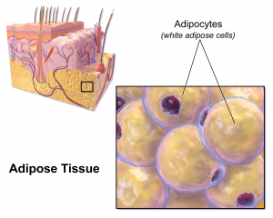
White fat stories calories and provides the body with insulation.
There are two types of fat in the human body: brown and white. Brown fat, the “good” fat, is rich in mitochondria, which gives it its brown appearance. Whereas white fat stores calories and acts as an insulator, mitochondria-rich brown fat burns energy to produce heat throughout the body and maintains body temperature. White fat, conversely, uses its stored energy to insulate the body and keep its temperature level. While all fat serves a purpose in the body, an excess of white fat cells causes obesity, a condition affecting one in three adults in the U.S. and the root cause of many potential health problems. Finding ways to convert white fat to brown opens a possibility of treating this problem naturally.
A new study in Scientific Reports proposes a clever way to convert fat types. Professor of Biomedical Engineering Samuel Sia, PhD, of the Columbia University School of Engineering and Applied Science, led a team which developed a method of converting white fat into brown using a tissue-grafting technique. After extracting and converting the fat, it can then be transplanted back into the patient. White fat is hard-wired to convert to brown under certain conditions, such as exposure to cold temperatures, so the trick for Dr. Sia’s team was finding a way to make the conversion last for long periods. The studies conducted with mice suggested that using these methods, newly-converted fat stayed brown for a period of two months.
Dr. Sia’s team will proceed to conduct further tests, especially on the subjects’ metabolism and overall weight after undergoing the procedure, and they hope that eventual clinical trials will result in new methods to treat or even prevent obesity in humans.
Cremins Lab Student Appointed Blavatnik Fellow
Linda Zhou is currently pursuing her MD/PhD in Genomics and Computational Biology under the supervision of Dr. Jennifer Phillips-Cremins.
The Perelman School of Medicine named Linda Zhou, a student in BE’s Cremins Laboratory, a Blavatnik Fellow for the 2018-2019 academic year. The selection process for this award is highly competitive, and Linda’s selection speaks to the excellent quality of her scholarship and academic performance. The fellows will be honored in a special ceremony at the Museum of Natural History in New York City.
Linda received her B.S. in Biophysics and Biochemistry from Yale University and is currently pursuing her M.D./Ph.D. in the Genomics and Computational Biology Program at Penn. “I am honored to be named a Blavatnik Fellow and am extremely excited to continue my graduate studies investigating neurological disorders and the 3D genome,” she said. “This support will be integral to achieving my long term goal of driving scientific discovery that will help treat human disease.”
Linda’s research is overseen by Penn Bioengineering Assistant Professor Jennifer Phillips-Cremins, PhD. “Linda is an outstanding graduate student,” said Dr. Cremins. “It is a true delight to work with her. She is hard working, intelligent, kind, and has extraordinary leadership ability. Her unrelenting search for ground-state truth makes her a shining star.”
The Blavatnik Family Fellowship in Biomedical Research is a new award announced by the Perelman School of Medicine in May of this year. This generous gift from the Blavatnik Family Foundation awards $2 million to six recipients in the Biomedical Graduate Studies Program at Penn for each of the next four years.
Growing Lungs in a Lab
As the demand for lung transplants continues to rise, so does the need for safe and effective transplanted lungs. Bioengineered lungs grown or created in labs are one way of meeting this demand. The problem – as is ever the case with transplants – is the high rate of rejection. The results of success are always better when cells from the patient herself (or autologous cells) are used in the transplanted organ.
Recently Joan Nichols, PhD, Professor of Internal Medicine, and Microbiology and Immunology, at the University of Texas Medical Branch at Galveston, successfully bioengineered the first human lung. Her latest study published in Science Translational Medicine describes the next milestone for Dr. Nichols’ lab: successfully transplanting a bioengineered lung into a pig.
These advances are possible due to Dr. Nichols’ work with autologous cells, continuing the trend of “on demand” medicine (i.e. medicine tailor for a specific patient) which we track on this blog. Dr. Nichols’ particular method is to build the structure of a lung (using the harvested organs of dead pigs in this case), de-cellularize the tissue, and then repopulate it with autologous cells from the intended recipient. This way, the host body recognizes the cells as friendly and the likelihood of acceptance increases. While further study is needed before clinical trials can begin, Dr. Nichols and her team see the results as extremely promising and believe that we are on the way to bioengineered human lungs.
Nanoparticles Combat Dental Plaque
Combine a diet high in sugar with poor oral hygiene habits and dental cavities likely result. The sugar triggers the formation of an acidic biofilm (plaque) on the teeth, eroding the surface. Early childhood dental cavities affect one in every four children in the United States and hundreds of millions more globally. It’s a particularly severe problem in underprivileged populations.
Dr. David Cormode is Assistant Professor of Radiology and Secondary Faculty in Bioengineering at Penn. His research includes Bioengineering Therapeutics, Devices and Drug Delivery and Biomaterials.
The flu virus is notoriously contagious, but there may be a way to stop it before it starts. In order for the influenza virus to successfully transport itself into the cells of a human host, it needs a certain protein called hemagglutinin which mediates its entry. By interfering with this vital ingredient, researchers can effectively kill the virus.
A new study in the Proceedings of the National Academy of Sciences discusses a method of disrupting the process by which this protein causes the virus to infect its host cells. This discovery could lead to more effective flu vaccines that target the flu virus at its root, rather than current ones which have to keep up with the ongoing changes and mutations of the virus itself. Indeed, the need for different vaccines to address various “strains” of the flu is moot if a vaccine can stop the virus from infecting people in the first place.
This breakthrough results from grants provided by the NSF, the Welch Foundation, and the NIH to Rice University and Baylor College of Medicine. Lead researchers José Onuchic, PhD, Harry C. and Olga K. Wiess Chair of Physics and Professor of Chemistry and BioSciences at Rice University; Jianpeng Ma, PhD, Professor of Bioengineering at Rice University and Lodwick T. Bolin Professor of Biochemistry at Baylor College of Medicine; and Qinghua Wang, PhD, Assistant Professor of Biochemistry at Baylor College of Medicine. Their team will continue to study the important role proteins play in how the flu virus operates.
People and Places
This week, we congratulate a few new leadership appointments in bioengineering. First, the Georgia Institute of Technology appointed Penn BE alumnus Andréas García, PhD, the new Executive Director of the Parker H. Petit Institute for Bioengineering and Bioscience. In addition to his new role, Dr. García is also the George W. Woodruff School of Mechanical Engineering Regents Professor. He conducts research in biomolecular, cellular, and tissue engineering and collaborates with a number of research centers across Georgia Tech. Dr. García graduated with both his M.S.E. and Ph.D. from the University of Pennsylvania’s Department of Bioengineering.
Secondly, the University of Minnesota Institute for Engineering in Medicine (IEM) named the Distinguished McKnight University Professor John Bischof, PhD, their new director. This follows Dr. Bischof’s recent position as interim director for the IEM. Dr. Bischof earned his Ph.D. in Mechanical Engineering at the University of California at Berkeley, and is currently a faculty member in both the Mechanical Engineering and Biomedical Engineering Departments at the University of Minnesota. Dr. Bischof holds the Carl and Janet Kuhrmeyer Chair in Mechanical Engineering.
At an earlier, but no less impressive, point in his academic career, Tanishq Abraham became the youngest person to graduate with a degree in biomedical engineering. The fifteen year old recently graduated summa cum laude from the University of California, Davis. As part of his graduating research, Abraham – a first-generation Indian-American – designed a device to measure the heart rates of burn victims. Abraham has already been accepted by U.C. Davis for his Ph.D. and plans to continue on to his M.D.
Finally, the work continues to create affordable and well-fitted prosthetics, especially for remote, rural, and underfunded areas both in the U.S. and abroad. Unfortunately, recent studies published by the Centre for Biomedical Engineering at the India Institute of Technology Delhi (IIT) demonstrate the uphill nature of this battle; stating that India alone contains over half a million upper limb amputees. To address this explosive population, researchers and entrepreneurs are using new bioengineering technologies such as digital manufacturing, 3D scanning and printing, and more. The best innovations are those that save time, resources, and money, without sacrificing quality in the prosthetic or patient comfort. Penn Engineering’s Global Biomedical Service (GBS) program similarly responds to this need, as each year students follow an academically rigorous course with a two-week immersive trip to China, where they learn how to create and fit prosthetic limbs for local children in conjunction with Hong Kong Polytechnic University.
Like many other fields, biomedical research is experiencing a data explosion. Some estimates suggest that the amount of data generated from the health sciences is now doubling every eighteen months, and experts expect it to double every seventy-three days by 2020. One challenge that researchers face is how to meaningfully analyze this data tsunami.
The challenge of interpreting data occurs at all scales, and a recent collaboration shows how new approaches can allow us to handle the volumes of data emerging at the level of individual cells to infer more about how biology “works” at this level. Wharton Statistics Department researchers Mo Huang and Jingshu Wang (PhD Student and Postdoctoral Researcher, respectively) collaborated with Arjun Raj’s lab in Bioengineering and published their findings in recent issues of Nature Methods and Proceedings of the National Academy of Sciences. One study focused on a de-noising technique called SAVER to provide more precise data from single cell experiments and significantly improves the ability to detect trends in a dataset, similar to how increasing sample size helps improve the ability to determine differences between experimental groups. The second method, termed DESCEND, creates more accurate characterization of gene expression that occur in individual cells. Together these two methods will improve data collection for biologists and medical professionals working to diagnose, monitor, and treat diseased cells.
Dr. Raj’s team contributed data to the cause and acted as consultants on the biological aspects of this research. Further collaboration involved Mingyao Li, PhD, Professor of Biostatistics at the Perelman School of Medicine, and Nancy Zhang, PD, Professor Statistics at the Wharton School. “We are so happy to have had the chance to work with Nancy and Mingyao on analyzing single cell data,” said Dr. Raj. “The things they were able to do with our data are pretty amazing and important for the field.”
Advancements in Biomaterials
This blog features many new biomaterials techniques and substances, and there are several exciting new developments to report this week. First, the journal of Nature Biomedical Engineering published a study announcing a new therapy to treat or even eliminate lung infections, such as those acquired while in hospital or as the result of cystic fibrosis, which are highly common and dangerous. Researchers identified and designed viruses to target and kill the bacteria which causes these infections, but the difficulty of administering them to patients has proven prohibitive. This new therapy, developed by researchers at the Georgia Institute of Technology, is administered as a dry powder directly to the lungs and bypasses many of the delivery problems appearing in past treatments. Further research on the safety of this method is required before clinical trials can begin.
A team at Harvard University published another recent study in Nature Biomedical Engineering announcing their creation of a tissue-engineered scale model of the left human heart ventricle. This model is made from degradable fibers that simulate the natural fibers of heart tissue. Lead investigator Professor Kevin Kit Parker, PhD, and his team eventually hope to build specific models culled from patient stem cells to replicate the features of that patient’s heart, complete with the patient’s unique DNA and any heart defects or diseases. This replica would allow researchers and clinicians to study and test various treatments before applying them to a specific patient.
Lastly, researchers at the Tufts University School of Engineering published in the Proceedings of the National Academy of Sciences on their creation of flexible magnetic composites that respond to light. This material is capable of macroscale motion and is extremely flexible, allowing its adaptation into a variety of substances such as sponges, film, and hydrogels. Author and graduate student Meg Li explained that this material differs from similar substances in its complexity; for example, in the ability for engineers to dictate specific movements, such as toward or away from the light source. Co-author Fiorenzo Omenetto, PhD, suggests that with further research, these movements could be controlled at even more specific and detailed levels.
CFPS: Getting Closer to “On Demand” Medicine
A recent and growing trend in medicine is the move towards personalized or “on demand” medicine, allowing for treatment customized to specific patients’ needs and situations. One leading method is Cell-Free Protein Synthesis (CFPS), a way of engineering cellular biology without using actual cells. CFPS is used to make substances such as medicine, vaccines, and chemicals in a sustainable and portable way. One instance if the rapid manufacture of insulin to treat diabetic patients. Given that many clinics most in need of such substances are found in remote and under-served locations far away from well-equipped hospitals and urban infrastructure, the ability to safely and quickly create and transport these vital substances to patients is vitally important.
The biggest limiting factor to CFPS is difficulty of replicating Glycosylation, a complex modification that most proteins undergo. Glycosylation is important for proteins to exert their biological function, and is very difficult to synthetically duplicate. Previously, achieving successful Glycosylation was a key barrier in CFPS. Fortunately, Matthew DeLisa, PhD, the Williams L. Lewis Professor of Engineering at Cornell University and Michael Jewett, PD, Associate Professor of Chemical and Biological Engineering at Northwestern University, have created a “single-pot” glycoprotein biosynthesis that allows them to make these critical molecules very quickly. The full study was recently published in NatureCommunications. With this new method, medicine is one step closer to being fully “on demand.”
People and Places
The Institute of Electrical and Electronics Engineers (IEEE) interviewed our own Penn faculty member Danielle Bassett, PhD, the Edwardo D. Glandt Faculty Fellow and Associate Professor in Bioengineering, for their website. Dr. Bassett, who shares a joint appointment with Electrical Systems Engineering (ESE) at Penn, has published groundbreaking research in Network Neuroscience, Complex Systems, and more. In the video interview (below), Dr. Bassett discusses current research trends in neuroscience and their applications in medicine.
Finally, a new partnership between Case Western Reserve University and Cleveland Clinic seeks to promote education and research in biomedical engineering in the Cleveland area. Cleveland Clinic Lerner Research Institute‘s Chair of Biomedical Engineering, Geoff Vince, PhD, sees this as an opportunity to capitalize on the renown of both institutions, building on the region’s already stellar reputation in the field of BME. Dozens of researchers from both institutions will have the opportunity to collaborate in a variety of disciplines and projects. In addition to professional academics and medical doctors, the leaders of this new partnership hope to create an atmosphere that can benefit all levels of education, all the way down to high school students.


 Michael Mitchell
 Speaker: Alison Pouch, Ph.D.
 Scientific claims are a foundation of scientific discourse. Extracting claims from scientific articles is primarily done by researchers during literature review and discussions. The enormous growth in scientific articles makes this ever more challenging and time-consuming. Here, we develop a deep neural network architecture to solve the problem. Our model an F1 score of 0.704 on a large corpus of expertly annotated claims within abstracts. Our results suggest that we can use a small dataset of annotated resources to achieve high-accuracy claim detection. We release a tool for discourse and claim detection, and a novel dataset annotated by experts. We discuss further applications beyond Biomedical literature.
 Scientific claims are a foundation of scientific discourse. Extracting claims from scientific articles is primarily done by researchers during literature review and discussions. The enormous growth in scientific articles makes this ever more challenging and time-consuming. Here, we develop a deep neural network architecture to solve the problem. Our model an F1 score of 0.704 on a large corpus of expertly annotated claims within abstracts. Our results suggest that we can use a small dataset of annotated resources to achieve high-accuracy claim detection. We release a tool for discourse and claim detection, and a novel dataset annotated by experts. We discuss further applications beyond Biomedical literature.