The Bioengineering Department seminar series kicks off for the fall semester in one week. We hope to see you there!
Jens Herberholz, Ph.D.
Speaker: Jens Herberholz, Ph.D.
Associate Professor, Department of Psychology, Neuroscience & Cognitive Science Graduate Program
Co-Director, Brain and Behavior Initiative (BBI)
University of Maryland College Park
Date: Thursday, October 3, 2019
Time: 12:00-1:30 pm
Location: Room 337, Towne Building
Title: “Developing Neuroengineering Solutions of Biomedical Relevance Using Crayfish as a Model System”
Abstract:
In my talk, I will first describe one of the main projects in my lab that investigates the underlying cellular-molecular mechanisms for changes in alcohol sensitivity of crayfish with different prior social experiences. In this context, I will explain why “simple” invertebrates may provide unique advantages for studying complex phenomena such as socially-dependent drug effects. Crayfish are inexpensive and easily maintained in the laboratory, and they have an accessible nervous system with large, identified neurons that link directly to behavior and can sustain many hours of experimental study. This allows for high precision and reproducibility and makes crayfish a suitable model not just for investigating neurobehavioral questions, but for developing and improving biomedical devices and tools. In the second part of my talk, I will illustrate two projects that are currently ongoing in collaboration with engineering colleagues at UMD. The first one aims to develop nanoparticles that wirelessly activate and record neural activity patterns using microwave signals. Preliminary data using individual neurons of the ex vivo crayfish nerve cord revealed that single action potentials can be robustly recorded by activating microwave signals in a nanoscale magnetic tunnel junction. The future goal of this project is to develop this technique for non-invasive monitoring and modulating of activity in brains of higher complexity. The second project interfaces the crayfish ex vivo ventral nerve cord and innervated hindgut with a multi-sensor 3D printed platform that contains cultured human gut cells and interchangeable colonies of microbiota. The physiological responses to serotonin release from cell cultures will be measured and quantified in crayfish neurons of the central and enteric nervous system and on corresponding hindgut motility with intracellular electrophysiology and motion tracking. The long-term goal is to develop a real-time, high-throughput discovery platform that allows detailed investigation of the cellular processes underlying the gut-brain axis.
Bio:
Dr. Jens Herberholz is an Associate Professor in the Psychology Department and the Director of the Neuroscience and Cognitive Science Program, an interdisciplinary, multi-departmental research and graduate training program at the University of Maryland, College Park. Dr. Herberholz received his PhD from the Technical University in Munich, Germany. His PhD work investigated the importance of mechanosensory signals during aggressive interactions in snapping shrimp. Following his PhD. he was a Postdoctoral Associate and Research Scientist at Georgia State University where he combined single-cell electrophysiology with behavioral analysis to study the neurobehavioral underpinnings of escape in crayfish. In his own laboratory, he continues to use crayfish as a primary animal model for research. Crayfish make complex behavioral decisions, and they feature an accessible nervous system with large, identifiable neurons, which allows for cellular and circuit-level analysis using neurophysiological, neuroanatomical, neurochemical, and neuroimaging techniques. His current research program focuses on identifying the structure and function of decision-making neural circuitry and understanding the interconnections between neural activity patterns and motor action in the context of aggression and predator avoidance. His most recent work addresses fundamental questions regarding the role of neurochemical inhibition, including the interplay between the neurocellular effects of alcohol and behavioral disinhibition, with the long-term goal of identifying how nervous system function is linked to adaptive and maladaptive behavioral output. Dr. Herberholz has published many peer-reviewed articles and conference abstracts as well as several book chapters on these topics; his research has been supported by the National Science Foundation (NSF), and featured by various media outlets. He is an Associate Editor for the journal “Behaviour”.
Penn Engineering is pleased to announce the names of the recipients of four scholarly chairs: Drs. Danielle Bassett, Russell Composto, Boon Thau Loo and Mark Yim. These are all well-deserved honors and we celebrate the privilege of having each of these scholars among us. Two of the recipients, Drs. Bassett and Composto, are members of the Bioengineering Department.
Danielle Bassett has been named the J. Peter Skirkanich Professor of Bioengineering.
Danielle Bassett, Ph.D.
Dr. Bassett is a Professor in the department of Bioengineering at the School of Engineering and Applied Science. She holds a Ph.D. in Physics from the University of Cambridge and completed her postdoctoral training at the University of California, Santa Barbara, before joining Penn in 2013.
Dr. Bassett has received numerous awards for her research, including an Alfred P Sloan Research Fellowship, a MacArthur Fellowship, an Office of Naval Research Young Investigator Award, a National Science Foundation CAREER Award and, most recently, an Erdos-Renyi Prize in Network Science to name but a few. She has authored over 190 peer-reviewed publications as well as numerous book chapters and teaching materials. She is the founding director of the Penn Network Visualization Program, a combined undergraduate art internship and K-12 outreach program bridging network science and the visual arts.
Dr. Bassett’s research is in the area of complex systems and network science, with applications to biological, physical and social networks. She examines dynamic changes in network architecture, the interaction between topological properties of networks, and the influence of network topology on signal propagation and system function. To learn more about Dr. Bassett and her research, please visit her faculty profile.
The J. Peter Skirkanich Professorship was established to honor J. Peter “Pete” Skirkanich, an alumnus, trustee and member of the School of Engineering and Applied Science Board of Overseers who also served as co-chair of Penn Engineering’s “Making History through Innovation” capital campaign and was a member of the University’s “Making History” steering committee. His generous support for Penn Engineering paved the way for Skirkanich Hall.
Russell Composto has been named the Howell Family Faculty Fellow in the School of Engineering and Applied Science.
Russell J. Composto, Ph.D.
Dr. Composto is a Professor in the department of Materials Science and Engineering at the School of Engineering and Applied Science with secondary appointments in Bioengineering and Chemical and Biomolecular Engineering. He joined Penn in 1990 after an appointment as a Research Scientist at Brookhaven National Laboratory and a postdoctoral fellowship at the University of Massachusetts. He is an alumnus of Cornell University, where he received his doctoral degree in 1987.
Dr. Composto is a member of a number of centers and institutes and is the director of Research and Education in Active Coatings Technologies (REACT) for human habitat, a Partnerships for International Research and Education (PIRE) project funded by the National Science Foundation (NSF) and the University of Pennsylvania. Dr. Composto is a previous recipient of the Provost’s Award for Distinguished PhD Teaching and Mentoring. He also serves at the Associate Dean for Undergraduate Education at Penn Engineering.
Dr. Composto’s research is in the area of polymer science and biomolecular engineering. His interests extend to polymer surfaces and interfaces, adhesion and diffusion, and nanocomposite polymer blend and copolymer films. His biomaterials work centers around manipulating the surface of polymers to elicit control over protein adsorption, as well as cell adhesion, orientation, and function, and he has an active research program at the interface of polymer science and biomolecular engineering, which combines block copolymer self-assemble as a basis for orienting stiff biological molecules. To learn more about Dr. Composto and his research, please visit his faculty profile.
The Howell Family Faculty Fellow was established to provide financial support to a faculty member in the School of Engineering and Applied Science. This faculty fellow helped launch the dean’s strategic goal to increase the School’s number of named, endowed faculty positions.
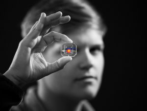
Michael Mitchell, Skirkanich Assistant Professor of Innovation in the Department of Bioengineering at the University of Pennsylvania, has received a Young Investigator Award from the Chinese Association for Biomaterials.
According to the Chinese Association for Biomaterials, “The CAB Young/Mid-Career Investigator Awards recognize the individuals who have successfully demonstrated significant achievements in the field of biomaterials research.”
The Chinese Association for Biomaterials was founded in 2015 at the Society for Biomaterials Annual Meeting. It is a non-profit professional organization that aims to facilitate exchange of research ideas and to promote collaboration among scientists in the fields of biomaterials research.
Mitchell joined the Department of Bioengineering at Penn in 2018 as Skirkanich Assistant Professor of Innovation. Previously, he was an NIH Ruth L. Kirschstein Postdoctoral Fellow with Institute Professor Robert Langer at the Koch Institute for Integrative Cancer Research at MIT. His research interests include biomaterials, drug delivery, and cellular and molecular bioengineering for applications in cancer research, immunotherapy, and gene therapy. Since joining Penn in 2018, Mitchell has received the NIH Director’s New Innovator Award, the Burroughs Wellcome Fund Career Award at the Scientific Interface, a Rising Star Award from the Biomedical Engineering Society, and the T. Nagai Award from the Controlled Release Society.
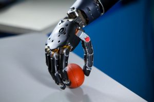
After nearly fifteen years of work, a new high-tech prosthetic arm from researchers at the University of Utah allows hand amputees to pluck grapes, pick up eggs without breaking them, and even put on their wedding rings. Named after Luke Skywalker’s robotic hand in the Star Wars saga, the LUKE Arm includes sensors that better mimic the way the human body sends information to the brain, allowing users to distinguish between soft and hard surfaces and to perform more complicated tasks. The arm relies heavily on an electrode array invented by University of Utah biomedical engineering professor Richard A. Normann, Ph.D., which is a bundle of microelectrodes that enable a computer to read signals from connected nerves in the user’s forearm.
But the biggest innovation in the use of these electrode arrays for the LUKE Arm is in the way they allow the prosthetic to mimic the sense of feeling on the surface of an object that indicates how much pressure should be applied when handling it. Gregory Clark, Ph.D., an associate professor of biomedical engineering at the University of Utah and the leader of the LUKE Arm project, says the key to improving these functions in the prosthetic was by more closely mimicking the path that this information takes to the brain, as opposed to merely what comprises that sensory information. In the future, Clark hopes to improve upon the LUKE Arm by including more inputs, like one for temperature data, and on making them more portable by eliminating the device’s need for computer connection.
Philly Voice Recognizes the Cremins Lab’s Innovations in Light-Activated Gene-Folding
While technological advancements over the past few decades have opened doors to understanding the topological structures of DNA, we still have far more to learn about how these structures impact and contribute to genome function. But here at Penn, the Cremins Laboratory in 3D Epigenomes and Systems Neurobiology hopes to fix that. Led by Jennifer E. Phillips-Cremins, Ph.D., members of the lab use light-activated dynamic looping (LADL) to better understand the way that genome topological properties and folding can affect protein translation. Cremins and her lab use this technique to force specific genome folds to interact with each other, and create temporary DNA loops that can then be bound together in the presence of blue light for certain proteins in the Arabidopsis plant. Using the data from these tests, researchers can better understand the genome structure-function relationships, and hopefully open the door to new treatments for diseases in which expression or mis-expression of certain genes is the cause.
Artificial Cells Can Deliver Molecules Better than the Real Thing
From pills to vaccines, ways to deliver drugs into the body have been constantly evolving since the early days of medicine.
Now, a new study from an interdisciplinary team led by researchers at the University of Pennsylvania provides a new platform for how drugs could be delivered to their targets in the future. Their work was published in the Proceedings of the National Academy of Sciences.
The research focuses on a dendrimersome, a compartment with a lamellar structure and size that mimic a living cell. It can be thought of as the shipping box of the cellular world that carries an assortment of molecules as cargo.
The scientists found that these dendrimersomes, which have a multilayered, onion-like structure, were able to “carry” high concentrations of molecules that don’t like water, which is common in pharmaceutical drugs. They were also able to carry these molecules more efficiently than other commercially available vessels. Additionally, the building block of the cell-like compartment, a janus dendrimer, is classified as an amphiphile, meaning it contains molecules that don’t like water and also molecules that are soluble in water, like lipids, that make up natural membranes.
“This is a different amphiphile that makes really cool self-assembled onions into which we were able to load a bunch of molecular cargos,” says co-author Matthew Good.
In a recent review of over 5,000 sleep studies, biomedical engineering researchers at the University of Texas at Austin found a connection between water-based passive body heating and sleep onset latency, efficiency, and quality. Using meta-analytical tools to compare all of the studies and patient data, lead author and Ph.D. candidate Shahab Haghayegh and his team found that a warm bath in the temperature range of 104-109 degrees Fahrenheit taken 1-2 hours before bed has the ability to improve all three considered sleep categories. This makes sense considering that our body’s Circadian rhythms govern both our sleep cycles and temperature, bringing us to a higher temperature during the day and a lower one at night during sleep. In fact, this lowering of body temperature before sleep is what helps to trigger the onset of sleep, so taking a warm bath and allowing your body to cool down from it before going to sleep enhances the body’s own efforts of naturally cooling down before we go to bed. With this new and comprehensive review, those who suffer from poor sleep quality may soon find solace in temperature regulation therapy systems.
People & Places
With the recent 50th anniversary of the first moon landing by Americans Neil Armstrong, Buzz Aldrin, and Michael Collins in 1969, ABC News looked back at one of the women involved in the project. Judy Sullivan was a biomedical engineer at the time of the project, and served as the lead engineer of the biomedical system for Apollo 11. In this role, she led studies on the astronauts’ breathing rates and sensor capabilities for the devices being sent into space to help the astronauts monitor their health. For the Apollo 11 mission and a lot of Sullivan’s early work at NASA, she worked on teams of all men, as women were often encouraged to become teachers, secretaries, or homemakers over other professions. Today, Sullivan says she’s thrilled that women have more career options to choose from, and wants to continue seeing more women getting involved in math and science.
We would like to congratulate Sanjay Kumar, M.D., Ph.D., on his appointment as the new Department Chair of Bioengineering at the University of California, Berkeley. Since joining the faculty in 2005, Kumar has received several prestigious awards including the NSF Career Award, the NIH Director’s New Innovator Award, the Presidential Early Career Award for Scientists and Engineers, and the Berkeley student-voted Outstanding Teacher Award.
We would like to congratulate Paul Ducheyne, Ph.D., a Professor in the Bioengineering Department and a Professor of Orthopaedic Surgery Research at Penn, on being selected for the International Award by the European Society for Biomaterials (ESB). The International Award is one of the ESB’s highest honors, recognizing scientists who have spent the majority of their careers outside of Europe. They are internationally recognized, have a high scientific profile, and have made major contributions to the field of biomaterials. Those nominated for the award typically also have had strong collaborations with the scientific community in Europe throughout their careers.
Beyond being a professor at Penn, Ducheyne is also the founder of XeroThera, a spin-out from Penn that develops novel concepts for tissue engineering and drug delivery based on his group’s twenty years of fundamental studies of sol gel-processed, nanoporous, oxide-based materials. XeroThera’s first product formulations focus on prophylaxis and treatment of surgical infections. A pipeline is being developed building from his group’s breakthrough data that demonstrate the utility of sol-gel synthesized silica-based nanoporous materials for therapeutic use. These materials may well represent a next generation of agents for delivery of drugs, including antibiotics, analgesics, and osteogenic and anti-inflammatory molecules.
In being selected for the International Award, Ducheyne joins only five previous recipients of it so far, a group of scientists that represents those at the top of the field in biomaterials worldwide. Ducheyne will give a presentation and award lecture for the ESB at its next annual meeting this September in Dresden, Germany. Read more about the ESB’s awards here and see the full list of 2019 awardees here.
The Department of Bioengineering at the University of Pennsylvania is excited to welcome César de la Fuente, Ph.D., as an Assistant Professor of Bioengineering. De la Fuente, who is also an Assistant Professor in the Department of Psychiatry and Microbiology in the Perelman School of Medicine and was recently named a Penn Presidential Professor, is the principal investigator of the de la Fuente Lab with current projects including the development of computer-made antibiotics, microbiome engineering technologies, and synthetic neuromicrobiology tools.
Dr. de la Fuente has wanted to learn the mysteries of the world around him from a young age, from the origins of life and human consciousness to how diseases can affect the body. His dream of understanding the building blocks of life began to take shape when he enrolled as a graduate student at the University of British Columbia to study microbiology, immunology, and protein engineering. After earning his Ph.D. in these subjects, de la Fuente went on to complete a post doctorate in synthetic biology and computer science at the Massachusetts Institute of Technology (MIT).
Recently, MIT recognized Dr. de la Fuente on its “35 Innovators Under 35” list, which honored de la Fuente as one of the world’s top 35 innovators and as a pioneer for his use of technology to improve antibiotics. Furthermore, GEN recently listed Dr. de la Fuente on its “Top 10 Under 40” list of young leaders in the life sciences, noting his development of transformative biotechnologies as a potential solution to antibiotic resistance. De la Fuente refers to this latest research as “Machine Biology,” a crossover of life and technology that “brings together elements of machines in order to computerize biological systems.”
His creativity in the merging of so many domains of science echoes throughout de la Fuente’s general approach to research and academia as well. While he emphasizes a thinking-from-the-ground-up approach, he also feels that “heterogeneous groups make better ideas,” and thus strives to maintain diversity in his lab — currently his entire lab is made up of international students and postdocs. In the future, de la Fuente hopes to extend his love of mentorship to the classroom in a course exploring the intersection of microbiology and synthetic biology, overlapping in a way similar to his research. We can’t wait for all of the innovation and creativity in engineering that de la Fuente will undoubtedly bring to our department.
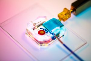
Rachel Young, a graduate student in Huh’s lab, holds up the new eye-on-a-chip device. The latest iteration of the lab’s eye-on-a-chip has a mechanical eyelid to simulate blinking, and was used to test an experimental drug for dry eye disease. By incorporating human cells into an engineered scaffolding, the eye-on-a-chip has many of the benefits of testing on living subjects, while minimizing risks and ethical concerns.
People who spend eight or more hours a day staring at a computer screen may notice their eyes becoming tired or dry, and, if those conditions are severe enough, they may eventually develop dry eye disease (DED). DED is a common disease with shockingly few FDA-approved drug options, partially because of the difficulties of modeling the complex pathophysiology in human eyes. Enter the blinking eye-on-a-chip: an artificial human eye replica constructed in the laboratory of Penn Engineering researchers.
This eye-on-a-chip, complete with a blinking eyelid, is helping scientists and drug developers to improve their understanding and treatment of DED, among other potential uses. The research, published in Nature Medicine, outlines the accuracy of the eye-on-a-chip as an organ stand-in and demonstrates its utility as a drug testing platform.
They collaborated with Vivian Lee, Vatinee Bunya and Mina Massaro-Giordano from the Department of Ophthalmology in Penn’s Perelman School of Medicine, as well as with Vivek Shenoy, Eduardo D. Glandt President’s Distinguished Professor in Penn Engineering’s Department of Materials Science and Engineering. Other collaborators included Woo Byun, Andrei Georgescu and Yoon-suk Yi, members of Huh’s lab, and Farid Alisafaei, a member of Shenoy’s lab.
Huh’s lab specializes in creating organs-on-a-chip that provide microengineered in vitro platforms to mimic their in vivo counterparts, including lung and bone marrow proxies launched into space this May to study astronaut illness. The lab has spent years fine-tuning its eye-on-a-chip, which earned them the 2018 Lush Prize for its promise in animal-free testing of drugs, chemicals, and cosmetics.
In this study, Huh and Seo focused on engineering an eye model that could imitate a healthy eye and an eye with DED, allowing them to test an experimental drug without risk of human harm.
The Huh lab’s eye-on-a-chip attached to a motorized, gelatin-based eyelid. Blinking spreads tears over the corneal surface, and so was a critical aspect to replicate in the researchers’ model of dry eye disease. cells. The cells of the cornea grow on the inner circle of scaffolding, dyed yellow, and the cells of the conjunctiva grow on the surrounding red circle. Artificial tears are supplied by a tear duct, dyed blue.
To construct their eye-on-a-chip, Huh’s team starts with a porous scaffold engineered with 3D printing, about the size of a dime and the shape of a contact lens, on which they grow human eye cells. The cells of the cornea grow on the inner circle of scaffolding, dyed yellow, and the cells of the conjunctiva, the specialized tissue covering the white part of human eyes, grow on the surrounding red circle. A slab of gelatin acts as the eyelid, mechanically sliding over the eye at the same rate as human blinking. Fed by a tear duct, dyed blue, the eyelid spreads artificial tear secretions over the eye to form what is called a tear film.
“From an engineering standpoint, we found it interesting to think about the possibility of mimicking the dynamic environment of a blinking human eye. Blinking serves to spread tears and generate a thin film that keeps the ocular surface hydrated. It also helps form a smooth refractive surface for light transmission. This was a key feature of the ocular surface that we wanted to recapitulate in our device,” says Huh.
For people with DED, that tear film evaporates faster than it’s replenished, resulting in inflammation and irritation. A common cause of DED is the reduced blinking that occurs during excessive computer usage, but people can develop the disease for other reasons as well. DED affects about 14 percent of the world’s population but has been notably difficult to develop new treatments for, with 200 failed clinical drug trials since 2010 and only two currently available FDA-approved drugs for treatment.
Huh’s lab has been considering the drug-testing potential of organs-on-a-chip since their initial conceptualization, and, because of its surface-level area of impact, DED seemed the perfect place to start putting their eye model to the test. But before they started a drug trial, the team had to ensure their model could really imitate the conditions of DED.
“Initially, we thought modeling DED would be as simple as just keeping the culture environment dry. But as it turns out, it’s an incredibly complex multifactorial disease with a variety of sub-types,” Huh says. “Regardless of type, however, there are two core mechanisms that underlie the development and progression of DED. First, as water evaporates from the tear film, salt concentration increases dramatically, resulting in hyperosmolarity of tears. And second, with increased tear evaporation, the tear film becomes thinner more rapidly and often ruptures prematurely, which is referred to as tear film instability. The question was: Is our model capable of modeling these core mechanisms of dry eye?”
The answer, after much experimentation, was yes. The team evoked DED conditions in their eye-on-a-chip by cutting their device’s artificial blinking in half and carefully creating an enclosed environment that simulated the humidity of real-life conditions. When put to the test against real human eyes, both healthy and with DED, the corresponding eye-on-a-chip models proved their similarity to the actual organ on multiple clinical measures. The eyes-on-a-chip mimicked actual eyes’ performance in a Schirmer strip, which tests liquid production; in an osmolarity test, which looks at tear film salt content; and in a keratography test, which evaluates the time it takes for a tear film to break up.
Having confirmed their eye-on-a-chip’s ability to mirror the performance of a human eye in normal and DED-inducing settings, Huh’s team turned to the pharmaceutical industry to find a promising DED drug candidate to test-drive their model. They landed on an upcoming drug based on lubricin, a protein primarily found in the lubricating fluid that protects joints.
“When people think of DED, they normally treat it as a chronic disease driven by inflammation,” says Huh, “but there’s now increasing evidence suggesting that mechanical forces are important for understanding the pathophysiology of DED. As the tear film becomes thinner and more unstable, friction between the eyelids and the ocular surface increases, and this can damage the epithelial surface and also trigger adverse biological responses such as inflammation. Based on these observations, there is emerging interest in developing ophthalmic lubricants as a topical treatment for dry eye. In our study, we used an lubricin-based drug that is currently undergoing clinical trials. When we tested this drug in our device, we were able to demonstrate its friction-lowering effects, but, more importantly, using this model we discovered its previously unknown capacity to suppress inflammation of the ocular surface.”
By comparing the testing results of their models of a healthy eye, an eye with DED, and an eye with DED plus lubricin, Huh and Seo were able to further scientists’ understanding of how lubricin works and show the drug’s promise as a DED treatment.
Similarly, the process of building a blinking eye-on-a-chip pushed forward scientists’ understanding of the eye itself, providing insights into the role of mechanics in biology. Collaborating with Shenoy, director of the Center for Engineering MechanoBiology, the team’s attention was drawn to how the physical blinking action was affecting the cells they cultivated to engineer an artificial eye on top of their scaffolding.
“Initially, the corneal cells start off as a single layer, but they become stratified and form multiple layers as a result of differentiation, which happens when these cells are cultured at the air-liquid interface. They also form tight cell-cell junctions and express a set of markers during differentiation,” Huh says. “Interestingly, we found out that mechanical forces due to blinking actually help the cells differentiate more rapidly and more efficiently. When the corneal cells were cultured under air in the presence of blinking, the rate and extent of differentiation increased significantly in comparison to static models without blinking. Based on this result, we speculate that blink-induced physiological forces may contribute to differentiation and maintenance of the cornea.”
In other words, human cornea cells growing on the scientists’ scaffold more quickly became specialized and efficient at their particular jobs when the artificial eyelid was blinking on top of them, suggesting that mechanical forces like blinking contribute significantly to how cells function. These types of conceptual advances, coupled with drug discovery applications, highlight the multifaceted value that engineered organs-on-a-chip can contribute to science.
Huh and Seo’s eye-on-a-chip is still just dipping its toes into the field of drug testing, but this first step is a victory that represents years of work refining their artificial eye to reach this level of accuracy and utility.
“Although we have just demonstrated proof-of-concept,” says Seo, “I hope our eye-on-a-chip platform is further advanced and used for a variety of applications besides drug screening, such as testing of contact lenses and eye surgeries in the future.”
“We are particularly proud of the fact that our work offers a great and rare example of interdisciplinary efforts encompassing a broad spectrum of research activities from design and fabrication of novel bioengineering systems to in vitro modeling of complex human disease to drug testing,” says Huh. “I think this is what makes our study unique and representative of innovation that can be brought about by organ-on-a-chip technology.”
This work was supported by the National Institutes of Health through grants 1DP2HL127720–0, R01EY026972 and K08EY025742–01, the National Science Foundation through grants CMMI:15–48571, and Research to Prevent Blindness.
Brian Chow, David Issadore, Dongeun (Dan) Huh, Linh Thi Xuan Phan, Amish Patel and Aleksandra Vojvodic
The School of Engineering and Applied Science has granted tenure to six faculty members, including three from the Department of Bioengineering.
Tenured faculty at Penn Engineering demonstrate teaching excellence and international leadership in their fields of study and research collaborations.
Brian Chow
Associate Professor in Bioengineering Chow’s research focuses on the discovery and engineering of photoreceptors and sensory proteins for manipulating and monitoring the physiology of genetically targeted cells, and the application of these tools to reveal principles of cellular dynamics. His work has advanced the rational design of light activated proteins and the use of optogenetic reagents to study cell signaling.
David Issadore
Associate Professor in Bioengineering Issadore’s research combines microelectronics, microfluidics, and nanomaterials to create miniaturized platforms for the diagnosis of disease. His work has the potential to radically change the way we diagnose and treat diseases by bringing the technologies out of the lab and directly to the point of care.
Dongeun (Dan) Huh
Associate Professor in Bioengineering Huh’s research aims to develop innovative bioengineering tools and technologies using biologically inspired design principles and micro- and nano-scale engineering techniques to create systems that mimic the structure and function of human physiological systems.
Linh Thi Xuan Phan
Associate Professor in Computer and Information Science Phan’s work focuses on making cyber-physical systems (CPS) safer, faster, and more secure, both by strengthening the theoretical foundations and by developing practical solutions. Her recent projects include a cloud platform with real-time capabilities, a new diagnosis technique for timing-related faults, and new ways to defend CPS against attacks from insiders and/or external attackers.
Amish Patel
Associate Professor in Chemical and Biomolecular Engineering Patel’s research strives to achieve a molecular-level understanding of solvation and transport in aqueous and polymeric systems, with applications ranging from the prediction of protein interactions to the design of advanced materials for water purification and energy storage. His group combines principles of statistical mechanics and liquid state theory with state-of-the-art molecular modeling and atomistic simulation techniques to study these biological, nanoscopic and polymeric systems.
Aleksandra Vojvodic
Associate Professor in Chemical and Biomolecular Engineering Vojvodic’s research focuses on theory and computation-driven materials design. Her lab uses computational frameworks to obtain fundamental understanding of surface and interface properties of complex materials that can be used to develop theoretical models for chemical transformations and energy conversion. These models have been used to predict new catalyst materials for several chemical reactions which have been experimentally synthesized and tested, validating the desired properties of the computationally predicted catalyst material.
We would like to congratulate Penn Bioengineering faculty members Brian Chow, Ph.D., Dongeun (Dan) Huh, Ph.D., and David Issadore, Ph.D., on all of their recent promotions to tenured positions as Associate Professors. Both Chow and Issadore taught the second half of the foundational course in the Penn Bioengineering undergraduate curriculum, Bioengineering Modeling, Analysis, and Design Laboratory, in which students form lab groups to complete modules in microfluidics, synthetic biology, bioelectrical signal analysis, and bioanalytical spectroscopy.
Brian Chow, Ph.D.
Outside of the classroom, Chow’s research focuses on the creation of dynamic input and output interfaces for cells through the use of optogenetics, synthetic biology, genomics, and device engineering. The Chow lab’s current projects include the exploration of functional diversity of photoreception, engineering optically active genetically encoded tools, and their applications in neuroscience and mammalian synthetic biology. His research is supported by the NIH and he is the recipient of a 2017 NSF CAREER Award. Chow also supports undergraduate innovations in research by hosting the annual Penn team for the International Genetically Engineered Machine (iGEM) competition, a program which he helped to create during his time as a graduate student at MIT. One group of Bioengineering students under Chow’s mentorship used the iGEM project as a springboard to create an accessible, open-source plate reader.
David Issadore, Ph.D.
The Issadore lab at Penn focuses on the use of microelectronics and microfluidics for medical diagnostics. In projects that combine elements of bioengineering, electrical engineering, chemical engineering, and applied physics, Issadore and his team use an interdisciplinary approach to create miniaturized low-cost platforms for disease diagnosis. His company Chip Diagnostics received the JPOD @ Philadelphia QuickFire Challenge Award last month. Earlier this year, Issadore taught the Penn Engineering course Appropriate Point of Care Diagnostics (APOC), which culminated in a service trip to Ghana (read blog posts written by participating students here). This fall, he will take over the core Bioengineering undergraduate course in Bioengineering Signals and Systems, which focuses on applications in ECG signaling, cochlear implants, and biomedical imaging.
Dan Huh, Ph.D.
Dr. Huh is the principal investigator of the BIOLines Lab at Penn, which is best known for its work on bioinspired engineering systems that Huh calls “organs-on-a-chip.” Using design and engineering principles based on microfluidics and biomimicry, the Huh lab creates microengineered systems that can reconstitute the structural and functional complexity of healthy and diseased human physiological systems in ways not possible using traditional cell culture techniques. His research has been featured in TEDx, and he has won several prestigious honors and awards including the Bernard Langer Distinguished Lectureship, Lush Prize, the McPherson Distinguished Lectureship, CRI Technology Impact Award, John J. Ryan Medal, Design of the Year Award and Best Product of the Year Award from London Design Museum, NIH Director’s New Innovator Award, and Analytical Chemistry Young Innovator Award. This fall, Huh will teach a graduate level course in biomicrofluidics that will cover the use of microfluidics for biomedical application.
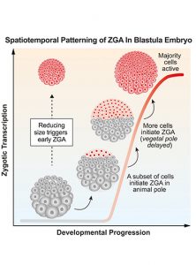
Awakening of the zygote genome over time as decreasing individual cell size triggers early embryo transcription. (Image: Hui Chen, Penn Medicine; Cell Press)
There is a transition during early development when an embryo undergoes biochemical changes, switching from being controlled by maternal molecules to being governed by its own genome.
For the first time, a team from the Perelman School of Medicine found in an embryo that activation of its genome does not happen all at once, instead it follows a specific pattern controlled primarily by the various sizes of its cells. The researchers published their results as the cover story in Developmental Cell.
In an early embryo undergoing cell division, maternally loaded RNA and proteins regulate the cell cycle. The genomes of the zygote—a term for the fertilized egg—are initially in sleep mode. However, at a point in the early life of the embryo, these zygotic nuclei “wake up” and expression from their genomes takes biochemical control over subsequent embryo development. But how an embryo “recognizes” when to undergo this transition has remained unknown.
“How an embryo ‘hands over’ control of development from mother to zygote is a fundamental question in developmental biology,” says senior author Matthew C. Good, an assistant professor of both cell and developmental biology and bioengineering. “Previously it was not appreciated that different regions of a vertebrate embryo can undergo genome activation at different times, or how directly cell size regulates the awakening of a zygote’s genome.”