A recent study published in Science Translational Medicine announces a discovery which could halt cartilage degeneration caused by osteoarthritis: “These researchers showed that they could target a specific protein pathway in mice, put it into overdrive and halt cartilage degeneration over time. Building on that finding, they were able to show that treating mice with surgery-induced knee cartilage degeneration through the same pathway via the state of the art of nanomedicine could dramatically reduce the cartilage degeneration and knee pain.” This development could eventually lead to treating osteoarthritis with injection rather than more complicated surgery.
Among a team of Penn Engineering and Penn Medicine researchers, the study was co-written by Zhiliang Cheng, Research Associate Professor in Bioengineering, Andrew Tsourkas, Professor in Bioengineering, and Ling Qin, Associate Professor of Orthopaedic Surgery in the Perelman School of Medicine and member of the Bioengineering Graduate Group. The lead author was Yulong Wei of the Department of Orthopaedic Surgery and the McKay Orthopaedic Research Laboratory.
(Left to right) Top row: Jennifer E. Phillips-Cremins, Rajan Jain, and Eric Joyce. Middle row: Melike Lakadamyali, Golnaz Vahedi, and Gerd Blobel. Bottom row: Bomyi Lim, Arjun Raj, and Stanley Qi.
Popular accounts of the human genome often depict it as a long string of DNA base pairs, but in reality the genome is separated into chromosomes that are tightly twisted and coiled into complex three-dimensional structures. These structures create a myriad of connections between sites on the genome that would be distant from one another if stretched out end-to-end. These “long range interactions” are not incidental — they regulate the activity of our genes during development and can cause disease when disrupted.
Now two teams of researchers at the Perelman School of Medicine at the University of Pennsylvania, each led by Jennifer E. Phillips-Cremins, associate professor and Dean’s Faculty Fellow in the Department of Bioengineering at the School of Engineering and Applied Science and of Genetics at the Perelman School of Medicine have been awarded grants totaling $9 million from the National Institutes of Health (NIH), as part of a major NIH Common Fund initiative to understand such 3D-genomic interactions.
The initiative, known as the 4D Nucleome Program, broadly aims to map higher-order genome structures across space and time, as well as to understand how the twists and loops of the DNA sequence govern genome function and cellular phenotype in health and disease.
N.B.: In addition to Phillips-Cremins, collaborators include Arjun Raj, Professor in Bioengineering and Genetics, and Bioengineering Graduate Group Members Melike Lakadamyali, Associate Professor in Physiology, and Bomyi Lim, Assistant Professor in Chemical and Biomolecular Engineering.
Since the country began shutting down in March, I have joined the majority of the world in calling the times “unprecedented”: The word, which I rarely used before the pandemic, is now a staple of my lockdown lexicon. In March, we all got the email that changed the trajectory of the rest of our semester and the school year. Since then, COVID-19 has been impacting lives here at Penn, around the nation, and the world. Hanging out with friends and family on Zoom, managing work and school from home, social distancing, wearing masks everywhere, and constantly washing hands have been the reality of our new normal for months.
It has been almost ten months since the World Health Organization declared COVID-19 a pandemic and this has posed a global crisis like nothing most of us have experienced in our lifetime. At Penn, the campus community including students and staff have rallied to keep each other safe, all while doing what is possible to ensure that lectures, teaching, and research are possible in ways that uphold the university’s mission of “strengthening the quality of education and producing innovative research and models of healthcare delivery by fostering a vibrant inclusive environment and fully embracing diversity.”
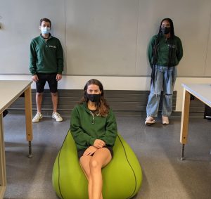
BE students Alexa Rybicki, Ifeoluwa Popoola, and Caitlin Frazee meet for BE 309 in the Gather.Town virtual lab space.
In Penn Engineering’s Bioengineering Department, the Stephenson Foundation Educational Laboratory & Bio-MakerSpace has been at the heart of ensuring that lab-based classes run as smoothly as possible given the circumstances. First off, during the summer, the lab launched a Slack site that not only kept students engaged and connected through fun, daily “Questions of the Day” but also gave them the opportunity to reach out to our staff and obtain their expertise for coursework and personal projects. The staff at the Stephenson Lab also supported and continue to support Senior Design students (BE 495) with their projects by ordering, receiving, packaging, arranging pickups, or mailing supplies needed to complete their Senior Design projects. In addition, class time takes place using Gather.Town to recreate our Bio-MakerSpace virtually. In other classes, video tutorials of some of the experiments students were missing out on were produced over the summer and made available to students so they could learn by seeing what the lab staff were doing in the videos. For the Bioengineering Modeling, Analysis, and Design (BE MAD) class (BE 309), in addition to videos, our lab Engineer, Michael Patterson, developed software through which students can enter design criteria and have experimental data emailed to them.
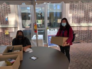
Picking up lab supplies outside in the Engineering complex
The staff at the lab also supported a Rehabilitation Engineering course (BE 514) taught by Michelle Johnson, Associate Professor in Physical Medicine and Rehabilitation and Bioengineering, by putting together supplies that enabled students in the class to reengineer toy bunny rabbits to be more accessible to children with disabilities. Optical Microscopy (BE 518), another Bioengineering course, taught by Christopher Fang-Yen, Associate Professor in Bioengineering and Neuroscience, offers students an introduction to the fundamental concepts of optics and microscopy. The staff at the lab put together kits and made them available for pickup by the students in the class.
In a time when the shape of education looks vastly different from what we anticipated this year, the Bio-MakerSpace has been instrumental in ensuring that students still have access to resources that make their learning experience an enriching one. In these unprecedented times, the lab has been able to encourage students to keep up and be engaged with their coursework while also fostering creativity in students, virtually and remotely. While we may not know what life after the pandemic will look like, one thing to be sure of is that the Stephenson Lab will always be a reliable place for Penn students to get support for personal projects and coursework when needed.
Solumtochukwu (Somto) Egbogais a Master’s Student in Bioengineering, graduating December 2020. She also is a student employee for the Stephenson Foundation Bioengineering Laboratory & Bio-MakerSpace.
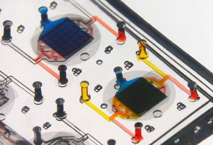
Huh’s organ-on-a-chip devices contain human cells, allowing for experiments that could not otherwise be practically or ethically performed.
Chlorine gas is a commonly used industrial chemical. It is also highly toxic and potentially deadly; it was used as a chemical weapon in both World War I and the Syrian Civil War and has led to multiple deaths from industrial accidents. Mixing certain household cleaners can also produce the toxic gas, leading to lasting lung injuries for which there are currently no effective treatments.
Now, researchers at Penn Engineering and Penn’s Perelman School of Medicine are collaborating with BARDA, the U.S. Office of Health and Human Services’ Biomedical Advanced Research and Development Authority, to address this need using their lung-on-a-chip technology.
The laboratory of Dan Huh, associate professor in the Department of Bioengineering, has developed a series of organ-on-a-chip platforms. These devices incorporate human cells into precisely engineered microfluidic channels that mimic an organ’s natural environment, providing a way to conduct experiments that would not otherwise be feasible.
Dan Huh, PhD
Huh’s previous research has involved using a placenta-on-a-chip to study which drugs are able to reach a developing fetus; investigating microgravity’s effect on the immune system by sending one of his chips to the International Space Station; and testing treatments for dry eye disease using an eye-on-a-chip, complete with a mechanical blinking eyelid.
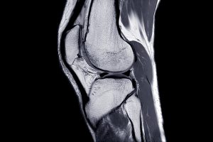
MRI Knee joint or Magnetic resonance imaging sagittal view for detect tear or sprain of the anterior cruciate ligament (ACL).
Using a magnetic field and hydrogels, a team of researchers in the Perelman School of Medicine have demonstrated a new possible way to rebuild complex body tissues, which could result in more lasting fixes to common injuries, such as cartilage degeneration. This research was published in Advanced Materials.
“We found that we were able to arrange objects, such as cells, in ways that could generate new, complex tissues without having to alter the cells themselves,” says the study’s first author, Hannah Zlotnick, a graduate student in bioengineering who works in the McKay Orthopaedic Research Laboratory at Penn Medicine. “Others have had to add magnetic particles to the cells so that they respond to a magnetic field, but that approach can have unwanted long-term effects on cell health. Instead, we manipulated the magnetic character of the environment surrounding the cells, allowing us to arrange the objects with magnets.”
In humans, tissues like cartilage can often break down, causing joint instability or pain. Often, the breakdown isn’t in total, but covers an area, forming a hole. Current fixes are to fill those holes in with synthetic or biologic materials, which can work but often wear away because they are not the same exact material as what was there before. It’s similar to fixing a pothole in a road by filling it with gravel and making a tar patch: The hole will be smoothed out but eventually wear away with use because it’s not the same material and can’t bond the same way.
What complicates fixing cartilage or other similar tissues is that their makeup is complex.
“There is a natural gradient from the top of cartilage to the bottom, where it contacts the bone,” Zlotnick explains. “Superficially, or at the surface, cartilage has a high cellularity, meaning there is a higher number of cells. But where cartilage attaches to the bone, deeper inside, its cellularity is low.”
So the researchers, which included senior author Robert Mauck, PhD, director of the McKay Lab and a professor of Orthopaedic Surgery and Bioengineering, sought to find a way to fix the potholes by repaving them instead of filling them in. With that in mind, the research team found that if they added a magnetic liquid to a three-dimensional hydrogel solution, cells, and other non-magnetic objects including drug delivery microcapsules, could be arranged into specific patterns that mimicked natural tissue through the use of an external magnetic field.
Beth Winkelstein, Megan Sperry, and Eric Granquist
Pain may be a universal experience, but what actually causes that experience within our brains is still poorly understood. Pain often continues long after the relevant receptors in the body have stopped being stimulated and can persist even after those receptors cease to exist, as is the case with “phantom limb” pain.
The exact experience an individual will have after a painful incident comes down to the complex, variable connections formed between several different parts of the brain. The inability to predict how those connections will form and evolve can make pain management a tricky, frustrating endeavor for both healthcare providers and patients.
Now, a team of Penn researchers has shown a way to make such predictions from the pattern of neural connections that begin to take shape soon after the first onset of pain. Though their study was conducted in rats, it suggests that similar brain imaging techniques could be used to guide treatment decisions in humans, such as which individuals are most likely to benefit from different drugs or therapies.
The study, published in the journal Pain, was led by Beth Winkelstein, Eduardo D. Glandt President’s Distinguished Professor in Penn Engineering’s Department of Bioengineering and Deputy Provost of the University of Pennsylvania, along with Megan Sperry, then a graduate student in her lab. Eric Granquist, Director of the Center for Temporomandibular Joint Disease at the Hospital of the University of Pennsylvania in the Department of Oral & Maxillofacial Surgery, and assistant professor of Oral & Maxillofacial Surgery in Penn’s School of Dental Medicine, also contributed to the research.
“Our findings provide the first evidence that brain networks differ between acute and persistent pain states, even before those different groups of rats actually show different pain symptoms,” says Winkelstein.
César de la Fuente a Presidential Assistant Professor in the Perelman School of Medicine’s departments of Psychiatry and Microbiology and Engineering’s department of Bioengineering, has racked up accolades for his innovative, computational approach to discovering new antibiotics.
Now, in his most recent study, de la Fuente has shown how these vital drugs might be derived from wasp venom.
The study, published in The Proceedings of the National Academy of Sciences, involved altering a highly toxic small protein from a common Asian wasp species, Vespula lewisii, the Korean yellow-jacket wasp. The alterations enhanced the molecule’s ability to kill bacterial cells while greatly reducing its ability to harm human cells. In animal models, de la Fuente and his colleagues showed that this family of new antimicrobial molecules made with these alterations could protect mice from otherwise lethal bacterial infections.
There is an urgent need for new drug treatments for bacterial infections, as many circulating bacterial species have developed a resistance to older drugs. The U.S. Centers for Disease Control & Prevention has estimated that each year nearly three million Americans are infected with antibiotic-resistant microbes and more than 35,000 die of them. Globally the problem is even worse: Sepsis, an often-fatal inflammatory syndrome triggered by extensive bacterial infection, is thought to have accounted for about one in five deaths around the world as recently as 2017.
“New antibiotics are urgently needed to treat the ever-increasing number of drug-resistant infections, and venoms are an untapped source of novel potential drugs. We think that venom-derived molecules such as the ones we engineered in this study are going to be a valuable source of new antibiotics,” says de la Fuente.
De la Fuente and his team started with a small protein, or “peptide,” called mastoparan-L, a key ingredient in the venom of Vespula lewisii wasps. Mastoparan-L-containing venom is usually not dangerous to humans in the small doses delivered by wasp stings, but it is quite toxic. It destroys red blood cells, and triggers a type of allergic/inflammatory reaction that in susceptible individuals can lead to a fatal syndrome called anaphylaxis—in which blood pressure drops and breathing becomes difficult or impossible.
Mastoparan-L (mast-L) also is known for its moderate toxicity to bacterial species, making it a potential starting point for engineering new antibiotics. But there are still some unknowns, including how to enhance its anti-bacterial properties, and how to make it safe for humans.
Brian Litt, professor in Engineering’s Department of Bioengineering and the Perelman School of Medicine’s departments of Neurology and Neurosurgery, has received a five-year, $5.6 million Pioneer Award from the National Institutes of Health, which will support his research on implantable devices for monitoring, recording and responding to neural activity.
The Pioneer Award is part of the agency’s High-Risk, High-Reward Research Program honoring exceptionally creative scientists. It challenges investigators to pursue new research directions and develop groundbreaking, high-impact approaches to a broad area of biomedical or behavioral science. Litt’s neurodevice research represents a new frontier in addressing a wide variety of neurological conditions.
In epilepsy, for example, these devices would predict and prevent seizures; in Parkinson’s patients, implants will measure and communicate with patients to improve mobility, reduce tremor and enhance responsiveness. Other implants might improve hearing or psychiatric symptoms by querying patient perceptions, feelings, and altering stimulation patterns algorithmically to improve them
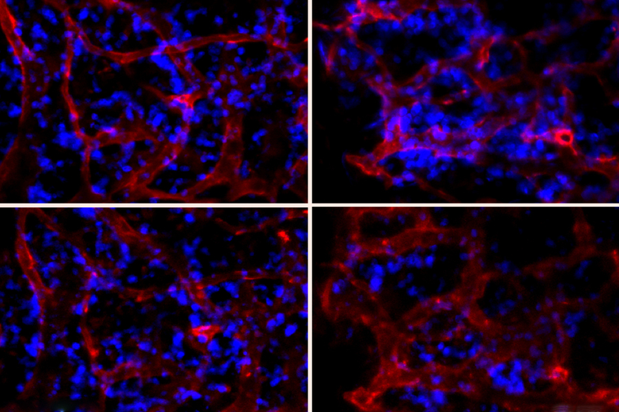
Using specialized nanoparticles, researchers from Penn Engineering and the Massachusetts Institute of Technology (MIT) have developed a way to turn off specific genes in cells of bone marrow, which play an important role in producing blood cells. These particles could be tailored to help treat heart disease or to boost the yield of stem cells in patients who need stem cell transplants.
This type of genetic therapy, known as RNA interference, is usually difficult to target to organs other than the liver, where nanoparticles would tend to accumulate. The researchers were able to modify their particles in such a way that they would accumulate in the cells found in the bone marrow.
In a recent Nature Biomedical Engineering study, conducted in mice, the researchers showed that they could use this approach to improve recovery after a heart attack by inhibiting the release of bone marrow blood cells that promote inflammation and contribute to heart disease.
“If we can get these particles to hit other organs of interest, there could be a broader range of disease applications to explore, and one that we were really interested in in this paper was the bone marrow. The bone marrow is a site for hematopoiesis of blood cells, and these give rise to a whole lineage of cells that contribute to various types of diseases,” says Michael Mitchell, Skirkanich Assistant Professor of Innovation in Penn Engineering’s Department of Bioengineering, one of the lead authors of the study.
Marvin Krohn-Grimberghe, a cardiologist at the Freiburg University Heart Center in Germany, and Maximilian Schloss, a research fellow at Massachusetts General Hospital (MGH), are also lead authors on the paper, which appears today in Nature Biomedical Engineering. The paper’s senior authors are Daniel Anderson, a professor of Chemical Engineering at MIT and a member of MIT’s Koch Institute for Integrative Cancer Research and Institute for Medical Engineering and Science, and Matthias Nahrendorf, a professor of Radiology at MGH.
Mitchell’s expertise is in the design of nanoparticles and other drug delivery vehicles, engineering them to cross biological barriers that normally block foreign agents. In 2018, he received the NIH Director’s New Innovator Award to support research on delivering therapeutics to bone marrow, a key component of this new study.
The researchers have shown they can deliver nanoparticles to the bone marrow, influencing their function with RNA silencing. At top right, the bone marrow is not yet treated with particles that turn off a gene called SDF1. At bottom right, the number of neutrophils (blue) decreases, indicating that they have been released from bone marrow after treatment. At left, treatment with a control nanoparticle does not affect the number of neutrophils before and after treatment.
Single cell sequencing aided researchers in identifying a previously undiscovered molecule in the brain.
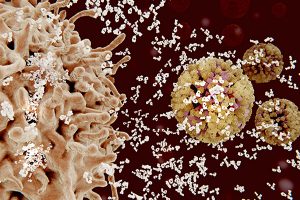
Chimeric antigen receptor (CAR) T cell therapy has revolutionized treatment of leukemia, lymphoma, and multiple myeloma. But some people who have received this treatment experience neurotoxicity, or damage to the brain or nervous system.
New research from a team led by Avery Posey, an assistant professor of systems pharmacology and translational therapeutics in the Perelman School of Medicine, provides evidence that this side effect may owe to a molecule in the brain that scientists previously didn’t know was there.
The work, published in the journal Cell, revealed that the protein CD19 is present in brain cells that protect the blood-brain barrier. Prior to the finding, scientists believed CD19 was only expressed on B cells, and the protein served as a target for certain forms of CAR-T therapy. The discovery may chart a path forward for new strategies to effectively treat cancer while sparing the brain.
“The next question is,” says Posey, “can we identify a better target for eliminating B cell related malignancies other than CD19, or can we engineer around this brain cell expression of CD19 and build a CAR T cell that makes decisions based on the type of cell it encounters—for instance, CAR T cells that kill the B cells they encounter, but spare the CD19 positive brain cells?”

 A recent study published in Science Translational Medicine announces a discovery which could halt cartilage degeneration caused by osteoarthritis: “These researchers showed that they could target a specific protein pathway in mice, put it into overdrive and halt cartilage degeneration over time. Building on that finding, they were able to show that treating mice with surgery-induced knee cartilage degeneration through the same pathway via the state of the art of nanomedicine could dramatically reduce the cartilage degeneration and knee pain.” This development could eventually lead to treating osteoarthritis with injection rather than more complicated surgery.